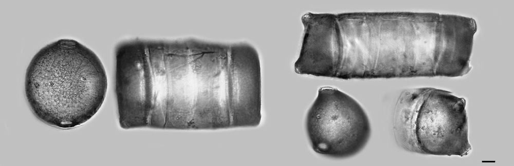
Fig. 1.
Figure 1 above is my nemesis the cylinder, valve view and girdle
view. The
currently accepted name for this diatom is Pleurosira laevis
(Ehrenberg) the synonym
Biddulphia laevis Ehrenberg is no longer accepted. The valve measures
64µm high by 61µm wide and the girdles length is
approximately 107µm. The diatom on the right may also be
Pleurosira but I am not certain. A little more research is needed.
Three views
are presented the upper is the girdle and the lower two are oblique
views of the valve. Difficult to stand this diatom on end given the
valve shape. The girdle length as pictured is approximately 128µm.
.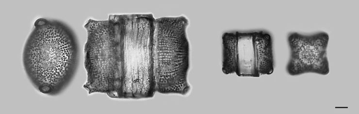
Fig. 2.
In Fig. 2 two fossil diatoms are
pictured. The diatom on the left, valve and girdle, is in older texts
Biddulphia. The
valve in long axis measures approximately 54µm and the length of
the girdle as pictured measures approximately 83µm. Biddulphia
according to algeBASE (http://www.algaebase.org/) is a "confused group
of 'biddulphioid' forms" and
states that some species "have been transferred to Odontella,
Pleurosira, Biddulphiopsis". I am inclined to identify this
diatom as Odontella but I may also be confused. The diatom on
the
right I thought when I first saw it was a pygmy Biddulphia
because unlike most observed it was small. Turning this diatom on end
was a pleasant surprise, it is Triceratium elegans (Greville) Grunow.
Its girdle measures
approximately 40µm in length and the valve is approximately
32µm.
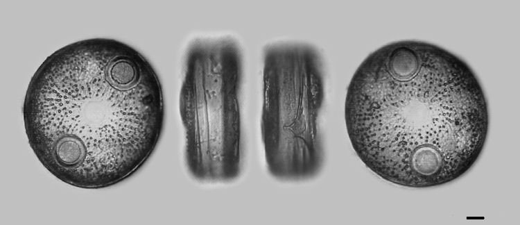
Fig. 3.
The fossil diatom in Fig. 3. is of the genus Auliscus. I have not
identified the species. The valve markings within this genus are quite
variable. This image depicts the entire diatom, valve,
girdle, girdle rotated
approximately 180 degrees, and the opposite valve. The two valves
though different are remarkably similar which is expected but nice to
observe first hand. The difference in the two
girdle views is interesting. The right girdle has unique markings
something like a pot overflowing. In A. Schmidt's Diatom Atlas, plate
31, fig. 15, Auliscus pruinosus var. sansibarica Grunow is depicted in
a stack or chain with this particular marking on each of the diatoms in
alignment. This representation suggests the marking could
have purpose? Diameter measures approximately 78µm.
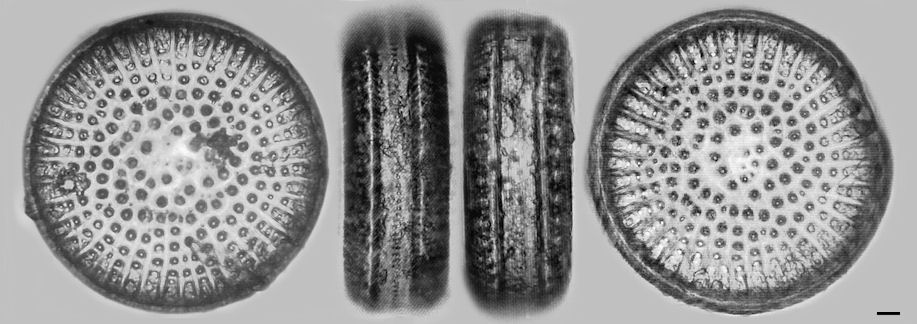
Fig. 4.
Fig. 4. is another fossil diatom genus Stictodiscus.
Pictured here is valve, two images of the girdle, one rotated
approximately 180 degrees, and the opposite valve. The alignment
markings found in Fig. 3 are not present on this diatom. The diameter
measures a healthy 116µm.
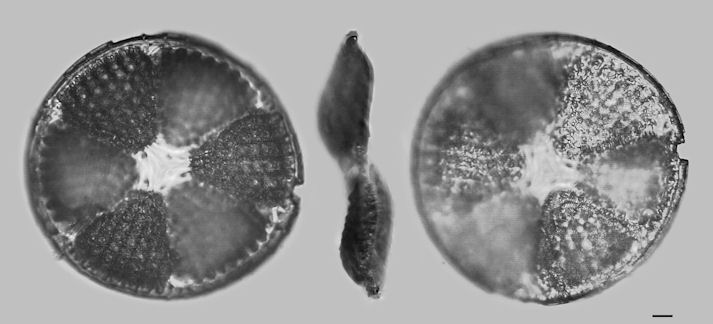
Fig. 5.
I wondered what the reverse side of a valve would look like. Fig. 5
depicts a valve high focus, side view of the valve, and high focus of
the reverse side of the same valve. This fossil diatom is Actinoptychus
bismarckii Schmidt.The genus Actinoptychus is described as being
comprised of
six or more undulating sectors or compartments, valve diameter
135µm. The
undulation is
apparent in the side view of this valve. Using the notch on the right
side of the valve as a reference it is easy to see which of the
alternating sectors are in focus.
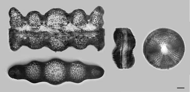
Fig. 6.
In Fig. 6 on the left is Biddulphia regina W. Smith. On top is the
girdle view and below is the valve view. On the right is Actinoptychus
senarius Ehrenberg, diameter 55µm, with the girdle view again
showing the defining undulations.
Actinoptychus is an interesting diatom found in the late Cretaceous
through recent times with an explosion of diversity in the Miocene
epoch.
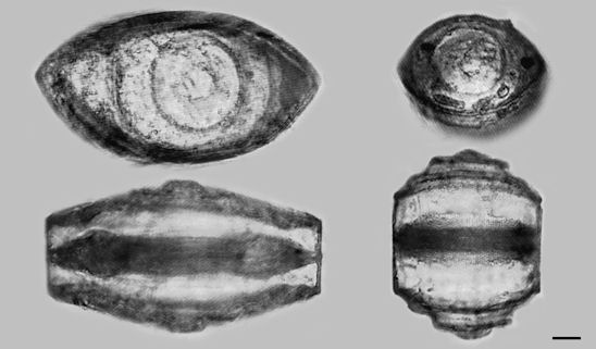
Fig. 7.
This last image, Fig. 7, is two examples of Goniothecium odontella. As
shown the width and height of the girdle can be quite varied. The
diatom on the right was a little difficult to stand for imaging the
valve, its top like shape guaranteed an oblique view. The width
of
the two valves are left 83µm and right 54µm.
The two diatoms in Fig. 1 are recent and the remaining diatoms Fig.
2 through Fig. 7 are fossil diatoms from the Miocene epoch. All the
diatoms are from Newport Beach, California. The bar in the lower right
corner is 10µm.
For instructions on sedimentation see 'sedimentation procedure' in
Frithjof A.S. Sterrenburg's article 'CLEANING
DIATOM SAMPLES'.
Comments to the
author are welcomed.
References :
Round, F.E., Crawford, R.M., Mann, D.G., "The Diatoms. Biology and
Morphology of the Genera", 1990, Cambridge University Press.
Schmidt, Adolf, "Atlas der Diatomaceenkunde", CD-ROM edition. [Editor's note: Available from Savona Books, UK.]
Van Heurck, Henri, 1885, "Synopsis des Diatomees de Belgique".
http://sdrc.lib.uiowa.edu/algae/vanheurck.
Vinyard, William C., 1979, "Diatoms of North America".
Wornardt, Walter W., 1967, "Miocene and Pliocene Diatoms from
California", Occasional Papers of the California
Academy of Sciences, No. 63. San Francisco.
Microscopy UK Front Page
Micscape Magazine
Article Library
© Microscopy UK or their contributors.
Published in the August 2010 edition of Micscape Magazine.
Please report any Web problems or offer general comments to the Micscape Editor,
via the contact on current Micscape Index.Micscape is the on-line monthly magazine of the Microscopy UK web
site at Microscopy-UK
© Onview.net Ltd, Microscopy-UK, and all contributors 1995 onwards. All rights reserved. Main site is at www.microscopy-uk.org.uk.