Notes on using the Sigma SD14 DSLR with Foveon X3 sensor on a microscope.
by David Walker, UK
The SD14 was the third digital SLR model offered by Sigma to use the Foveon X3 CMOS sensor technology. The sensor has three layers of detectors allowing each photosite to record R,G,B colour information, whereas the Bayer sensor typically uses an RGBG filter array to capture the colour information. Bayer sensors also typically have an anti-aliasing filter to reduce the Moiré effect (and potentially limiting the finest detail recorded), whereas the Foveon sensors in the Sigma models do not.
Sigma's cameras with the Foveon sensor have gained many devotees who often remark on the certain 'look' of the images with spatial depth and fine colour detail akin to film. The SD14 and latest SD15 use a 4.7 million x 3 layer detector sensor. There has been much debate on how the Foveon sensor's pixel count equates to a Bayer sensor's pixels and the relative merits of each type. The modest feature set of the 'SD' DSLR range, some performance / image quality issues and initial release pricing cf competitors' models prompted rather mixed reviews online and in the photo press. More than most cameras, probably largely because of its sensor, it seems to be a range that can polarise opinion; but it's refreshing that a maker goes against the 'multi megapixel / multi-menu' crowd in choice of sensor and camera design.
As a microscopy enthusiast, the sensor has retained a particular interest as wondered if it had merits over the Bayer type for photomicroscopy. Such imagery can have unusual aspects cf normal photographyfine detail in one colour (e.g. stained sections, fluorescence studies), the images may be of a single or very limited colour range (e.g. green for optimum imagery with achromats or phase, or using deep blue to maximise resolution of diatoms). With a Bayer sensor only one in four pixels records blue or red, green is only recorded by half the pixels; clever algorithms are required to extrapolate the missing colour information to produce the final image; unlike the Foveon sensor. The Sigma DSLRs seem to offer a consumer camera with a sensor recording the maximum amount of colour information. Some professional dedicated microscope cameras avoid using a Bayer sensor by adopting a monochrome sensor and taking successive images with R, G, B filters to combine into a colour image (or use a sensor for each colour).
My particular interest is in large on screen detailed imagery and also wondered if resizing the more modest 4.7 Mpixel Foveon images had benefits over resizing the typical 12 Mpixel image from a modern Bayer sensor.
With a long 'Nikon legacy', I've adopted Nikon digital SLRs and didn't pursue the Sigma DSLR route. However, the value of digital cameras plummets within a few years and the SD14, released in 2007, can be bought on eBay regularly for less than a quarter of its original cost. When a mint, boxed example came up on eBay, my curiosity got the better of me and decided to try one, in the knowledge that its depreciation was near complete and could be resold if it didn't suit for little loss.
I haven't to date found any articles describing use of the Sigma DSLRs for microscopy apart from a few comments and images shared in forums, so tentatively offer my own experiences below. These are very much from the approach of a 'hands on' microscopy enthusiast, with a simple assessment procedure - if two DSLRs each with a Bayer and Foveon sensor were available to capture a given image, which offered to me the best potential for capturing the detail sought and with a good 'image capture workflow'?
Only features of the SD14 relevant to microscopy are remarked on, its potential and features for normal photography have been extensively reviewed both online and in magazines. The camera was used on a Zeiss Photomicroscope III, using Zeiss 10X Kpl W eyepiece as projection lens. A selection of results were compared with the author's Nikon D5000, a typical current budget DSLR with APS Bayer sensor.
Extended operating notes for the SD14 are offered in the Appendix but a summary of more key aspects is below.
Coupling to microscope - a
T2 to Pentax PK
bayonet adapter (PK and Sigma SA bayonet almost the same) with 35mm SLR third party
photomicro' T2 adaptor. Foveon sensor has a 1.7x crop factor cf. 1.5x of an APS
Bayer.
Camera operation - The simple single screen menus and external
controls suited photomicrography.
Shutter release / vibration reduction - The mirror lock
up feature 'UP' feature can be used with wired remote if a chosen shutter speed
problematic.
Focussing - SD14 optical viewfinder usable for contrasty
subjects with gross features. The model predates extensive adoption by makers of live view*. Exact focus for low contrast subject requires accurate parfocality
with parallax eyepiece and/or post image appraisal and focus adjustment. (*Live view also not a feature in the latest model the SD15.)
Exposure - Metering
works in aperture priority 'A' mode. Exposure compensation readily set. Although author prefers manual metering on DSLRs.
Image
assessment - the camera's low resolution 150k LCD screen was good enough
with zoom in to check
for critical sharpness and quality of contrasty subjects, less suited for tricky subjects.
Camera connected to PC - The camera is
unable to transfer images on the fly automatically
after capture and camera does not work with USB cable inserted.
No remote control software is available.
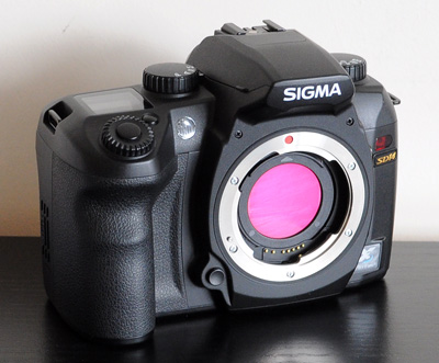
The Sigma SD14, like the earlier SD9 and SD10 has a 'dust protector' / 'hot mirror' just behind the lens mount. This is in a click fit mount on the SD14 and removable with care. It's a pity others makers haven't adopted this approach; other makers usually attach the UV/IR filter on the sensor placing any dust deposited near the focus plane. Removing the filter also offers the opportunity for near infrared / UV work without a permanent specialist modification to the camera.
The Sigma SD14 mounted to a Zeiss Photomicroscope III with Indian made 35 mm T2 SLR adapter. A Zeiss 10x Kpl W eyepiece on short collar was used for projection. The exposure mode dial and righthand mirror lock feature 'UP' is readily available if camera mounted as shown.
Shutter can be adjusted with lower left dial round shutter button. Shutter speed '1/4' in this case shown in LCD.
Aperture is preset to '1.0' to retain metering with lens off.
In manual 'M' a +/- 3.0EV indicator will show if exposure needs adjusting; although author prefers post capture image check / histogram for exposure determination.
A remote wired shutter is attached on the right, the third party models work well for ca £10 and about third price of Sigma's. Mirror lock is supported on this remote.
A T2 Pentax PK adaptor was used as recommended by others (see Appendix), but recently noticed that Sigma SA T2 mounts are now available (check US eBay).
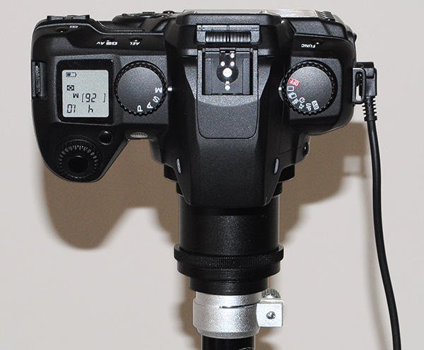
As Sigma forum members have noted, the Nikon DR-3 right angle finder fits well on the viewfinder (from below). The author tried this, but didn't find the 2X mag option eased focussing and preferred to keep the optics simple and use the camera viewfinder directly.
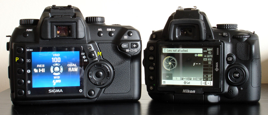
The Sigma SD14 (left) compared with the author's Nikon D5000 budget DSLR (downgraded from an earlier Nikon D300; half the price, same image quality, articulated screen, plus HD video).
Sigma SD14: The menu screen shown is the only operating menuto set ISO, image quality / format and white balance. The menu is shown with button M, play P button after capture will display image captured with histogram and zoom in functions. The simple layout and dial functions enable relatively easy use when camera facing down and inverted screen on a scope, if the camera is at a comfortable working height. As with other DSLRs without articulated screen, it was awkward
on the author's high Zeiss PMIII, i.e requires standing on a chair to check post capture image quality.
Nikon D5000: A busy menu which is rather fiddly to use although the articulated screen will face the menu forward when camera on a scope. The author uses tethered to a PC though for full remote control, live view and post image assessment.
Image protocol:
Both cameras exposed for highlights, i.e tonal balance tail as close to maximum as possible, images saved as RAW. Files processed in Photoshop Elements 7.0. Any colour cast corrected with white background, tonal balance adjust, resized with bicubic sharpening.
To completely remove any camera vibration differences, electronic flash was used (half silvered mirror on Photomicroscope base, SunPak 2400 flash with six power settings.)
Meaningful image comparisons between cameras are notoriously difficult, especially with photomicrographs as can be tonally flat and brightfield images from author's experiences invariably need some work up. Some comparisons are included but image quality can be as much dependent on choice of RAW converter, post image processing as native sensor properties. Cameras also differ in their default sharpness, contrast etc. SD14 were left on default. Nikon D5000 sharpness increased from default 4 to 6, as Nikon known to set camera's softer cf some other makers.
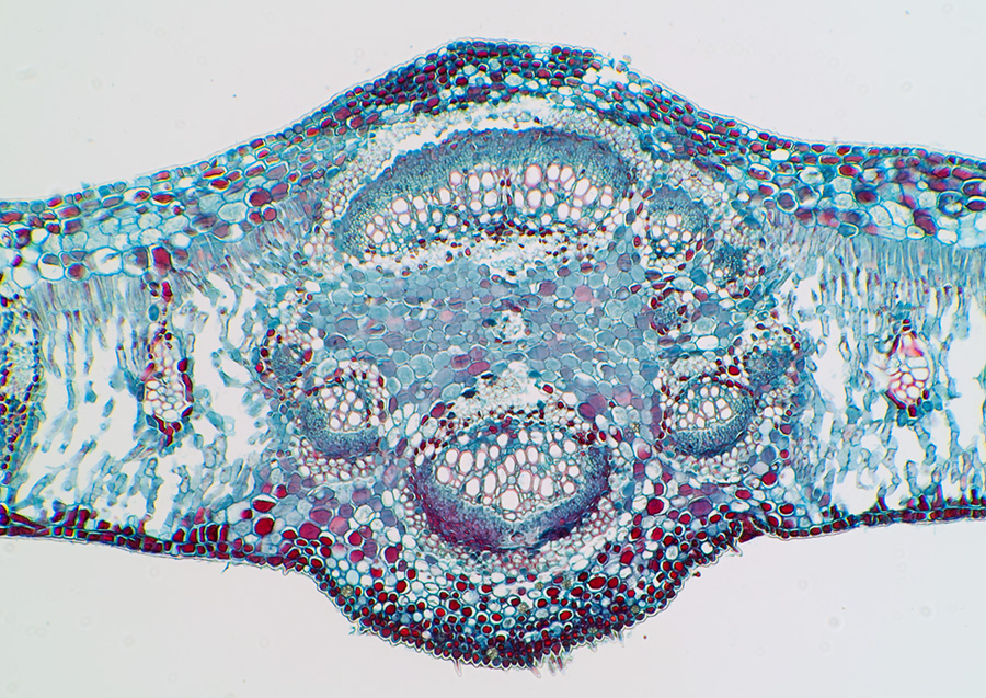
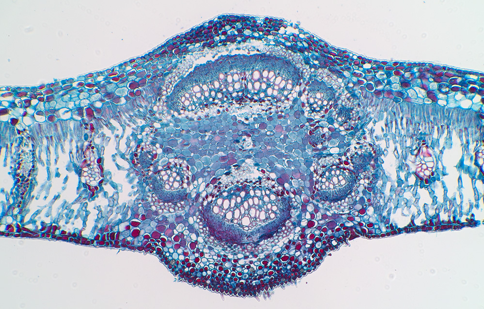
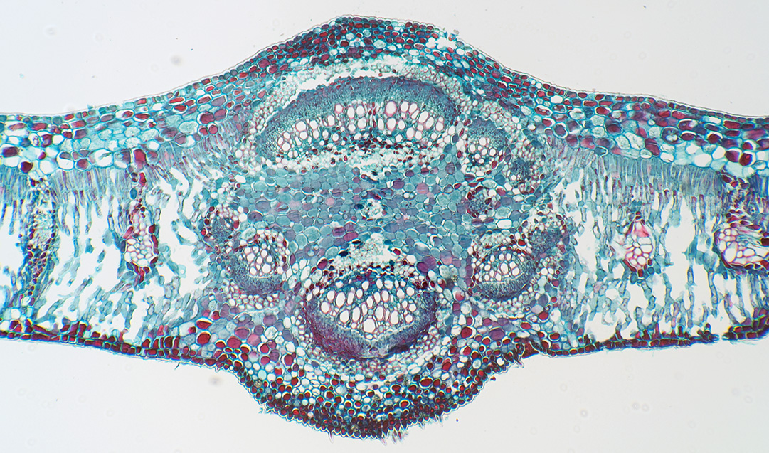
Typical stained botanical section
Biosil sea grape T/S leaf: Zeiss planapo 4/0.16 objective, electronic flash.
Contrasty subjects with well defined images such as these could be focused with the optical viewfinder, although had set up for parfocality with bino' head to check.
Sigma SD14, native ISO 100. Photoshop Elements 7.0 RAW plug-in.
Sigma SD14, image as above but using the bundled Sigma Photo Pro 4.0 RAW software.
The author preferred Photoshop Elements RAW conversion.
The blue is more intense and perhaps more pleasing, but the lighter blue is the more accurate to the slide subject.
Nikon D5000, from RAW, native ISO 200.
Typical stained histological section
Rat testis, iron haematoxylin and eosin, NBS course slide made by author: Zeiss planapo 10/0.32 objective, electronic flash.
This gives a flat image out of both cameras, so differences could equally be tonal levels adjustment chosen
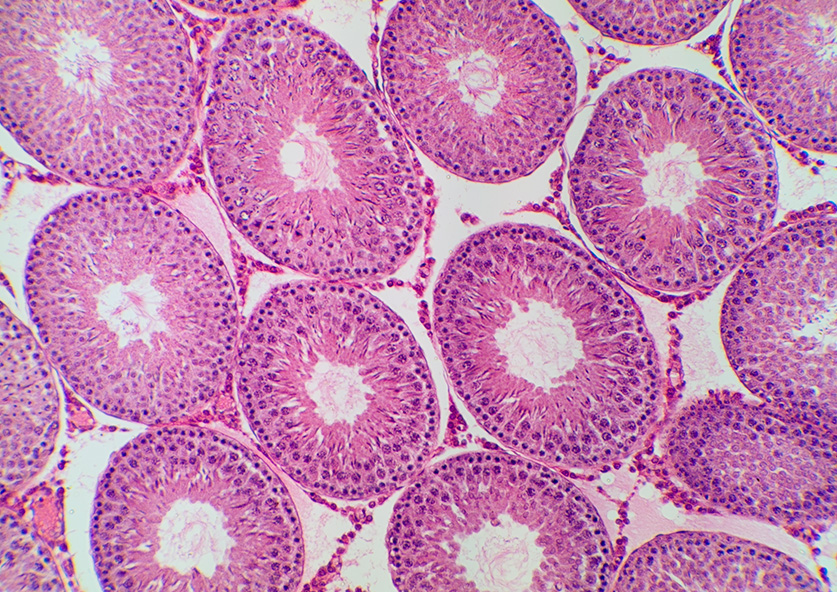
Sigma SD14, from RAW, Photoshop Elements 7.0
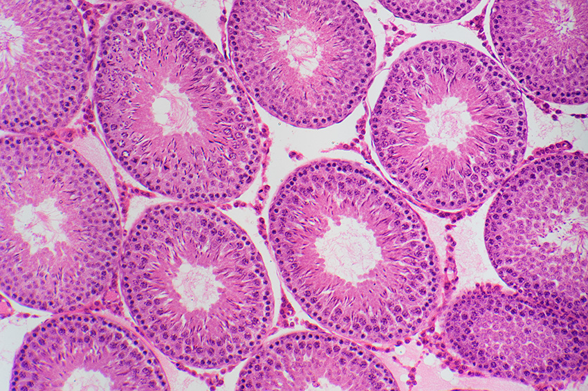
Nikon D5000, from RAW, Photoshop Elements 7.0.
Typical cross polarisers subjects. 'Crystals of Sodium and Quinine' (?), unnamed Victorian slide
Zeiss planapo 10/0.32 objective, electronic flash.
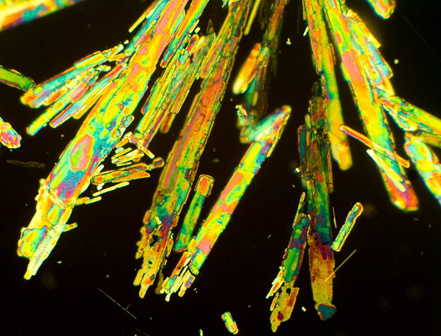
Sigma SD14, out of camera image. Image stacking would benefit as some crystals in slightly different focus planes.
Typical cross polarisers subject with lambda tint plate. Santonine, unnamed Victorian slide.
Zeiss planapo 10/0.32 objective, electronic flash.
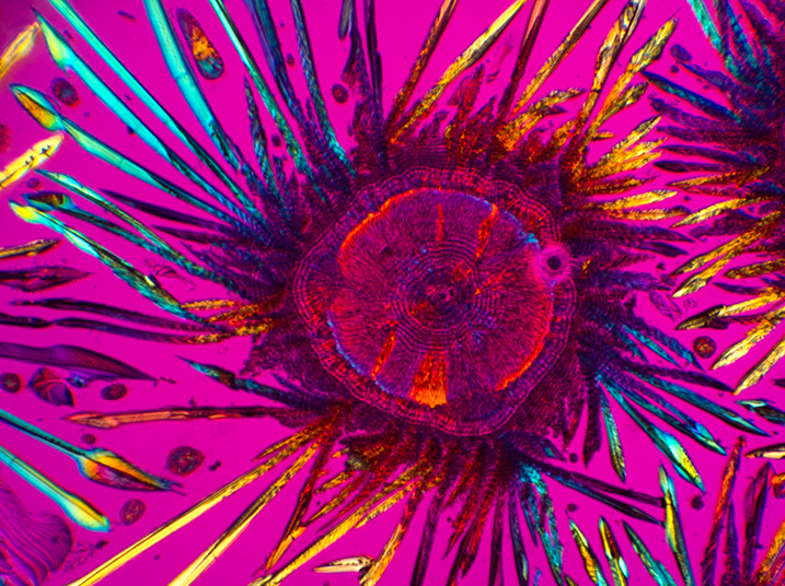
Sigma SD14, shadow and highlight level adjusted.
Phase contrast with monochromatic light. Diatom Cymbella gastroides in Styrax, Watson slide.
Zeiss Neofluar 40/0.75 objective with phase. electronic flash.
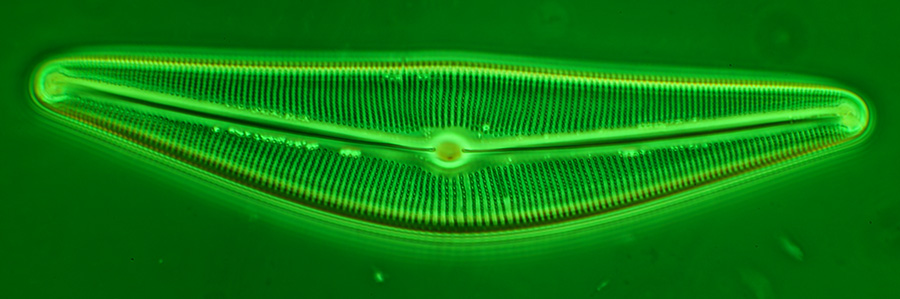
Sigma SD14, from RAW, Photoshop Elements 7.0. The camera's dynamic range copes well with the sometimes blown out haloes of a phase subject.
Low light, long exposure trial.
Zeiss 2.5/0.08 plan objective, 100W quartz halogen lamp, Zeiss III RS epi head.
Palate of whelk, Victorian slide, 'J&TJ'. No noise reduction set.
Exposure 30 secs, ISO 100. This is the longest exposure that can be set on the SD14. At default ISO, colours are good, rich blacks and good detail.
Left and right, crop of same subject taken at ISO 800 6 secs and ISO 1600 3 secs, respectively. There is a marked progressive loss of colour information at higher ISOs, inspection of image detail also shows colour noise artifacts. The Sigma SD14 is weak in this respect cf many modern Bayer sensor cameras, where high ISO performance is superior.
Dust protector filter removed. Near infra red trial.
Agabus water beetle. Zeiss 1x planachro objective. 100W quartz halogen set to 6V, IR 760 nm filter. Exposure 1/250th ISO 100.
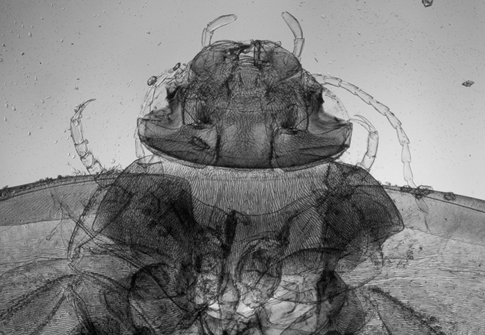
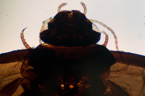
Left: Sigma SD14, near infra-red. Image converted to monochrome. The sensor is usefully sensitive to near IR with filter removed allowing transmission through dense exoskeletons of subjects such as insects to reveal detail. Images are rather flat tonally out of camera in near IR and requite tonal
balance adjustment. The sensitivity extends to at least 850 nm but
no observed benefit for this sort of subject. (
Photo' quality IR filters of various wavelngth cut-offs, are very cheap nowadays <£10 in 52 mm mount from China on eBay).
The SD14 was unable to set a white balance at 720 nm or higher wavelength, so images have a strong purple hue, reducing tonal range.
Right: Brightfield image in visible light taken with Nikon D5000, the dense exoskeleton and extreme contrast range prevents little internal detail being seen.
Even lighting at this ultra low mag on a compound microscope is tricky.
Comments to date
The Sigma SD14's size and external controls made it a pleasing camera to handle; the almost single screen menu system was a refreshing change after the multitude of menu features offered on many DSLRs. This simplicity also suited camera use when mounted on a vertical phototube (if camera is at a good working height). For photomicroscopy, some features were welcomed, such as retaining metering and offering mirror lock (unlike budget Nikon models I'm familiar with). Unlike some DSLR optical viewinders it didn't show artifacts that prevented focussing of contrasty subjects. For more demanding subjects, viewfinder focussing was tricky and an accurate parallax focussing setup is preferred. Although the author had such a setup, I had problems at times confirming the exact focus of subjects such as low contrast diatom frustules.
Preliminary assessment of the camera with its near IR / UV filter removed shows good sensitivity and potentially useful for this type of microscopy, although the filter is delicate and can easily be broken. The lack of live view makes IR/UV image focus tricky and a cheap monochrome security camera or microscope camera with IR/UV filter removed is much easier to use in this respect if a large pixel count isn't required.
If fuss-free photomicrography and post capture workflow is sought, the Sigma SD14 isn't ideal and at times find the lack of immediate critical image assessment frustrating because of lack of USB functionality when camera in use. A DSLR with live view fed to a PC screen (or viewed on camera's rear LCD) and if desired remote software control is a model much easier to use in this respect. But as commentators have remarked for Sigma Foveon camera use in normal photography, the Foveon sensor cameras can require time and patience to get the best out of them; results to date suggest that the SD14 is certainly capable of good photomicrography at normal ISO settings.
From trials to date of creating large screen images, my main interest, rather than prints, I haven't noticed any obvious benefit of the Foveon sensor over the Bayer for photomicrography, as remarked earlier, there are too many other parameters also affecting the final image look. The author is investigating a more accurate parallax setup to use for more demanding subjects where differences may be more evident, e.g. subjects with single colour fine detail near the limit of resolution of an objective, or whether results show that the higher pixel count of modern Bayer sensors negates any potential benefit of the Foveon sensor.
Comments to the author are welcomed.
Footnote added September 2010: The recent announcement by Sigma of their latest SD1 digital SLR with APS sized Foveon X3 sensor (1.5x crop), and 15.2 x 3 million photosites has attracted favourable comment in the online photo communities. This should be a very interesting camera for photomicrography with its potential ability to capture very high levels of detail in all colours and lack of anti-aliasing filter. But as remarked for the trials to date with the Sigma SD14, the SD1 needs to offer features like USB tethering to PC with auto image transfer and ideally live view and remote control software to bring it up to par with DSLRs which offer these features and which have proved invaluable for photomicroscopy.
Selection of resources
'Foveon X3 Technology Direct Image Sensors' - the maker's illustrated article on the sensors and data sheets.
'Sigma SD14 Resolution: 14 MP? 4.6 MP? How does the SD14 stack up against high end cameras like the Canon EOD 5D?' - a fascinating illustrated comparison by Mike Chaney.
Sigma SD14 - The Compendium - an invaluable compilation of 'User Tips' by Øyvind Strøm on all aspects of the Sigma SD14. The present author found this very valuable when learning how to use the camera to best effect.
'Infrared and ultraviolet imaging with a CMOS sensor having layered photodiodes' by David L. Gilblom, Sang Keun Yoo. A paper which includes details of the sensor's spectral characteristics.
Sigma Cum Laude - conversion kits for the Sigma SD14 to major camera maker's mounts.
Appendix: Extended operating notes.
Coupling to microscope - For practicality in a domestic environment, I prefer the direct coupling method rather than independent camera support. Sigma SA to T2 mounts don't seem to be readily available* but the SA mount is remarked to be near identical mechanically to the Pentax PK mount, so a PK T2 mount was used. The fit was rigid, with some force a small amount of rotational play was present and slight axis play as remarked by other users. *August 2010: At least one US camera dealer selling on eBay now offers them ($15 offer price accepted, 'Buy it Now' $25).
The sensor is slightly smaller than an APS sensor with a 1.7x rather than 1.5x sensor crop factor cf 35 mm. One of the shorter 35 mm microscope adapters was used to partly compensate for this.
Camera presentation and use vertically - The pentaprism facing user was most convenient to allow access to the top panel control dials and top LCD display. The limited feature set of the Sigma DSLRs has been criticised by some and welcomed by others. I fall in the latter category; after wading through endless screen menus to seek functions on even the budget DSLRs, what a refreshing experience this camera was to use. Apart from initial setup menus, there is a single screen to access the key features of ISO, white balance, image size and quality. Other features are on the dials and buttons. This simplicity suited vertical use on a microscope.
Shutter release / vibration reduction - The mirror lock up feature 'UP' is readily set on the top plate dial, but like some other makers' models, it cannot be used with the delayed release option (2 sec or 10 sec) which is on the same dial. A socket is provided for Sigma's wired remote release, but a cheaper third party release was used which cost about £10. The first press locked up shutter, then release pressed again for the exposure. Of DSLRs handled from Fuji, Nikon, Olympus and Canon this was subjectively one of the quietest shutters used, perhaps suggesting a light action. To date I've used electronic flash to remove any vibration issues; a series of shutter speed trials with/without mirror lock for each objective will be carried out in due course to assess any potential vibration issues for quartz halogen lamp use.
Focussing - Some cameras handled in past eg a Finepix S3 Pro gave odd artefacts in camera viewfinder on author's microscope, preventing reliable focusing. Although the SD14 viewfinder is quite coarse it did allow quite accurate focussing and successfully focussed eg diatom frustules by this method. There is no live view*, so permanent critical focussing would rely on setting up a very accurate parfocality with bino' eyepieces or adjustment after image assessment. *(This camera, released in 2007, predates widespread adoption of live view, although also not offered on the new SD15.)
Exposure - The camera with lens off will meter and set shutter speed in aperture priority 'A' mode. Although author did not assess the metering reliability as much prefer manual metering for microscopy by assessing the histograms post image capture. The top LCD display includes metering info' albeit upside down on a 'scope, (f1.0 has to be set first, a procedure that wasn't obvious from manual but learnt from forum discussions). In manual 'M' mode the meter can also be used to monitor correct metered exposure. Compensation of exposure of up to+/-3EV in increments can also be readily set which is handy for tricky subjects. In this respect it is more versatile than author's Nikon D5000 which like all Nikon's budget DSLRs to date offers no metering with lens off.
Workflow: image capture, and assessment
Isolated
camera - The procedure adopted was: focussing in eyepiece, image capture,
assessment of exposure on camera LCD from R,G,B histogram, detailed image check
on camera LCD using zoom and scan function. Although the screen is a very low
res. by modern standards 150,000 pixel 2.5 inch, it was good enough to check
for critical sharpness and quality.
The camera is slow to process images for display after exposure, particularly as the exposure time increases.
Camera connected to PC - The camera can be connected to a PC via supplied USB cable which can recognise the camera's compact flash card as external drive. Sigma's supplied Photo Pro software can transfer the images for screen assessment and work up. Unfortunately unlike other maker's DSLRs used to date, it is unable to transfer images on the fly automatically after capture. In fact the camera does not work with USB cable inserted. So author's workflow was to take a sequence of images, assess them provisionally on camera LCD, then connected camera to PC for image transfer.
Published in the August 2010 edition of Micscape.
Please report any Web problems or offer general comments to the Micscape Editor .
Micscape is the on-line monthly magazine of the Microscopy UK web site at Microscopy-UK
©
Onview.net Ltd, Microscopy-UK, and all contributors 1995
onwards. All rights reserved.
Main site is at
www.microscopy-uk.org.uk.