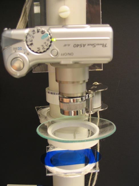
Optical Bench
| The Hunt for Leptodora by Howard Webb (St. Louis, MO, USA) |
Leptodora is the only native (to North America) predatory cladocera (water fleas), and the largest of the native species. Two other predatory species are found in the Great Lakes (Ercopagis pengoi ["fish-hook water flea"]and Bythotrephes longimanus ["spiny water flea"]), but these are invasive species. While larger than other daphnia (up to 1/4 inch), they are much harder to see. Other than the single dark eye, the rest of the body looks like transparent glass threads.
I first encounted Leptodora several years ago at Table Rock Lake in southwest Missouri. It was not until two days after I had filtered the samples and made slides, that I noticed something unusual in one of my culture bottles of 'leftovers'. Initially I thought it was a Chaoborus (of which there was one in the bottle), as it was of similar size and was only visible as a transparent reflection of light. It took me a full half hour to find and remove the Leptodora from a liter of water; at times while searching, I thought I was only seeing light reflections off of my glasses, or might just be imagining its presence.
Leptadora are too large for decent photomicroscopy using my compound microscope. Even with my lowest setting (10x objective and 10x eye piece), they more than fill the view; add the zoom of the digital camera to avoid vignetting, and at best I end up with a photo montage of various body parts.
I have tried lower power photography with my dissecting microscope, but so far I have not been able to get the correct lighting. My LEDs that will give me a shutter speed of 1/2000 sec with direct lighting will only give about 1/30 sec with indirect lighting. The speeds are two slow to avoid problems of vibration which blur the picture.
To add to the problem, my favorite mountant, PVAG (polyvinyl alcahol & glycerin) does not work well with large soft bodied organisms. Almost as soon as I added the mountant to the slide, the moisture inside the Leptodora began to be pulled out, and the thin carapace began to buckle. The final state is a poor representation of the live specimen.
The challenge was to find a way to take lower power images of a live specimen.
The images in this article could be better from several standpoints. Flushing the slides with bottled water would have removed a lot of the dirt particles. The hand lens has several optical flaws that show up as uneven focus. The point that I am trying to make is that with very little cost and equipment it is possible to decently document a lot of observations under make-shift conditions. It doesn't require an expensive microscope or even the best of initial images to still capture a lot of details. It is more important to get out there and enjoy observing than to worry whether your pictures will or will not win the Nikon contest.
I was going to Kentucky Lake for a week this summer, and knew it had a population of Leptadora. Besides the usual gear of kayak and plankton nets, I decided to try and build an 'optical bench' for larger scale photography. On occasions I have held a hand-lens in front of the camera for low magnification. This works, but I needed something that would hold the lens steadier than my hand.
I managed to collect for about a half hour for five evenings, and found one Leptadora on each of two occasions.
The 'optical bench' consists of scrap PVC pipe and a few other parts. A piece of Lexan is attached to the top as a camera mount. I like Lexan as it is easy to cut, drill and tap. A 1/4 inch bolt is the standard camera tripod adapter threading, so I use them about everywhere. I sliced rings from PVC couplings and split them to slide easily on the pipe. These split rings are used to hold slides, watch glasses, filters and lenses. Focusing is done by sliding the 'stage' up and down. This is crude, but turned out to be very effective. For most of this project I used a 16x doublet hand lens in front of the camera, which gave me full body images of the Leptadora.
|
|
|
Optical Bench
|
|
|
|
Makeshift working area
|
I picked up an EasyCAP video to USB adapter (it was cheap on eBay, but
only works with Windows XP) so I can watch my camera output on my
laptop. It is easier than trying to look at the camera back
for focusing. A limitation of video output is that the
resolution is lower than what the camera actually records. I
focus till the screen looks the sharpest, and have the pleasure of
seeing more detail in the pictures.
My LED lights and camera tend to have some strange interactions where
the image brightness is not steady (it turns out I had the wrong power adapter). Combined with the low
contrast of the Leptadora, the initial images often have poor
contrast. That is where Photoshop comes in handy, there is
often more detail in the image that a little manipulation can
bring it out.
This is why cover slips are necessary. I sucked off much of
the water to keep the Leptadora from moving around, and the surface
tension turned the water into a lens. The dark spots are
where the water was stretched higher than other places.
|
|
|
Leptadora - in watchglass
|
|
|
|
Leptadora - no cover slip
|
Here I used a coverslip. I used fragments of a coverslip as
'spacers', but there was still not enough room so that the coverslip
pressed down on the Leptadora and distorted it some.
|
|
|
Leptadora - with cover slip
(but some flattening)
|
|
|
|
Leptadora - original poor image
|
|
|
|
Leptadora - after Photoshop (auto correction), note added 'noise'
|
|
|
|
Leptadora - colored filters are always an option
|
|
|
|
Leptadora - after Photoshop of blue filter (auto correction)
|
|
|
|
Leptadora, cleaned up image
|
|
Leptadora - cleaned up image
|
|
|
|
Leptadora - close up of eye, showing mountant distortion
|
Location: Kentucky Lake, KY (37.002, -88.245).
Environmental
Conditions:
Water temperature:86F
Depth: up to 50 ft
Doublet Magnifier 16x from Carolina Biological
Microscope: Bausch
& Lomb monocular, 10x ocular, 4x, 10x and 40x objectives.
Illumination: Luxeon K2 LED
Camera: Canon A540 (6 Megapixel)
Software: Photoshop Elements
Comments to the author are welcomed.
Published in the August 2010 edition of Micscape Magazine.
Please report any
Web problems or offer general comments to the Micscape
Editor,
via the contact on current
Micscape Index.
Micscape is the
on-line monthly magazine of the Microscopy UK web
site
at http://www.microscopy-uk.org.uk/