Victorian Rambles - Part III
A selection of prepared slides of crystals.
by David Walker, UK
 Old microscope slides can be enjoyed on many levels, and this occasional series is a random dip into the author's modest collection to illustrate some hopefully interesting slide subjects and brief aspects of their history. Different lighting techniques
are explored to show their relative merits where appropriate.
Old microscope slides can be enjoyed on many levels, and this occasional series is a random dip into the author's modest collection to illustrate some hopefully interesting slide subjects and brief aspects of their history. Different lighting techniques
are explored to show their relative merits where appropriate.
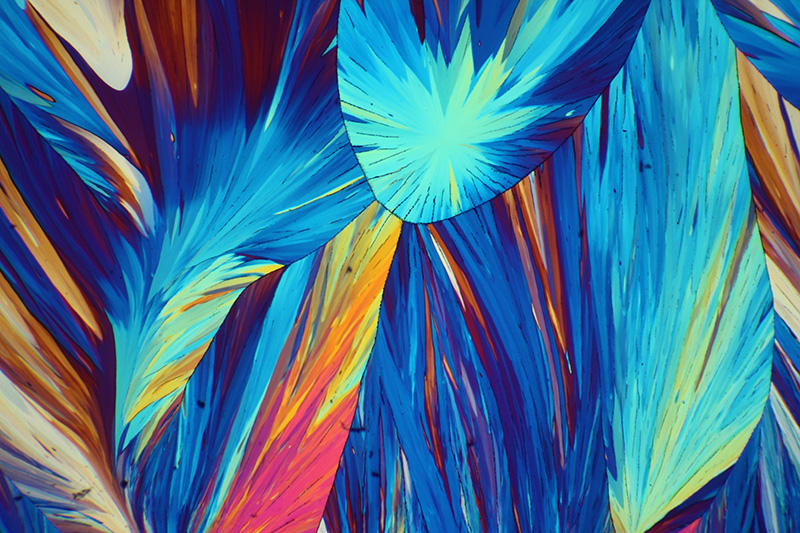
Crystals have a perennial appeal to study under the microscope, particularly when they are birefringent and respond to crossed polarising light. Nineteenth century slides of chemicals specifically for crossed polar study are quite common and below are a selection. Chemicals have been chosen that haven't featured before on Micscape as a number of contributors have shared striking images of
a wide variety. For example Brian Johnston's striking 'The colourful world of chemical crystals' series also see the Micscape Library 'Techniques - lighting-polar - crystals' section.
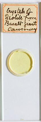
"Crystals of zeolite from basalt - Giant's Causeway"
This slide (shown right) is undated and mounter unknown but from its style is probably late 19th or early 20th century. The Giant's Causeway in Northern Ireland is world famous with its striking, predominantly hexagonal, interlocking basaltic columns formed in an ancient volcanic eruption. The type of zolite isn't stated but the National Museum of Northern Ireland lists nine zeolites associated with the formation and gives a summary of why they are formed with lava.
Zeolites are hydrated aluminosilicates and ca. 40-50 occur naturally but over 100 synthetic forms are known. Their microporosity of molecular dimensions are widely exploited in many applications. I have a fond affection for synthetic zeolites as used to work in the field of zeolite synthesis and their use for selective adsorption and catalysis in the petrochemicals industry.

Zeiss 6.3X planachromat with crossed polars and annular oblique (COL). A homemade annular stop for the Zeiss phase condenser gives COL at low powers which can impart extra three dimensionality to some subjects.
Three salts of platinocyanide and palladocyanide complexes
These slides are also anonymous but by the same mounter. Salts of platinocyanides and palladocyanides are often seen as subjects of older crystal slides because they can adopt attractive forms as shown below. The platinocyanide anion is divalent i.e. [Pt(CN)4]2- and forms a planar complex.
In an early Micscape article "On samples. slides and ... 'surfing'" I remarked on how researching often unassuming slides on the Web may lead to fascinating lines of further reading. The barium platinocyanide slide is an excellent example, as was previously unaware of the key role this salt played in the history of physics. It was the fluorescence of a barium platinocyanide coated surface some distance from the covered electrical discharge tube which Wilhelm Röntgen was using that led to his discovery of X-rays. The exact details of this experiment as reported from various sources vary somewhat and are discussed in Otto Glasser's biography of Röntgen. Surfaces treated with barium platinocyanide were already in use at the time for detecting the none visible parts of the electromagnetic spectrum. My slide of barium platinocyanide fluoresces when illuminated with a near UV LED torch (shown below).
In Paul Frame's 'Tales from the atomic age' he notes that early samples of the salt contained radium as an impurity and weakly self fluoresced; thus it was feasible for scientists of the time to have 'seen' radium. Frame demonstrated this self fluorescence by using an early sample of the salt.
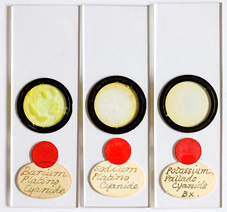

Three salts of platinum and palladium cyanide complexes. The right hand image shows the barium platinocyanide fluorescing under a near UV LED torch.
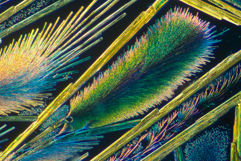
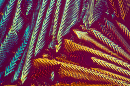
Barium platinocyanide, 6.3X objective, crossed polars and COL. Long needles and plant-like forms of smaller crystals are typical,
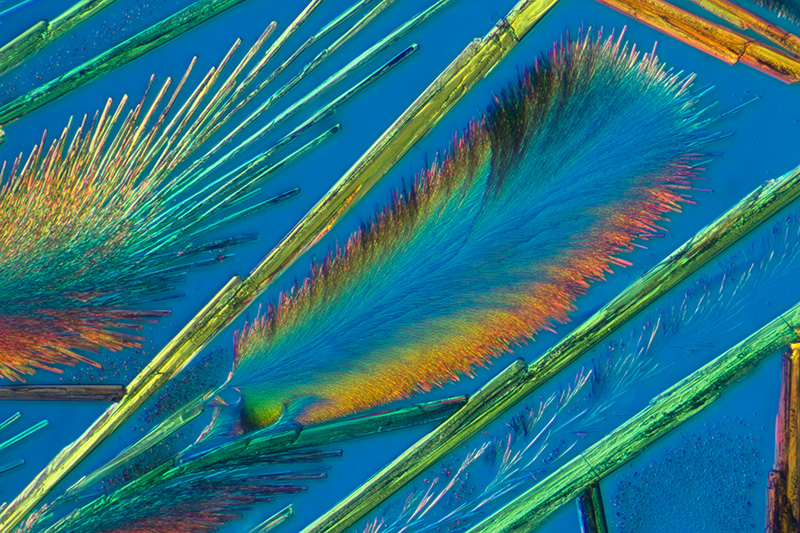
As above but with first order tint plate (blue).
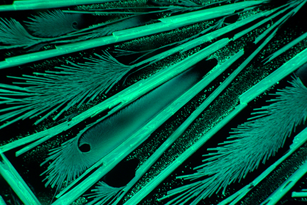
Barium platinocyanide using a Leitz NPL 6.3/0.20 objective showing fluorescence using epi near UV light. ISO 200, 5 sec exposure, using Zeiss III RS 'UV filter/mirror set' with the small but usable near UV component of a 100W quartz halogen lamp.
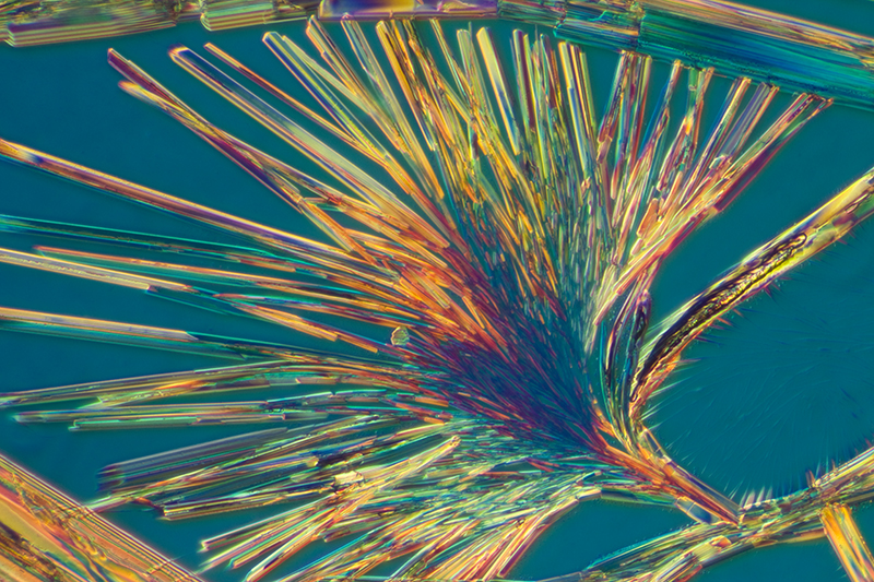
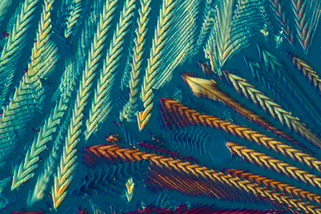
Sodium platinocyanide, 6.3X objective, crossed polars with blue tint plate and COL. Similar crystal forms to the barium salt but with larger plant-like forms as shown above.
Potassium palladocyanide,6.3X objective, crossed polars with COL. This salt shows more complex interlocking structures.
As above plus a first order tint plate (red).
Santonin
Santonin / santonine is a quite common slide 'For the Polariscope'. Wikipedia has a fascinating article on the complex organic chemical where it notes that it can be extracted from santonica, the dried flower heads of certain plants from the Artemisia genus which includes wormwood. It was used as an antihelminthic i.e. for the treatment of parasitic worms. Great care
was needed in its use to avoid potentially lethal side effects and more modern drugs have replaced it. I have two antique examples of the slide, one unnamed with typical green / red papering and another sold under the ubiquitous 'C.Baker' label.
Santonin as a prepared slide was popularised more recently by the late Eric Marson of Northern Biological Supplies (NBS). He sold a 'Microslide to show crystal growth' of santonin where it was advised to gently melt (m.pt 174C) and study the crystal regrowth. An example of an NBS slide is shown and also have an example prepared by myself when attending his well known slide making courses at Belstead House in Suffolk.
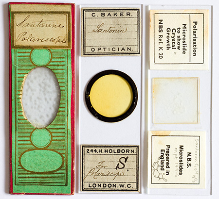
Two antique santonin slides and the recent NBS example sold to show crystal growth. 'For Polariscope' was often stated when the chemical or other subject was suitable to study with crossed polarising light.
|
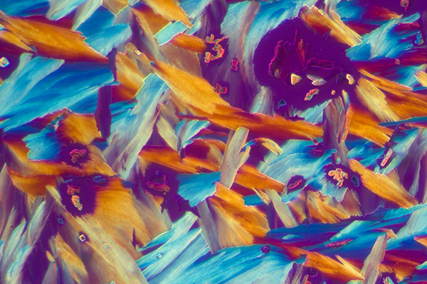
The sequence of five images below show santonin (Baker slide), first between crossed polars and then the effect of adding one of the following plastic retarders: l/4, l/2, l (530 nm) or l (560 nm) from Knight Optical..
The full wave plate with the traditional tint of a 'first order red' has a specific use in polarised light microscopy e.g. of rocks and minerals, but as a modifier in 'artistic' polarised light imagery, the colour may jar as a background colour with some subjects. I often prefer the full wave plate which creates a gentler blue.
Retarders for this sort of work don't have to be the highest optical quality, as they can be sat on the polariser beneath the stage and approximately aligned at 45 degrees to the plane polarised light. A variety of plastic sheets from around the home can also be tried, e.g. the cellophane that wraps greetings cards.
|
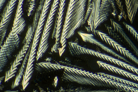
Crossed polars only
|
|
Crossed polars with l/4
|
Crossed polars with l/2
|
|
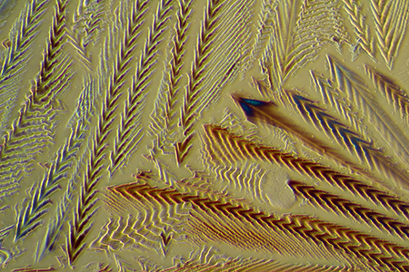
Crossed polars with l 530 nm gives a red tint background.
|
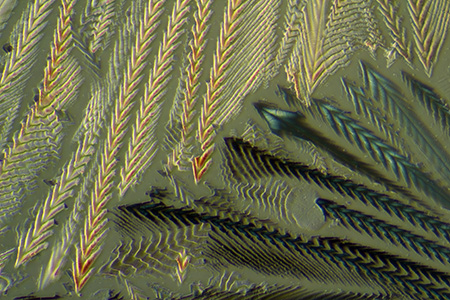
Crossed polars with l 560 nm gives a blue tint background.
|
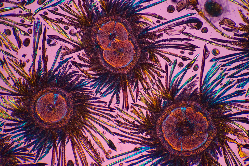
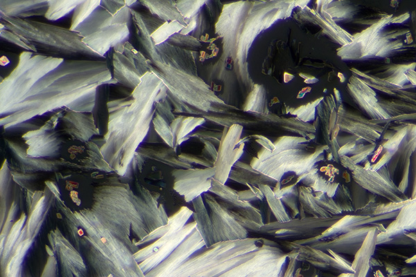
Papered santonine slide, 2.5X objective, crossed polars with red tint plate and COL This slide has attractive micro-crystalline forms surrounded by larger needles. The polars have been slightly 'uncrossed' to give a paler background tint.
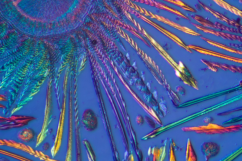
Papered santonin slide, 6.3X objective, crossed polars, blue tint plate and COL. Close up of one of the 'organic forms'.
For comparison, this is the NBS slide of santonin that has been remelted a few times and gives different continuous plates in contrast to the two antique slides of the same chemical shown above. Zeiss 2.5X objective, crossed polars with blue tint plate.
One of the limitless appealing aspects of crystal slides is that every slide of the same chemical is different, with 'worlds within worlds' when each slide is examined under the microscope.
Comments to the author are welcomed.
Equipment: Zeiss Photomicroscope III with Zeiss 10X Kpl W eyepiece on small collar as projection eyepiece using a Canon 600D DSLR body. Recent Canon DSLRs are particularly suited for photomicroscopy as by default or as an option they have an electronic first curtain shutter (EFSC) in live view mode which ensures exposures are essentially free from camera sourced vibration (see Charles Krebs' informative article on EFSC on Canon DSLRs). Mirrorless removable lens cameras with EFSC include the Sony NEX 5N and the NEX 7.
©
Microscopy UK or their contributors.
Published
in the August 2012 edition of Micscape.
Please
report any Web problems or offer general comments to
the
Micscape
Editor
.
Micscape
is the on-line monthly magazine of the Microscopy UK web
site at
Microscopy-UK
©
Onview.net Ltd, Microscopy-UK, and all contributors 1995
onwards. All rights reserved.
Main site is at
www.microscopy-uk.org.uk.