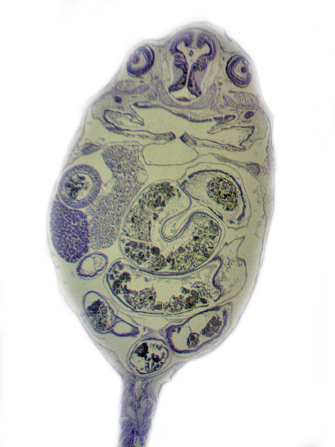
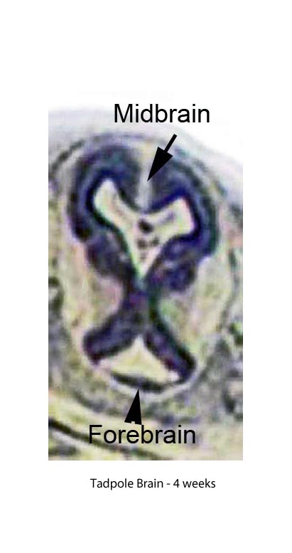
Tadpole 4 weeks
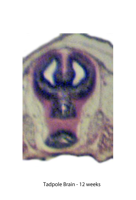
Tadpole 12 weeks
Tadpole 17 weeks

|
Sections Showing the Development of the Tadpole by Mike Morgan, UK |
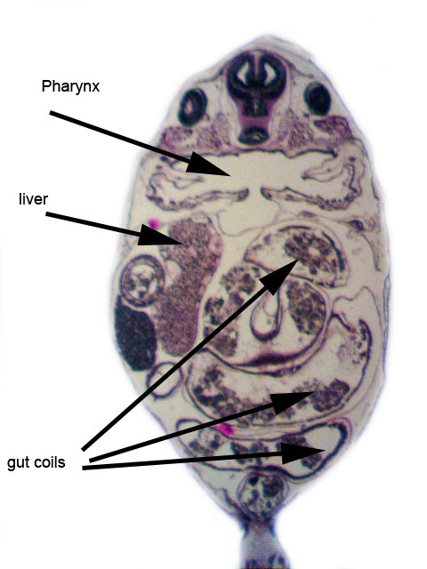
It is interesting to observe the changes both in the brain and the gut of the tadpole in the process of metamorphosis.
The sections shown are from 4 weeks to 17 weeks development.
(Paraffin Wax Sections, cut at 15µ, see Appendix for details of the preparation protocol.)
Each section is not fully annotated as the same structures appear in each.
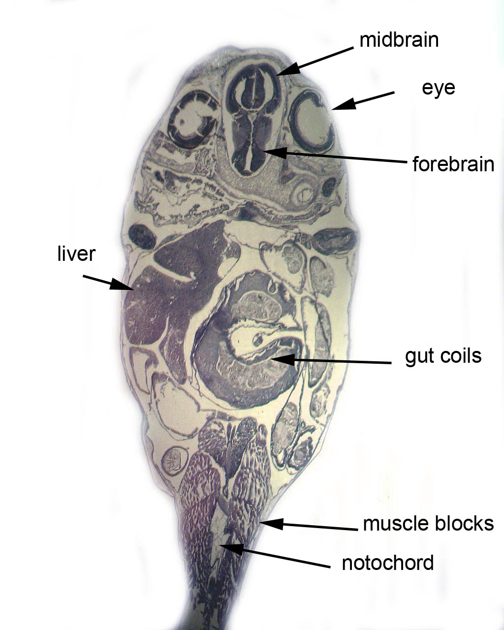
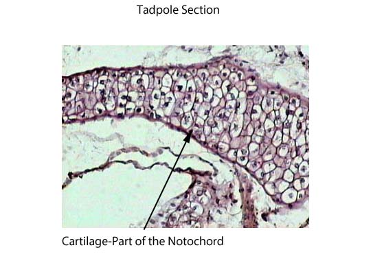
I include a section, which shows the notochord sitting under the neural tube of the hindbrain. This is a cartilaginous structure that all chordate embryos possess. The last vestiges are the intervertebral discs in the adult.
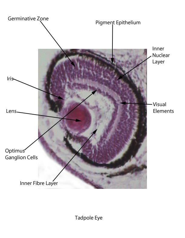
Also included is a section of the eye of the tadpole, which shows most of the structures present.
Gradually the tail of the tadpole disappears in the process of Apoptosis or programmed cell death, a process stimulated by the thyroid gland of the tadpole.
Many thanks to Dr Harry Isaacs of York University for his interest and help in identifying various parts of the anatomy in the sections.
Comments to the author Mike Morgan are welcomed.
Appendix - preparation protocol
Tadpoles were dispatched by immersion in 70% alcohol.
Fixed in 10%
formalin for 24 hours.
Sections were cut at 15 micron thickness and
Haupt's medium was used to adhere them to the slide.
Sections were
stained with Haematoxylin/Eosin.
Haupt’s
Medium (Section
Adhesive)
Gelatin
1.0 gm.
Distilled water
100 ml.
Dissolve at 30°C and add 15 ml
glycerol and 2 gm. of phenol crystals.
Stir, cool and filter.
Placing the slides in a desiccator in formalin vapour for
24 hours will also ensure the sections adhere to the slides.
|
|
|
|
|
|
|
|
|
|
|
|
Published in the August 2012 edition of Micscape.
Please report any Web problems or offer general comments to the Micscape Editor .
Micscape is the on-line monthly magazine of the Microscopy UK web site at Microscopy-UK
©
Onview.net Ltd, Microscopy-UK, and all contributors 1995
onwards. All rights reserved.
Main site is at
www.microscopy-uk.org.uk.