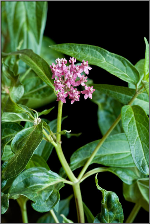
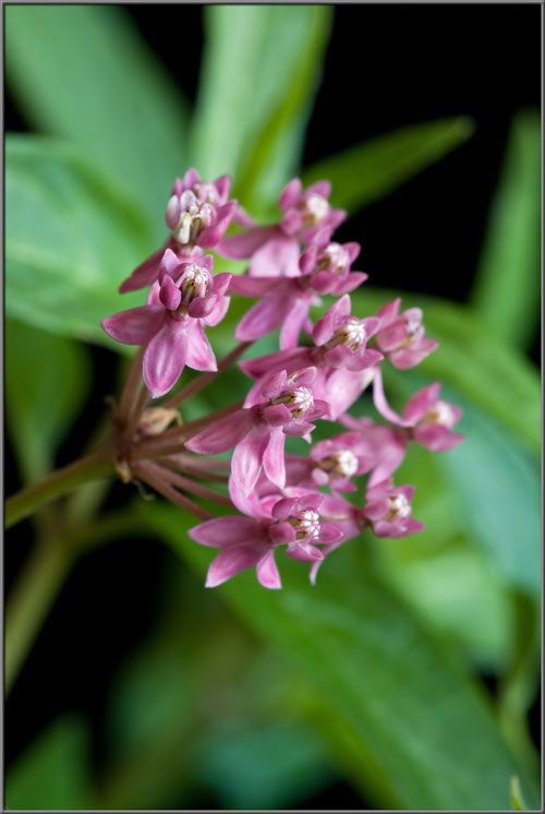
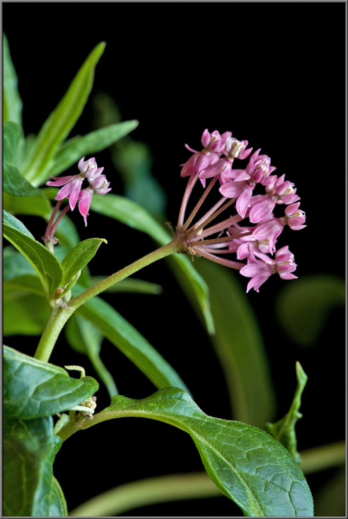
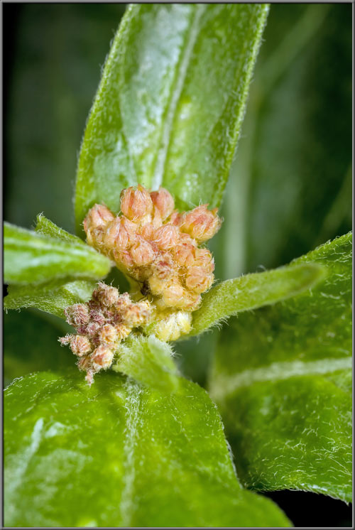
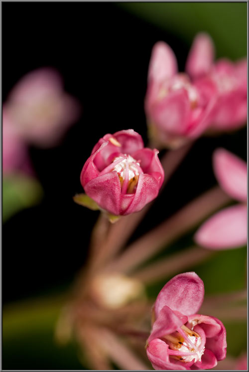
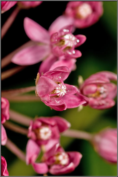
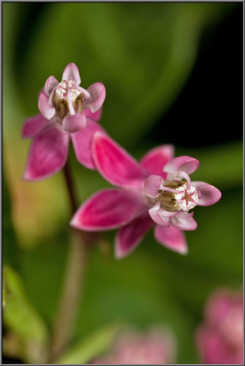
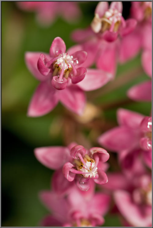
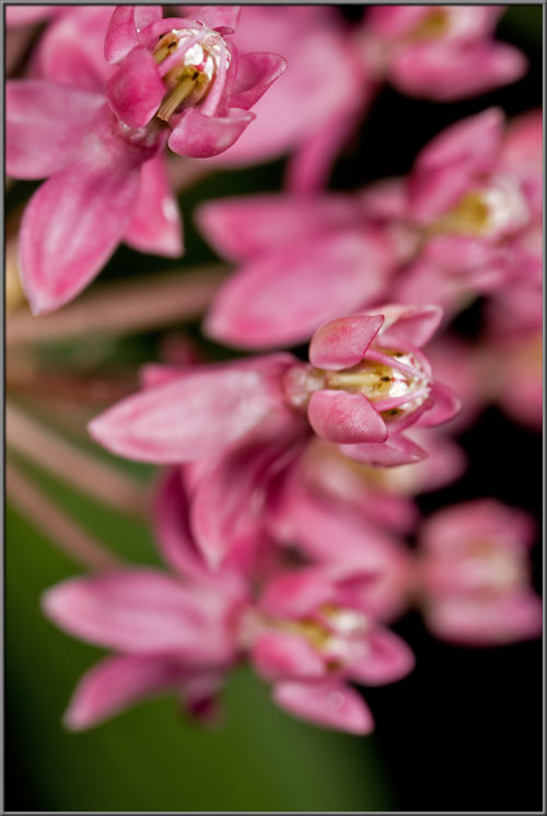
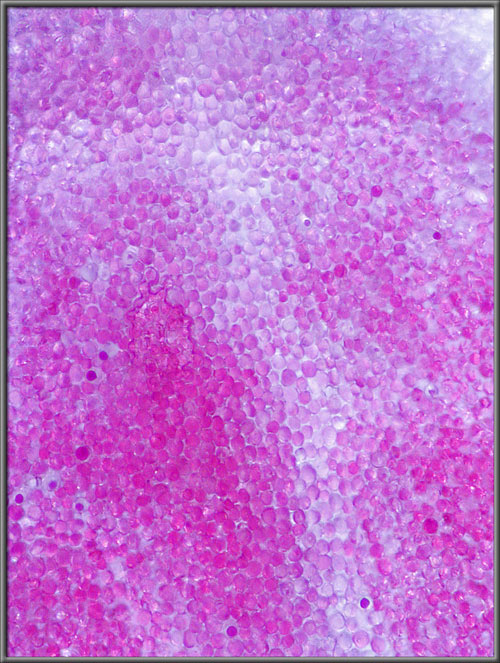
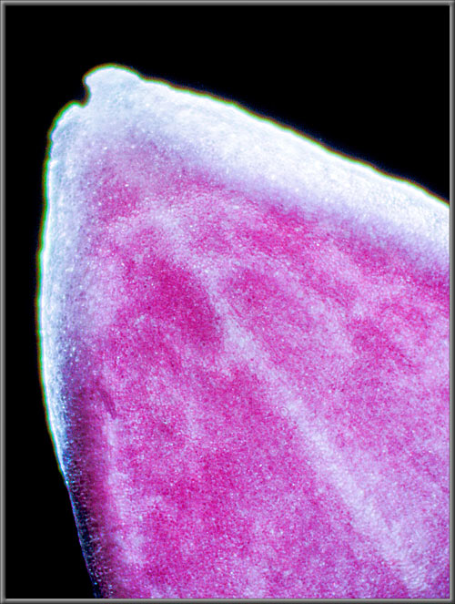
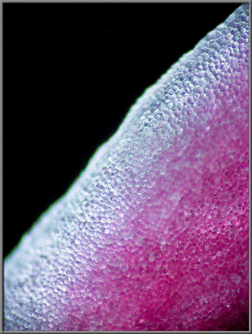
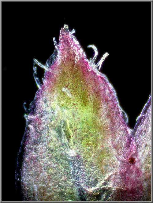
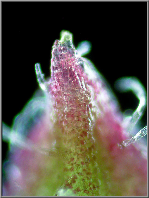
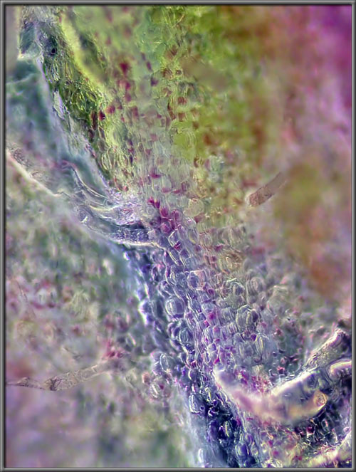
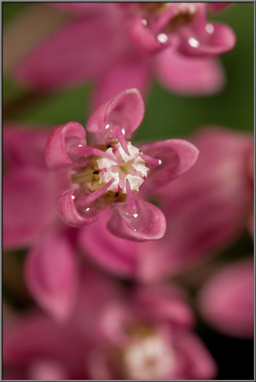
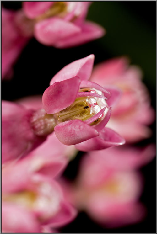
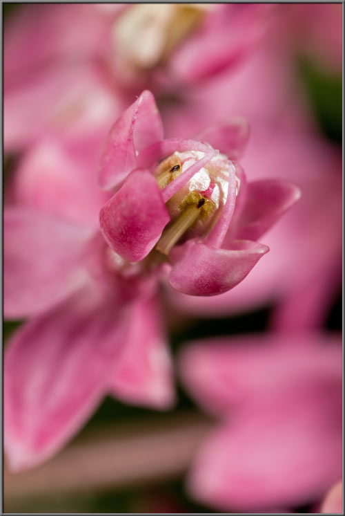
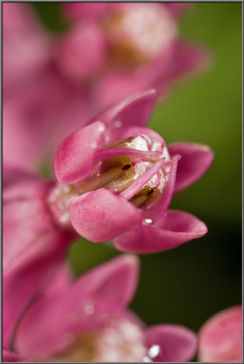
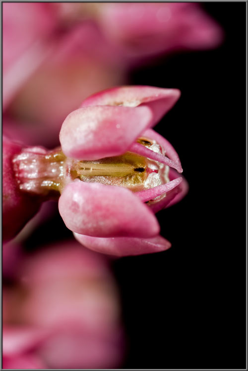
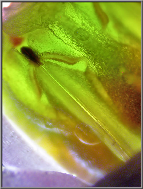
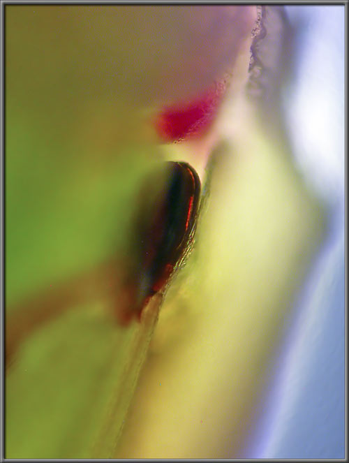
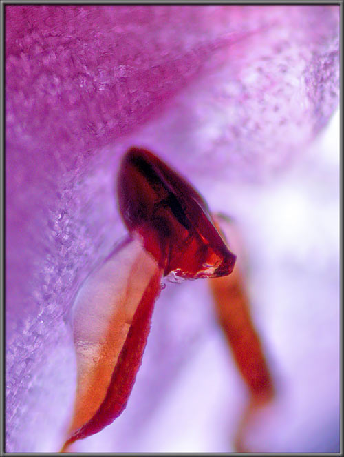
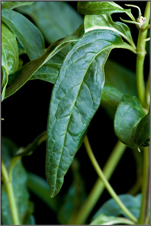
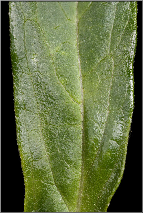
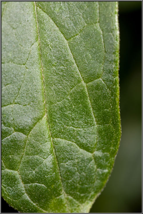
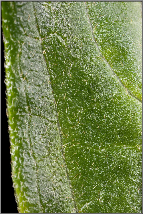
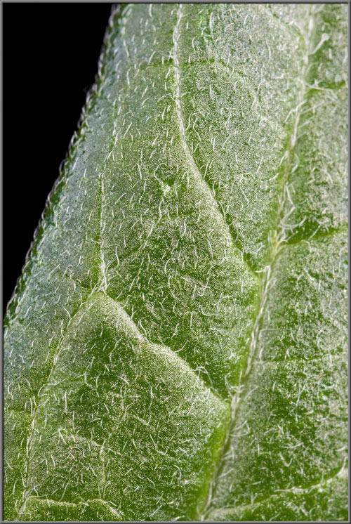
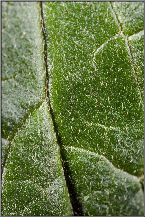
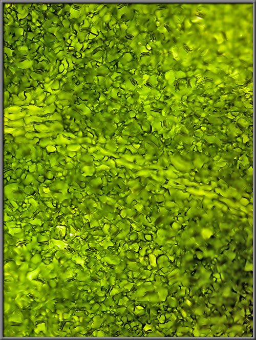
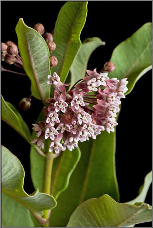
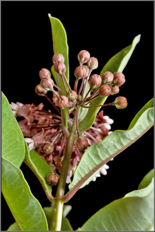
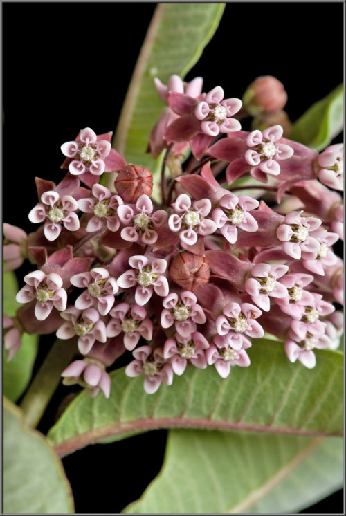
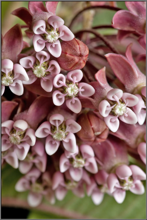
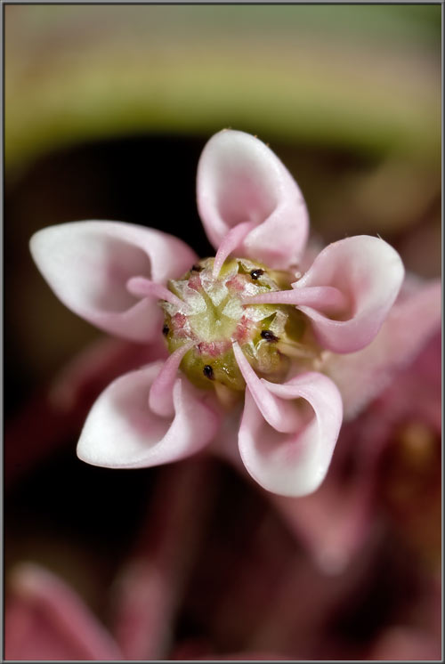
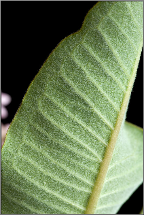
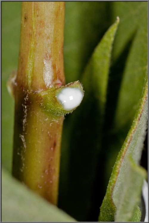
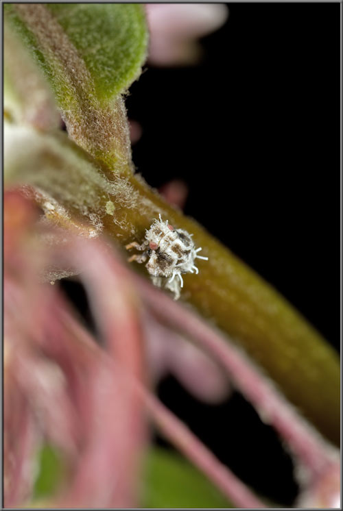
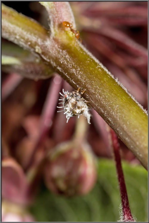
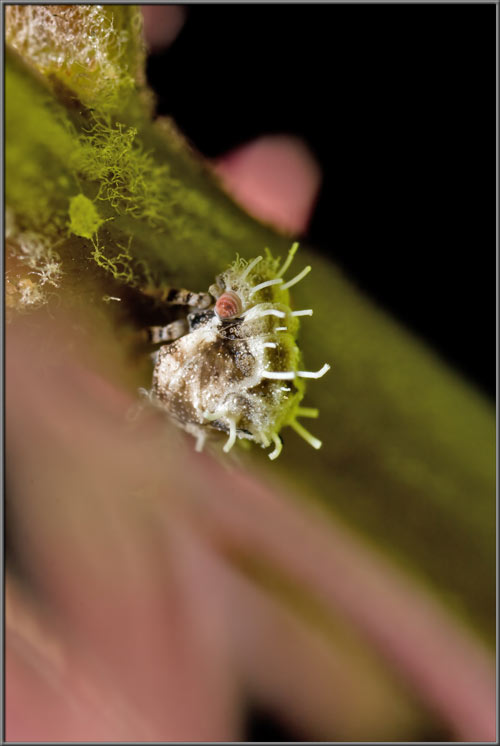
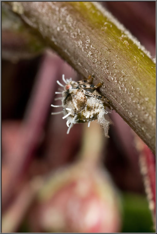
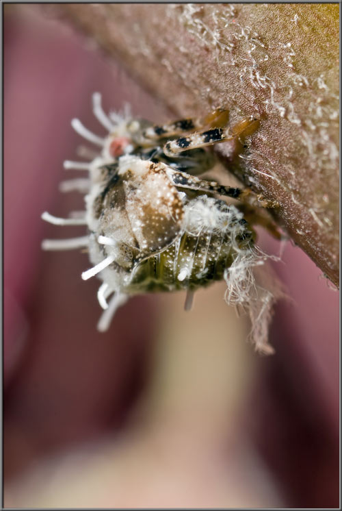
A Close-up View of a

Swamp Milkweed Cultivar
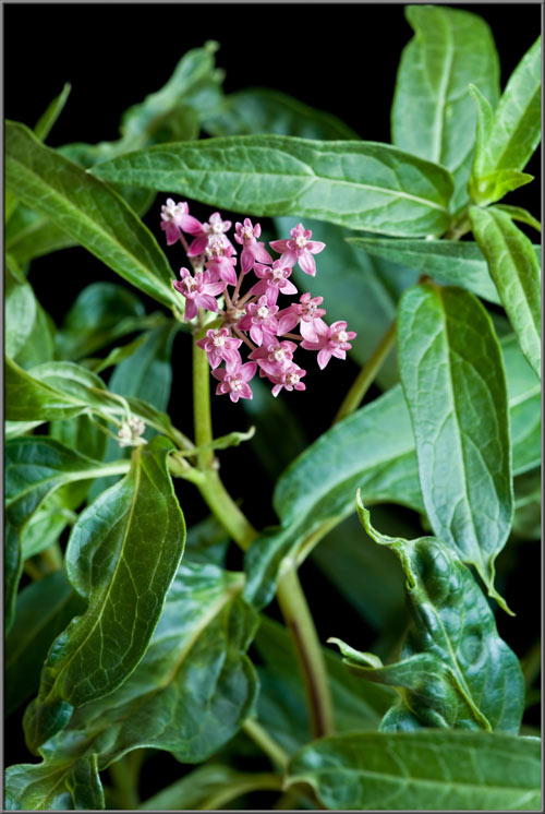
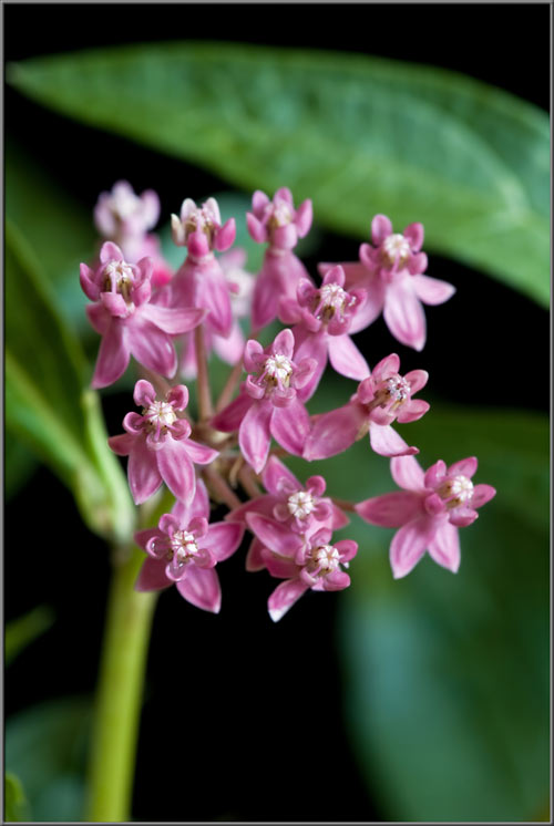
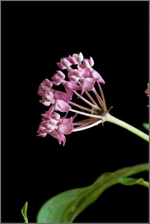
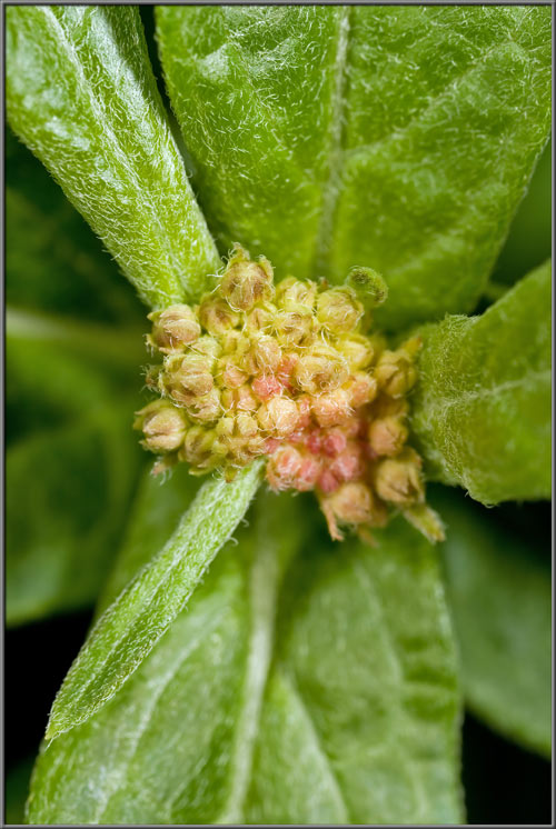
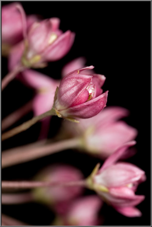
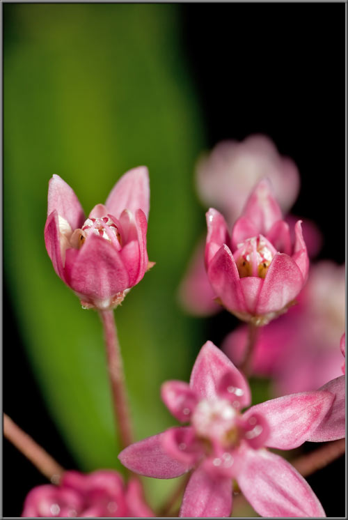
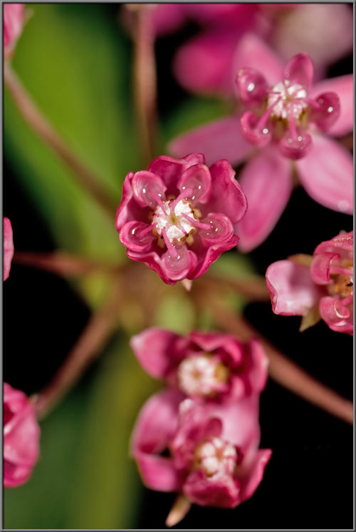
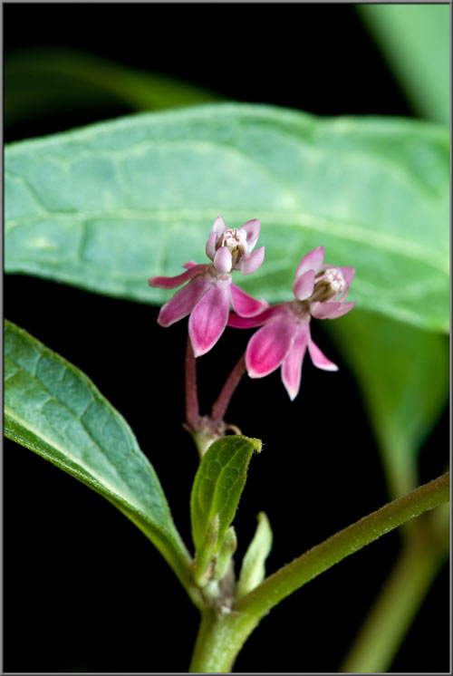
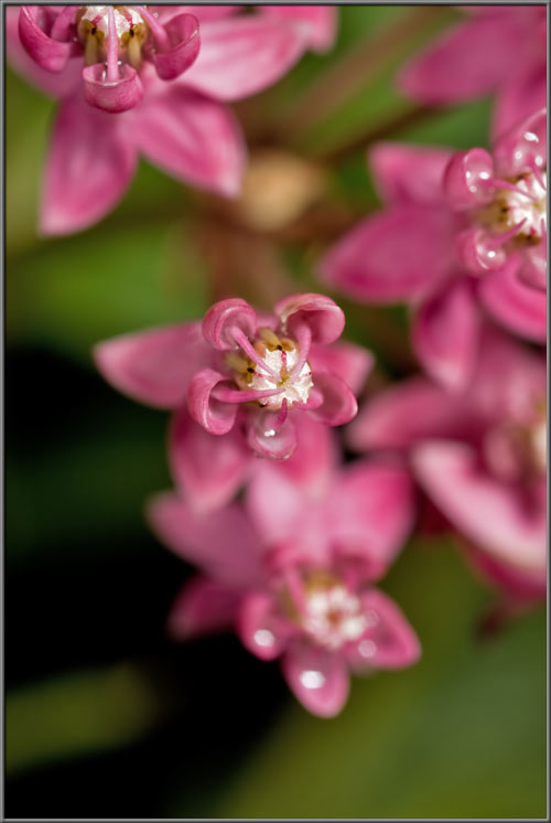
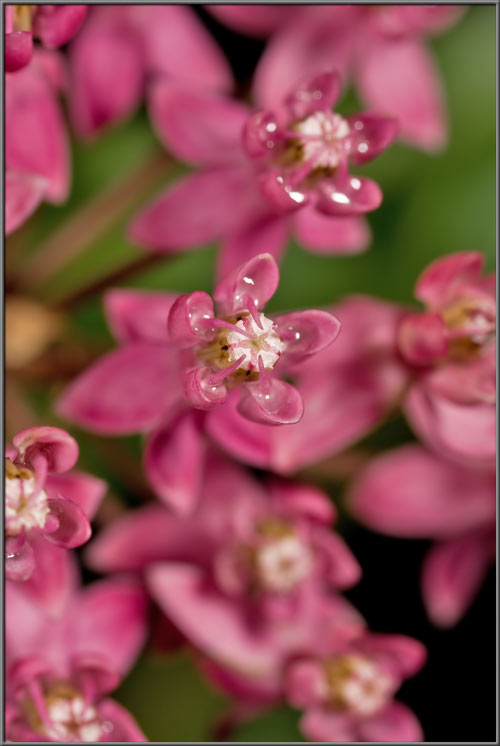
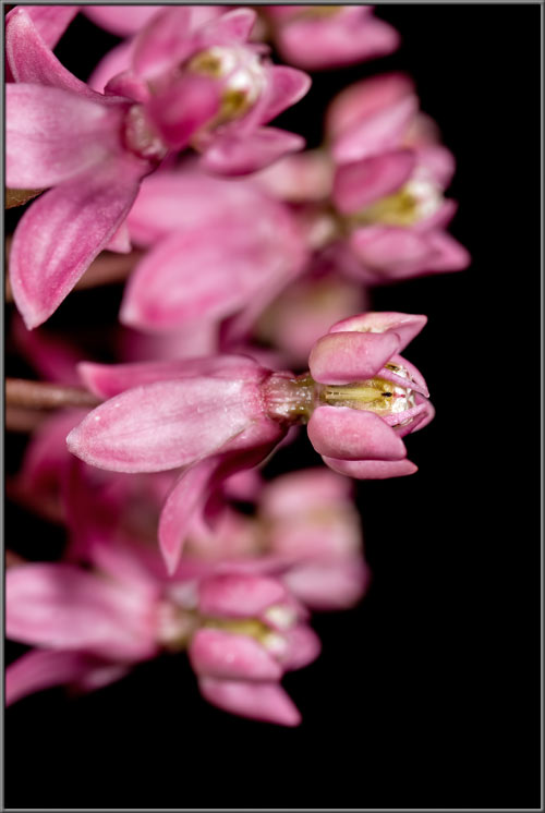
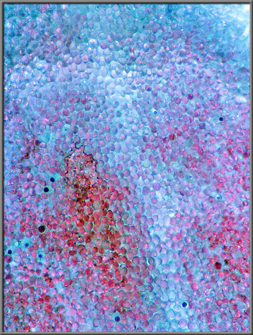
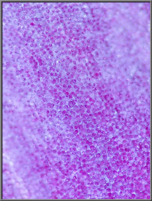
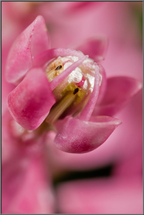
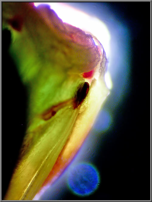
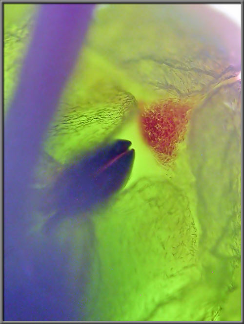
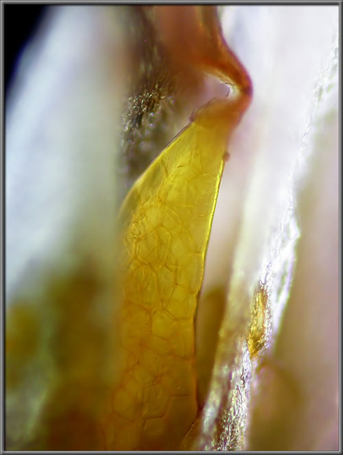
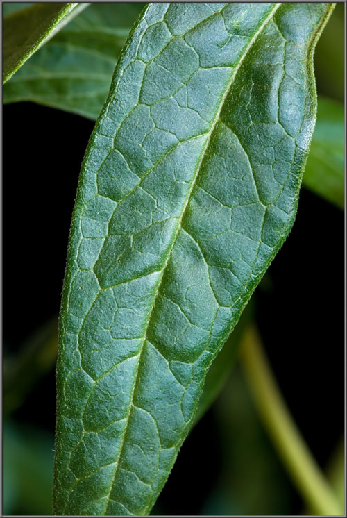
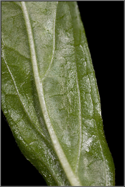
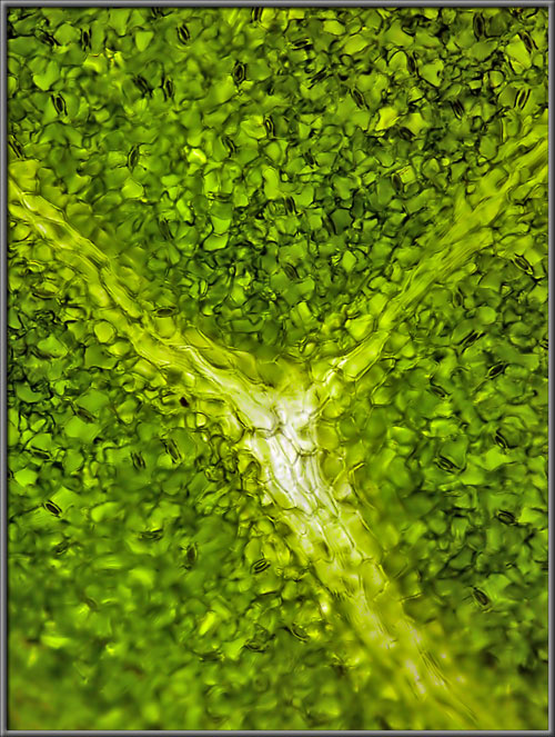
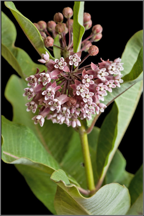
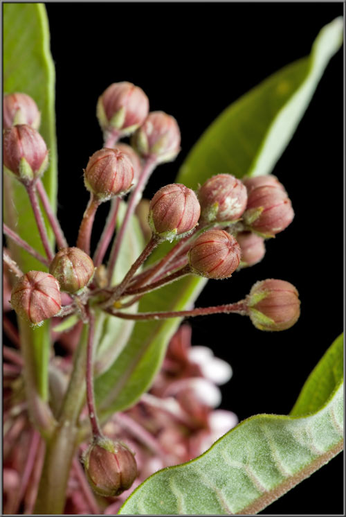
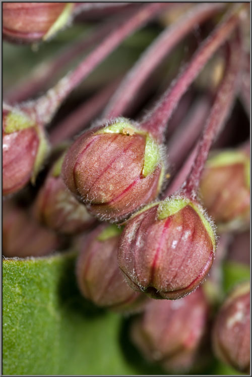
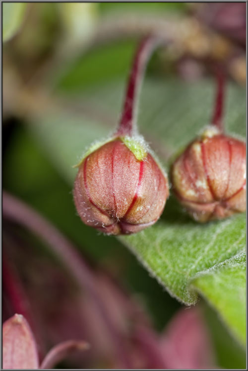
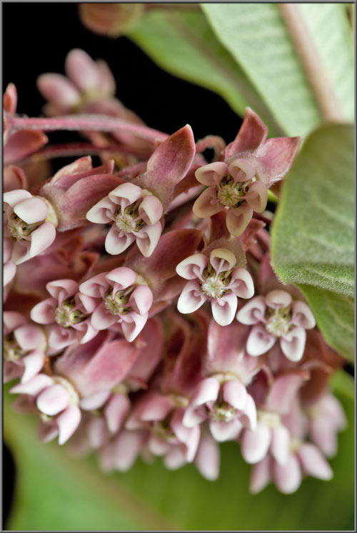
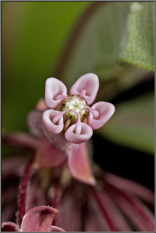
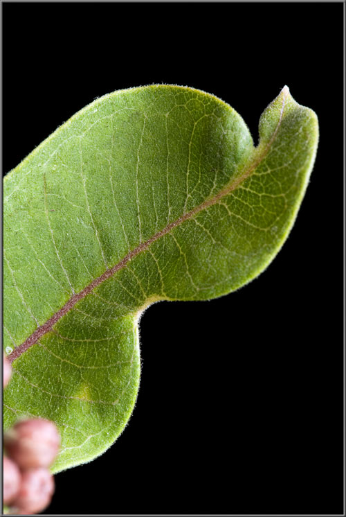
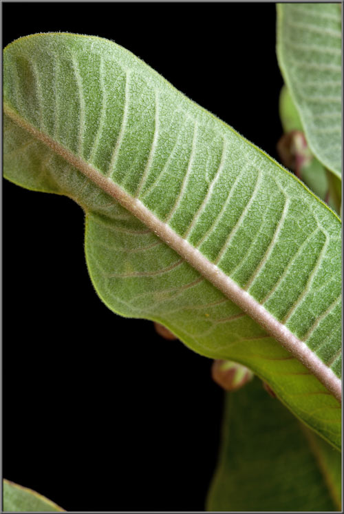
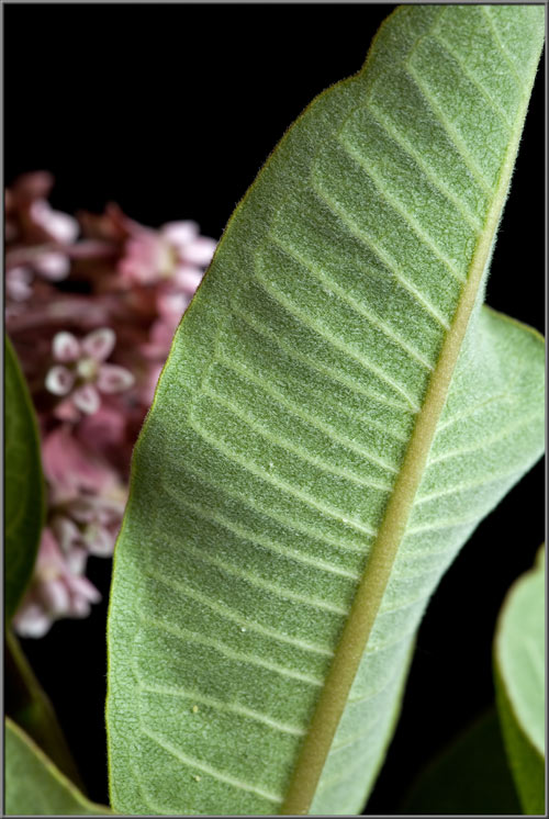
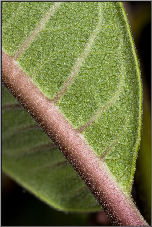
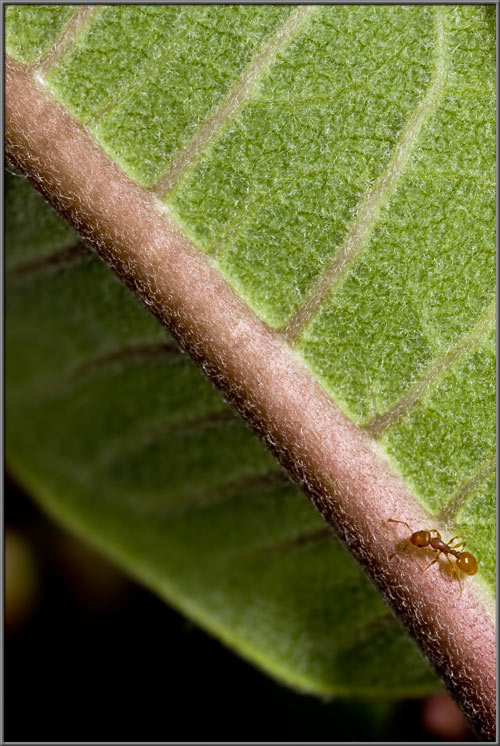
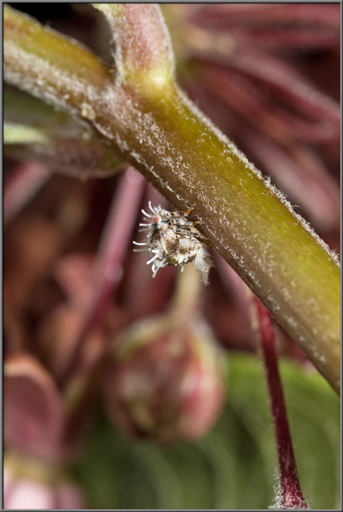
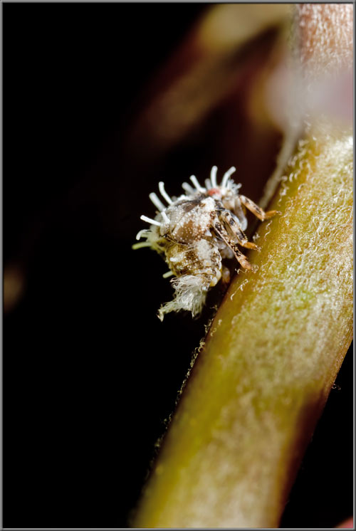
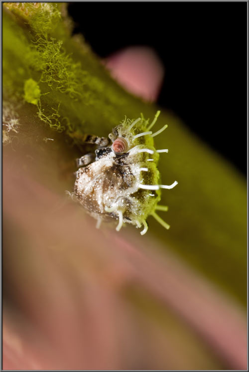
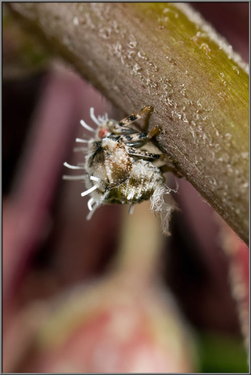

Asclepias incarnata 'Soul Mate'


by Brian Johnston   (Canada)


Although I have seen images of the native North American plant Swamp Milkweed in wildflower books, I have never come upon one in real life.  When this plant turned up at my local greenhouse it appeared to offer a chance to compare it with the ubiquitous Common Milkweed Asclepias syriaca.  The plant studied here of course, is a cultivar, and not the actual wildflower.  ‘Soul Mate’ grows to more than a metre in height, with tall, branching stems.  The plant is large, but its flowers are certainly not!  Flower diameters of approximately 6 mm are typical.

Swamp Milkweed’s flowers are mildly fragrant (vanilla-like), and deep rose-pink in colour.  Leaves are glossy green, lance-shaped (lanceolate), and unlike Common Milkweed, are not hairy.


Closer views of a group of flowers reveal each flower’s narrow waist, lower ring of five bent-back (reflexed) petals, and upper ring composed of what look like petals, but are actually referred to as corona limbs, or hoods.  This upper ring (facing the viewer in the images) is called the ‘crown’ of the flower.


Flowers are connected by their relatively long stalks to a common point at the top of a stem.  Such an arrangement is called an umbel.


The sequence of images below shows the development of extremely early stage buds.  As yet there is no indication of the colour of the final flowers.


Notice that when a more mature bud finally begins to bloom, its five lower petals are bent up to enclose the crown structure.


This arrangement is easier to see in the first of the two images below.


Strangely, in some blooming flowers, the top crown structure is not deep pink in colour as it is in most blooms.  (At this stage the outer ring of petals mentioned above has ‘reflexed’ or bent back to form the flower’s lower ring (the corolla)).


If we look down at the top of a flower, it is obvious that Milkweed is unusual.  At its centre is a white disk which in this species is the stigma.  It is referred to as the ‘stigmatic disk’.  Surrounding it are five red ‘petaloid appendages’ called ‘hoods’.  From the base of each of these hoods extends a curved ‘horn’.  The five hoods and horns collectively form the flower’s ‘corona’.  Remember from earlier that the five, pointed, bent-back (reflexed) petals at the base of the flower form its ‘corolla’.  In many of the images, a liquid can be seen glistening in the cup-like section of each hood.  This is the flower’s nectar, used to attract insects in order to accomplish fertilization.


If you look carefully between the hoods, there are obviously structures located just beneath the stigmatic disk.  These structures play a part in the flower’s fertilization, and will be looked at in greater detail later in the article.


If the tip of one of the flower’s reflexed petals is examined under a microscope, it is apparent that the cells at its edge have no pigment.  The central section appears to be light pink with many darker pink spots in a random pattern.


Two identical views taken with higher magnification show one of these large spots.  The image on the left is true-colour, while that on the right is false-colour, because Photoshop’s ‘Levels’ function was utilized to increase contrast.  The third image shows similar area.


Cells near the edge of a petal are unpigmented.


If a petal is pulled from the flower, and its point of connection to the flower is examined under the microscope, short hairs can be seen growing from the tissue.


In the images that follow, look for the tiny, black, oval structure positioned between the hoods, just under the stigmatic disk.  Between this black structure, called the ‘corpusculum’, and the flower’s waist, there runs a very narrow slit called the ‘stigmatic slit’.  Attached to the oval, black corpusculum by means of two tiny threads called ‘translators’, are two of the flower’s pollen masses.  Unlike flowers that produce dust-like pollen, the Milkweed produces sticky elongated lumps of waxy pollen called ‘pollinia’.  When an investigating insect accidentally has one of its legs slip into the stigmatic slit, it may catch the corpusculum, and as the insect removes the leg, it may pull away the corpusculum and its two pollinia.  Later the insect may fly to another flower and the pollinia may come in contact with the stigma, resulting in fertilization.  Weak insects may have a leg permanently trapped in the stigmatic slit, and may remain trapped until they die.  Others may pull away from the flower, leaving the leg stuck to the slit.


Photomicrographs follow that show some of the structures mentioned above.  The two narrow strucures that form an inverted V from the bottom of the black corpusculum are the translators.  The stigmatic slit can be seen clearly in the second and third images.


Views of the corpusculum and translators from different angles follow.


Here is an image showing the base of one of a flower’s translators (brown), and its point of connection to a pollinium (yellow).


Mature Swamp Milkweed leaves are lance-shaped (lanceolate), and glossy dark green on their upper surfaces. 


Young leaves are lighter in colour, and their upper surfaces are dotted with very short, fine hairs.


Two views of the underside of one of these younger leaves show the prominent longitudinal vein that runs its length.


Photomicrographs showing a leaf’s underside can be seen below.


For the sake of comparison, I brought in a Common Milkweed plant (Asclepias syriaca), to photograph.  (Note:  A  more detailed study of this plant can be found in one of my earlier articles.)  Although this plant is a weed, and the one studied above is not, I much prefer the Common Milkweed plant.  Both the flowers, and leaves are larger, and the muted colour pallet, is to me, more elegant.  (I guess there’s no accounting for taste!)


Like Swamp Milkweed, Common Milkweed in the bud-stage has its four ‘real’ petals temporarily bent up to protect the flower’s reproductive structures during their development.  Also note the green, pointed sepals at the bud’s base that are missing in Swamp Milkweed.


Structurally, the flowers of both species are identical.  They differ only in size (Common about four times larger than Swamp), and colouration.


This is a test.  Can you identify in the images below, the stigmatic disk, hoods, horns, a corpusculum, and a stigmatic slit?


Common Milkweed’s leaves are not shiny on their upper surfaces, and their shape is more oval than lanceolate.


The leaves’ undersides are much hairier than those of the earlier species, and here the hairs are dense and downy in nature.


Closer views of the prominent vein on the underside of a leaf, and nearby tissue explain why the structure feels ‘soft’ to the touch.


Although when cut, both plants exude toxic sap, in the Common Milkweed it is much ‘milkier’ than in the Swap Milkweed.


While taking the photographs I noticed two insects investigating the plant.  The first was a common ant.


The second was the strangest insect that I have ever seen!  In fact, it was so slow moving that I first thought it to be a bit of fluff that had been blown onto the plant by the wind.  Only when I saw that it had legs, and eyes was I forced to change my mind.  It looks like an alien insect in a science fiction film! 


If you can identify the insect, I would appreciate hearing from you.  It’s appropriate I think, that such a strange insect should be found on an equally strange plant!


Photographic Equipment


The low magnification, (to 1:1), macro-photographs were taken using a 13 megapixel Canon 5D full frame DSLR, using a Canon EF 180 mm 1:3.5 L Macro lens.

A 10 megapixel Canon 40D DSLR, equipped with a specialized high magnification (1x to 5x) Canon macro lens, the MP-E 65 mm 1:2.8, was used to take the remainder of the images.

The photomicrographs were taken using a Leitz SM-Pol microscope (using a dark ground condenser), and the Coolpix 4500.


A Flower Garden of Macroscopic Delights

A complete graphical index of all of my flower articles can be found here.


The Colourful World of Chemical Crystals

A complete graphical index of all of my crystal articles can be found here.


 All comments to the author Brian Johnston are welcomed.

Microscopy UK Front Page
Micscape Magazine
Article Library


© Microscopy UK or their contributors.

Published in the August 2013 edition of Micscape.
Please report any Web problems or offer general comments to the Micscape Editor.
Micscape is the on-line monthly magazine of the Microscopy UK web
site at Microscopy-UK  


© Onview.net Ltd, Microscopy-UK, and all contributors 1995 onwards. All rights reserved. Main site is at www.microscopy-uk.org.uk with full mirror at www.microscopy-uk.net .