|
Re-processing Old Images by Richard L. Howey, Wyoming, USA |
My computer images files are as cluttered as my lab and so I recently decided to go look at some of the archived images and see how many I could eliminate and then do some reorganization. However, I quickly discovered that there were some interesting images that had only been marginally processed before (or, in some cases, not at all) and that many of my earliest images were of a ridiculously small size. Why I did that I now have no explanation for. As a consequence, I decided to take a few of the images which intrigued me and experiment with re-processing and enhancing them. I quickly discovered two important facts: 1) not all images are worth attempting to re-process (obvious, in one sense, but not always immediately discernible due to the second fact), 2) digital imaging technology is extraordinary in that it can capture detail that is not immediately evident and is sometimes revealed only after considerable tweaking. Here I don’t mean that one goes in and alters the image as such, but rather uses the means of the graphics program to reveal content that is indeed there, but for a variety of reasons may be hidden or obscured. Clearly, there are also reasons to go in and alter parts of an image; for example, I frequently eliminate unnecessary clutter around the central object by homogenizing the background. My perspective is that the image should present the main content with the least distraction and noise.
In this essay, I want to concentrate on showing you the kinds of improvements that are possible and a few examples where re-processing the image is simply not worth the time and effort or other cases where it’s very difficult to decide which image is better. In most types of cases, I will use a “before” and “after” format of presentation so that you can easily compare them. The original image will always appear on the top and the re-processed image directly below it.
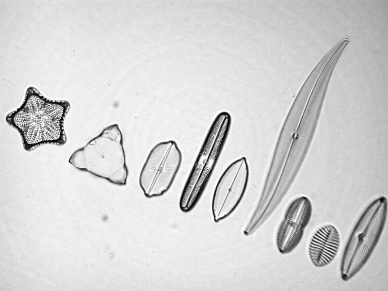
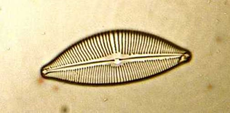
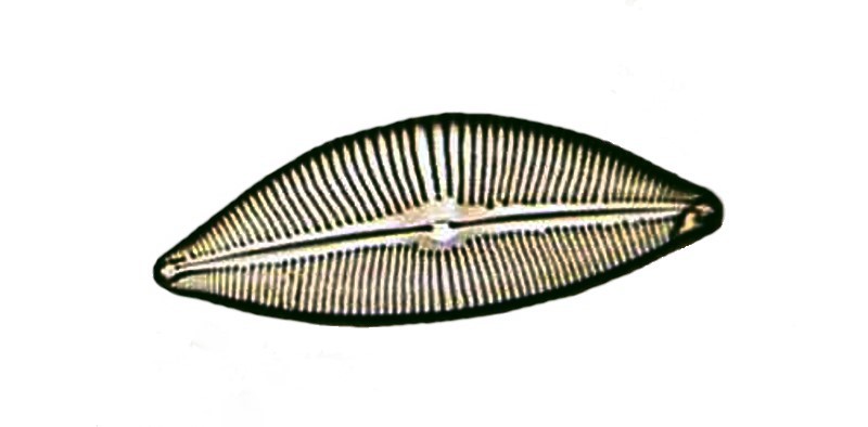
Let’s begin with a test slide of diatoms. The original image appears first and the only thing I have done to it is cut the size to make it fit a reasonable format.
The re-processed image was treated with the “focus” effect, “level”, “contrast”, and then I “cleaned” the background to get rid of any “noise” that distracted from the diatoms.
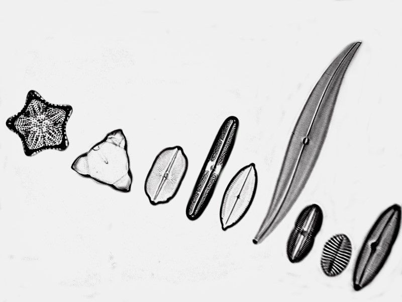
As you can see the diatoms are slightly sharper and there is less noise to distract the eye. So, in this case, the image was definitely worth re-processing.
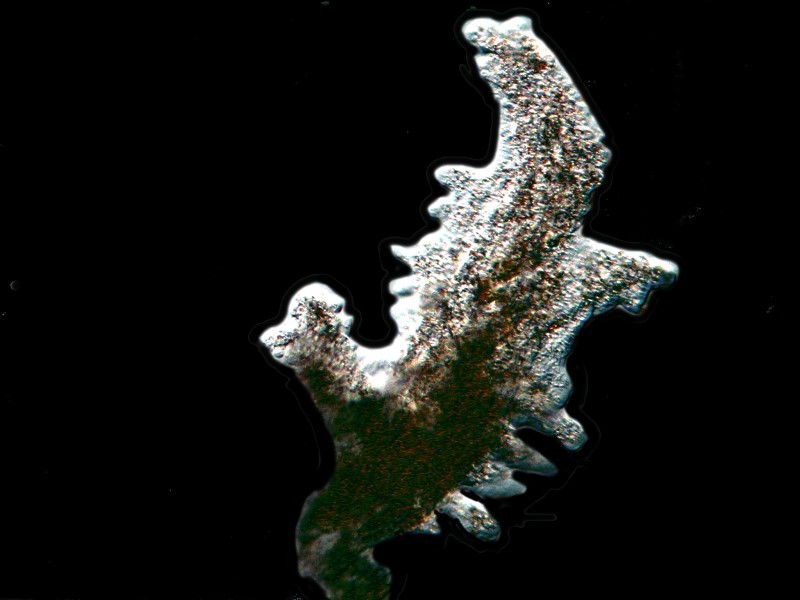
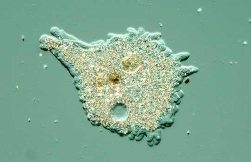
The next set is more ambiguous. This Amoeba proteus was taken with Nomarski Differential Interference Contrast (DIC). There are elements to recommend this image as it does have a kind of clarity and detail, particularly in the upper half of the organism.
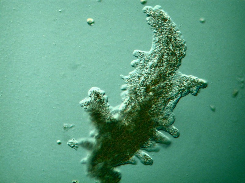
The re-processing was fairly dramatic in this case and involved shifting the color balance of the organism, increasing contrast, and radically altering the background which creates a sense of slightly more dimensionality in the amoeba which, in my view, justifies the effort of re-processing.
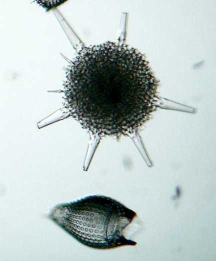
Next, let’s look at a couple of radiolaria. I’ll show you first the original ridiculously small image [432 x 72] (as I said above, I don’t know why I ever did this) and then the original image in what has now become my standard format for Micscape [800 x 600].

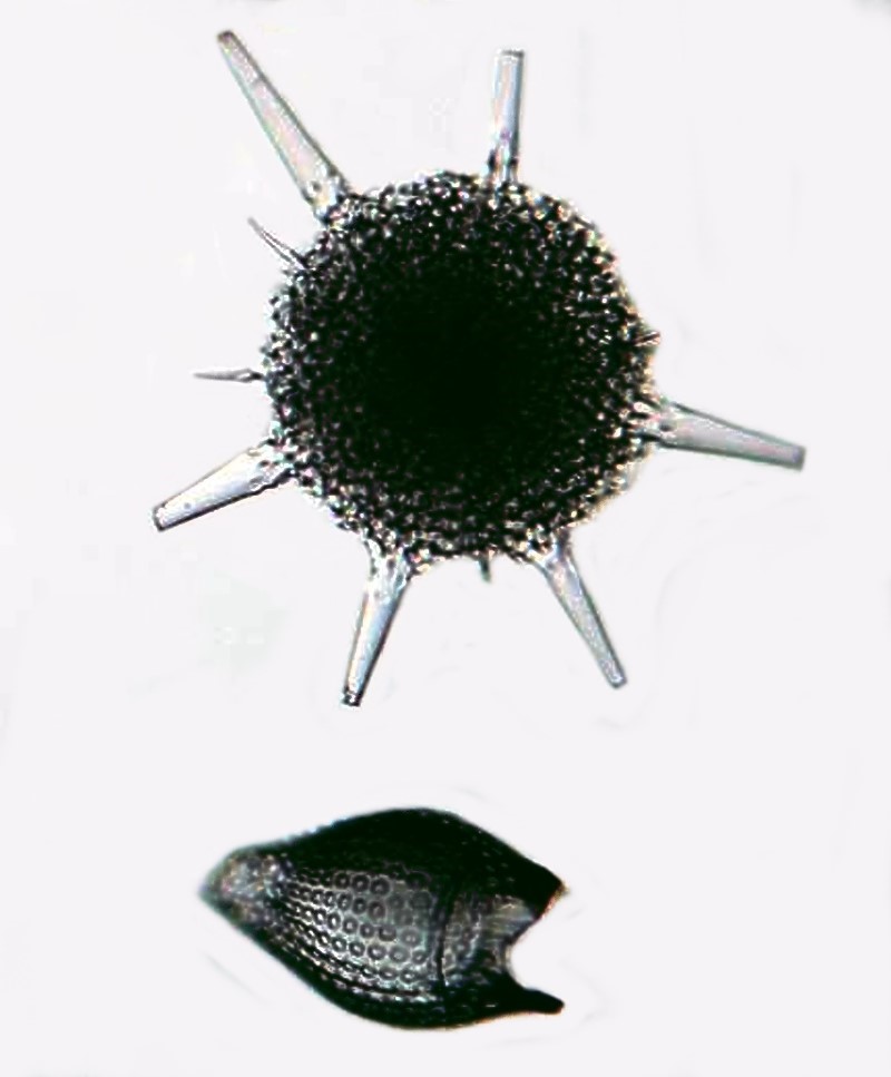
In re-processing, I used the “focus” function, increased contrast, changed the color balance and cleaned the background using white to further increase the contrast. You will notice that in this image the “spikes” have a more vitreous appearance which is desirable since the “test” of the shell is siliceous.
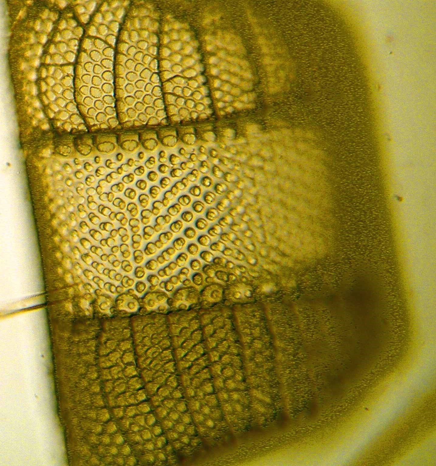
Another diatom reveals a different sort of problem. This form is relatively thick and thus focus becomes a problem. At the time this was taken, I had no “stacking” software. As you can readily see, the original image is quite fuzzy.
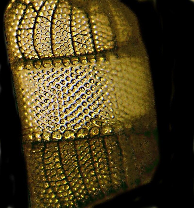
However, by using a variety of effects, including “Unsharp Mask”, I was able to achieve a much more pleasing image, but one that is certainly deficient now compared to the results one can obtain using stacking software.
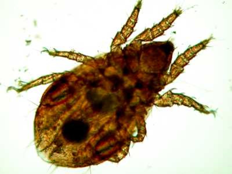
Next up is a tiny mite. In the un-processed image, the image is again rather fuzzy and the “noise” detracts.
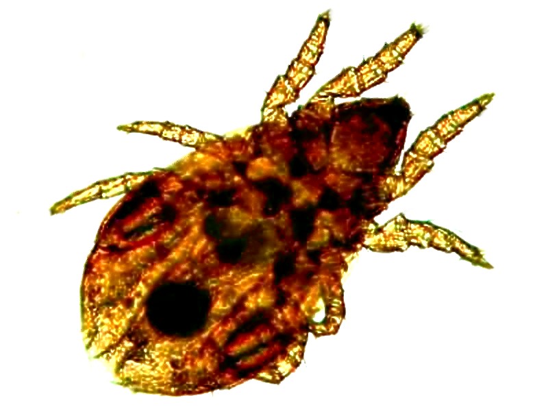
Using the focus functions and cleaning the background improves the image considerably. Both the body and the appendages are sharper and one can see more detail. This is another case where, due to the depth of field of the subject, stacking software would be a distinct advantage.
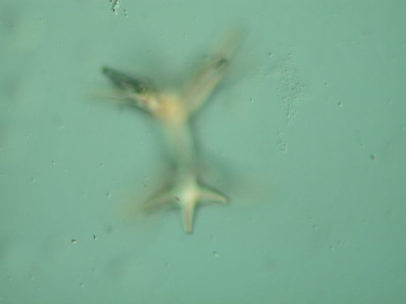
Just to show you how much difference stacking software can make, I’ll show you an example of a glass sponge spicule. My poor ancient memory tells me that about 60 images were “stacked” to get the final result. This particular spicule is “spiky” and thus has considerable depth of field. This first image is the beginning one and then the next is the final result.
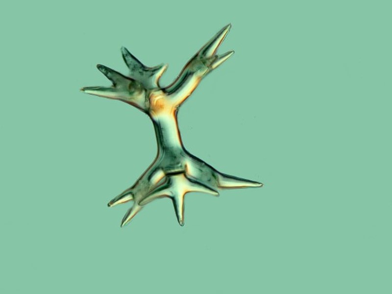
This type of software is a great boon to the microscopist who is dealing with “thick” specimens.
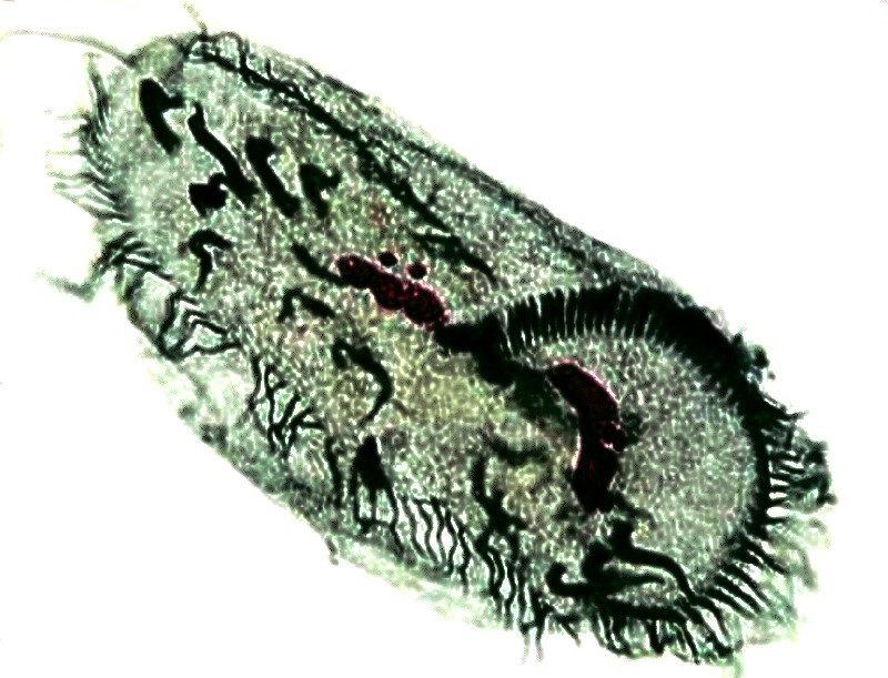
Some images of protists can be significantly improved by re-processing as is the case for this image of another amoeba. There is also an advantage in terms of the image having been taken using DIC.
The image is reasonably good to begin with.
However, by sharpening the focus, the image shows enhanced detail, and enhanced depth of field, and the alteration of background increases contrast.

Sometimes, simply altering the background to enhance contrast can improve an image considerably as we can see with this diatom.
At other times, tweaking an image too much can result in a degradation of the image. In this case, we have a nicely stained specimen of the hypotrich Stylonichia and in the first case, I used only focus function.
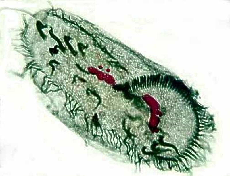
The second image was re-processed using a variety of functions and clearly to the detriment of the image. So, one needs to know when to stop trying to “enhance” and not be so fussy as to over process. Sometimes it is tempting to work out a processing routine using a fixed series of functions and then apply that in a rather mechanical fashion. In general, I think this is a rather poor approach and I think it’s much preferable to treat each image as something unique and to experiment and work out which functions truly enhance that particular image.
This is a clear example of re-processing gone overboard.
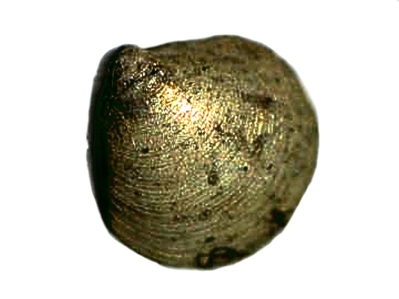
Next, is an example of a tiny clam shell which I subjected to a silver staining process and clearly this image needs enhancement.
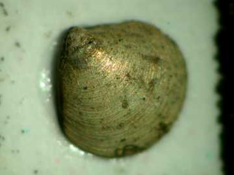
A somewhat better focus was acquired and contrast was increased by altering the background.
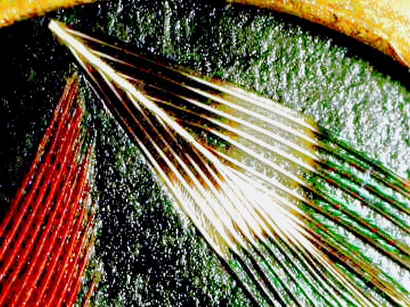
A very nice result is sometimes attained by using the “level” function and changing the color balance. In this original image of a feather, the colors are somewhat muted.
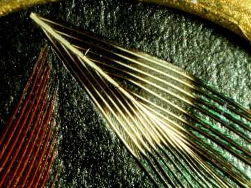
However, when re-processed the colors are more vivid.
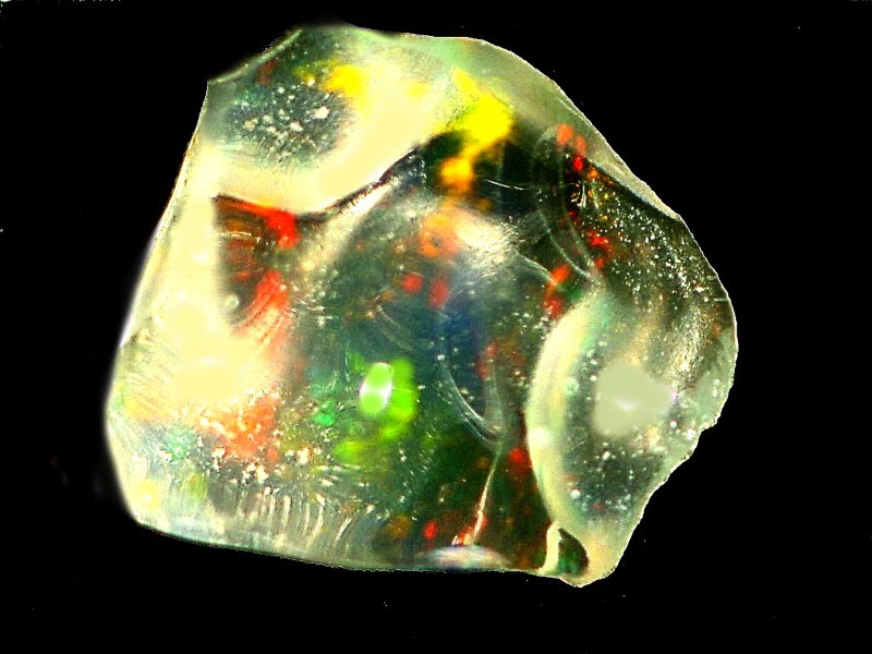
Some types of specimens present special challenges and in this case it is a small fragment of opal. Because it is colorful, translucent, and highly reflective it is a difficult subject to photograph.
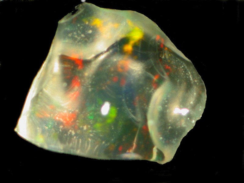
However, with a bit of processing, it becomes an attractive and interesting image.
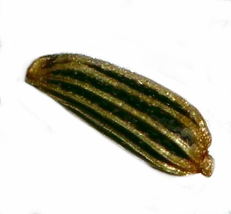
An extensive set of images could be used to examine a wide variety of seed types. I will show you one example here. Again, stacking software could improve it further, but none of the images taken here utilized that since, at that time, I had no such software available.
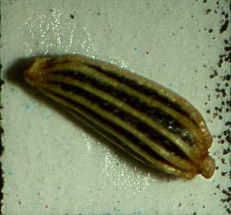
Nonetheless, the computer graphics software does transform this into a presentable image.
In the process of undertaking this investigation into archived images, I discovered many which could be discarded, but more importantly, many which could be re-processed and used in the future to provide soporific articles for Micscape readers, because remember, sleeping pills are bad for you.
All comments to the author Richard Howey are welcomed.
Editor's note: Visit Richard Howey's new website at http://rhowey.googlepages.com/home where he plans to share aspects of his wide interests.
Microscopy UK Front
Page
Micscape
Magazine
Article
Library
Published in the August 2016 edition of Micscape Magazine.
Please report any Web problems or offer general comments to the Micscape Editor .
Micscape is the on-line monthly magazine of the Microscopy UK website at Microscopy-UK .
©
Onview.net Ltd, Microscopy-UK, and all contributors 1995
onwards. All rights reserved.
Main site is at
www.microscopy-uk.org.uk .