|
An Assortment of Marine Invertebrates From the Philippines Part 3: Miscellaneous Groups by Richard L. Howey, Wyoming, USA |
In this segment, I want to look at some specimens other than echinoderms, specimens which over the years have at one time or another captivated my interest. We’ll begin with sponges. It used to be that people were quite familiar with sponges as they have many household uses. Now, however, we have a variety of synthetic sponges and sponge substitutes which we use for cleaning, scrubbing, and even bathing. Nonetheless, I’ll assume that you already know what a traditional household sponge looks like and we’ll take a look at some of the less typical sponges. This first example looks almost like a small piece of sculpture and it’s certainly something you wouldn’t want to use for bathing as it’s composed of silica, in other words, glass.
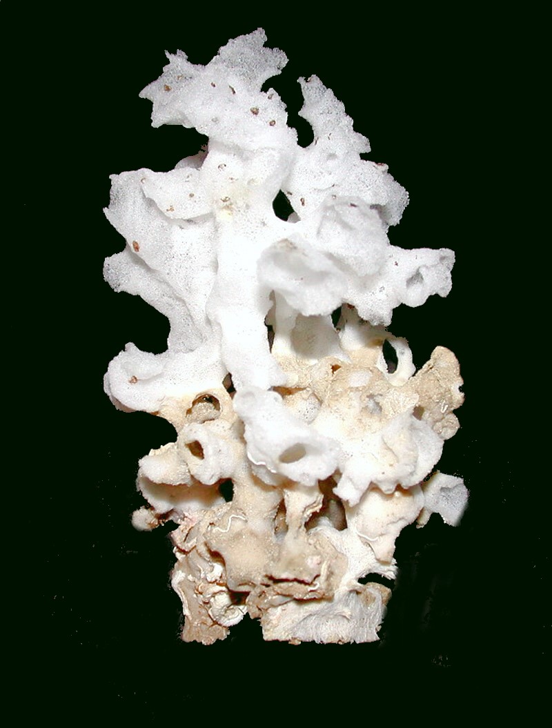
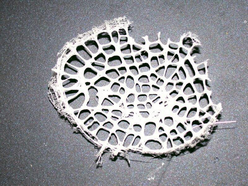
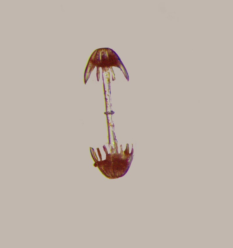
As it turns out some of the oddest and most beautiful sponges are what are called “glass sponges” or hexactinellids due to a prominent 6 pronged type of spicule, a tiny “skeletal” structure composed of silica. Perhaps the most famous one is the Venus Flower Basket or Euplectella aspergillum. Its skeleton is a beautiful piece of architecture and it has some remarkable characteristics, so I will show you several aspects of this remarkable creature. First off, it should be noted that it is not a single animal, but it is colony consisting of thousands of cells. First, here is an image of the whole skeleton of the colony.
From this view, one can’t get an idea of the most interesting features and so let’s look at some closeups. First off, a closeup on the central area of the “tube”.
Next, a closeup at the top where you can get a side view of the osculum or “sieve plate” on the top of the tube.
And here is one of the osculum which was damage in separating it from the tube.
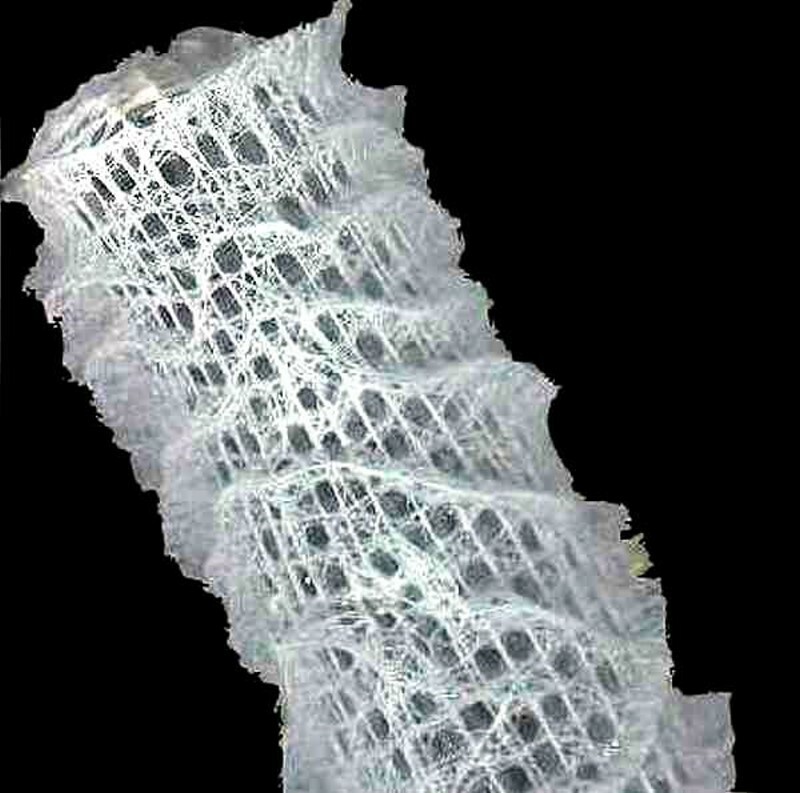
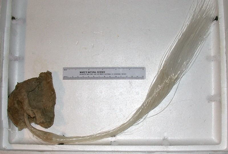
In previous views of the tube, we could see ridges of spicules going up the side at a steep angle like a staircase designed by an inebriated architect. In the image below, we learn 2 interesting things: 1) the central column is defined by a series of circular rings and 2) we can see some long “threads” rising up from the base and lying along side the column. This creates the wrong impression. When these sponges are collected, they are almost always bundled in this manner for shipment; however, in their natural habitat, these long thread for a kind of anchor and extend down into the detritus. These glass threads are optically superior to any fiber optic we can yet create. In the substrate, a wide variety of shells of forams, diatoms, pteropods, etc. become attached to this “anchor” and thus it becomes an interesting object of investigation in and of itself.
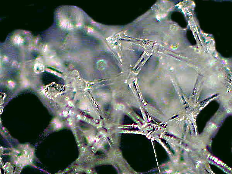
If we take a tiny bit of the tube and examine it under the microscope, we discover yet another level of complexity, for it becomes evident that this skeleton consists of thousands upon thousands of fused glass spicules.
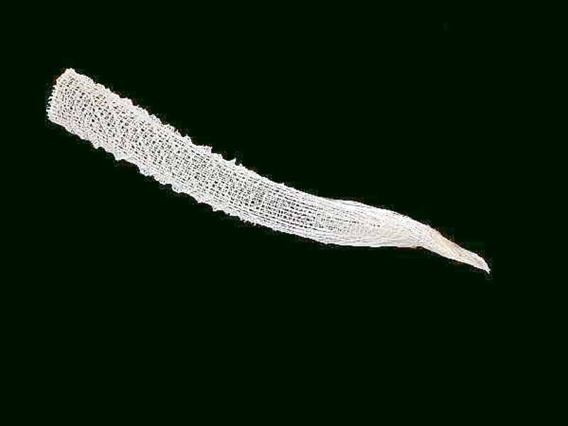
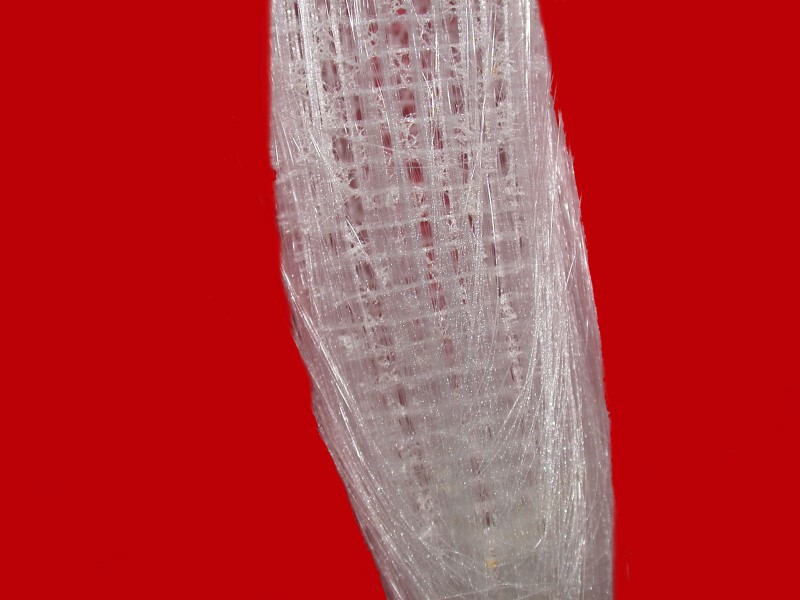
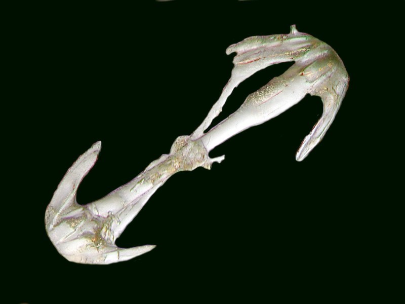
Glass sponges present many complicated puzzles which researchers have been investigating using highly sophisticated techniques, including Scanning Electron Microscopy to try to better understand the processes involved in the creation of these spicules and fibers. There are some other species of glass sponges that have anchor fibers even longer and possibly more complex than those of Euplectella. One is Hyalonema, which I call the “sponge with a tail”. It is also known as the glass rope sponge. I’ll let you have a look for yourselves.
The fibers are a bit over 18 inches in length and have extraordinary optical properties which I have discussed in other Micscape essays on Hyalonema and Euplectella. The fibers of another species Monoraphis chuni can grow up to 10 feet and a rather remarkable claim has been made that this sponge can live up to 11,000 years! (Needless to say, this a matter of some debate.) Researchers at the Max Planck Institute at Potsdam-Golm have been investigating the ultra-microstructure of these fibers. (Follow this link to learn more.)
The variety of spicules in these glass sponges is quite impressive and I’ll show you just a few examples here.
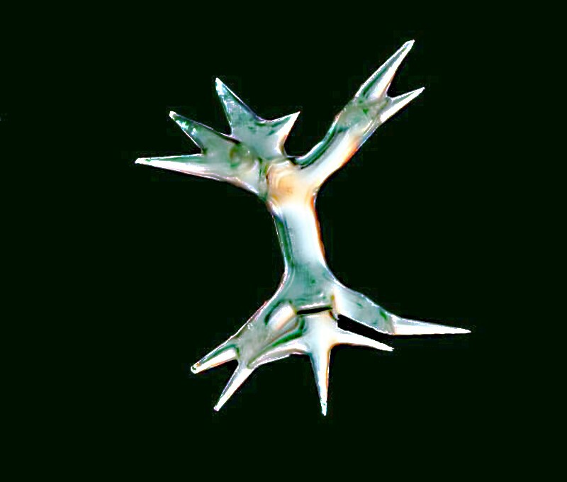
Here are 2 images of the so-called “dumbbell spicule”.
Next is what I call the “Christmas tree spicule”.
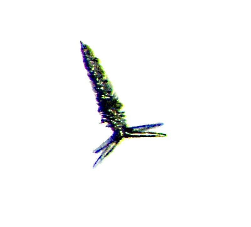
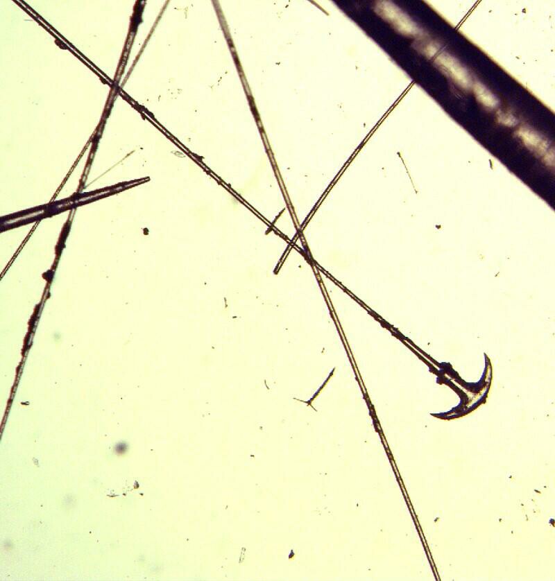
Then an anchor-shaped spicules on a long glass fiber.
And finally, a spicule which looks rather like a micro-version of some exotically dangerous Japanese throwing weapon.
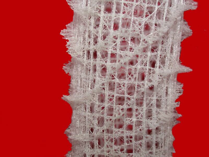
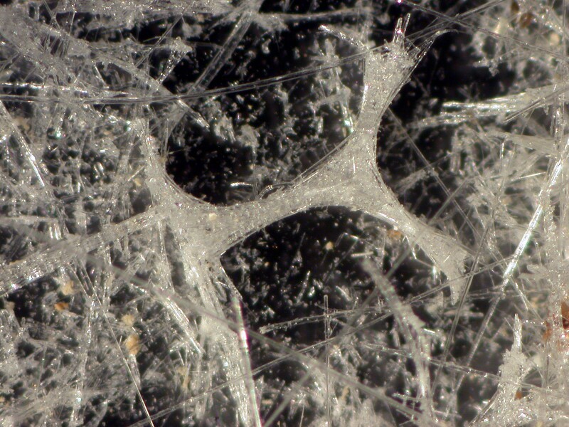
As I mentioned, glass sponges fuse spicules to form highly intricate lattices as you can see in the image below.
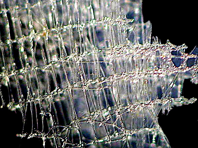
If we look even closer, we can see the joints criss-crossing and the construction looks a bit like it had been made out of glass Tinkertoys.
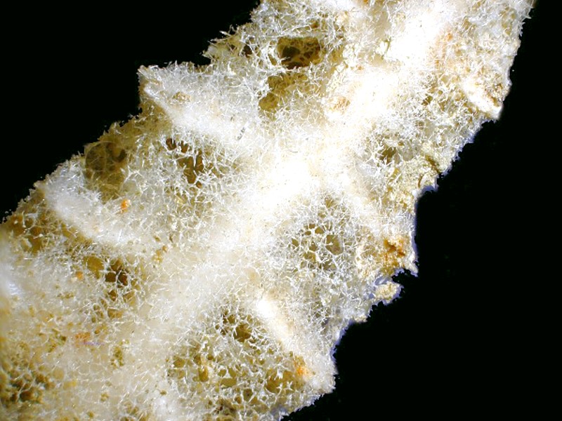
Sponges are incredibly adaptable and can be found in surprising niches such as the one below that has “colonized” the outer surface of a “thorny” sea urchin spine.
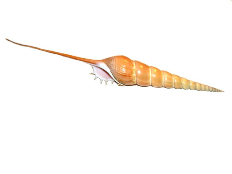
One area of collecting which I have tried to minimize is that of the molluscs, largely because there are so many beautiful shells that one could easily become overwhelmed. However, there are certain ones that are irresistible and I’ll show you 4 that I think fit into that category.
The first might be thought of as a unicorn gastropod; it is the Tibia fusus which I find intriguingly graceful.
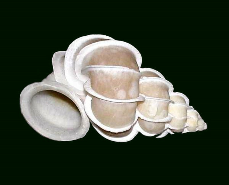
Next is the miniature “spiral staircase” of Wentletrap derived from a Dutch word. It is a small shell, but has a kind of highly appealing elegance and perhaps it’s just because of the derivation of its name, but it does for some reason make me thing of M.C. Escher.
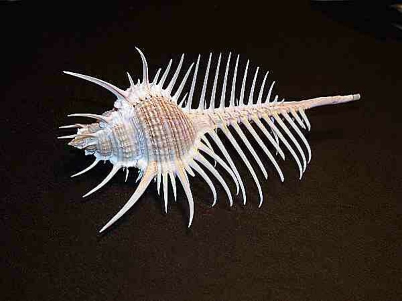
A very aristocratic contribution to any collection is the Venus Comb murex. It rather reminds me of a scaled-down version of one of Theo Jansen’s Strandbeest.
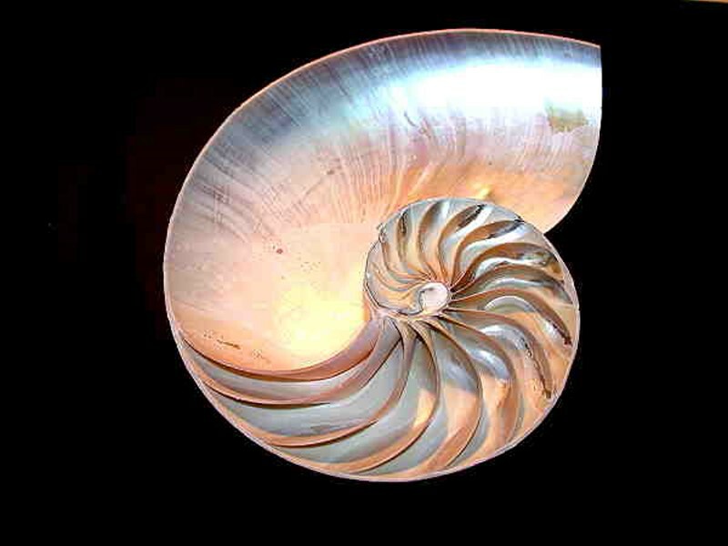
And no collection would be complete without a sliced Nautilus shell. Here we encounter one of the most direct and pleasing intersections of Nature and Mathematics and Art. This gorgeous spiral can be described by the Fibonacci sequence which generates a logarithmic spiral. The sequence consists of 1,1, 2, 3, 5, 8, 13..., that is after the initial “1s” each number is generated by the sum of the two preceding numbers. Claims have been made for the descriptive Fibonacci sequence in relation not only to certain shells, but to the florets of certain flowers especially sunflowers, pine cones, spiral nebulae, and the uncoiling tendrils of certain plants, especially ferns. (Follow this link and this to learn more.)
And now, here is the sliced Nautilus shell in all its delicacy.
We are slowly learning how much there is yet to know from marine organisms about a wide range of phenomena that can provide us with clues to develop new approaches to the solution of complex problems, such as, the creation of better fiber optics. Another example is from studies of the “lowly” barnacle which Darwin spent eight years studying and produced 4 books on them. One of the intriguing aspects of the “rock barnacles”, which anyone who has visited a rock beach is familiar with and may even have the vestiges of a few scars from a slippery fall in their area, is the extraordinarily strong “glue” by means of which they attach themselves to their substrate. It is very difficult to pry them loose without breaking them. This adhesive has been studied for among other possible uses as a aid in dentistry. There is charming story about Darwin’s son, George, visiting the house of a friend and being told that his father didn’t have a study, whereupon George asked: “But where does your father do his barnacles?”
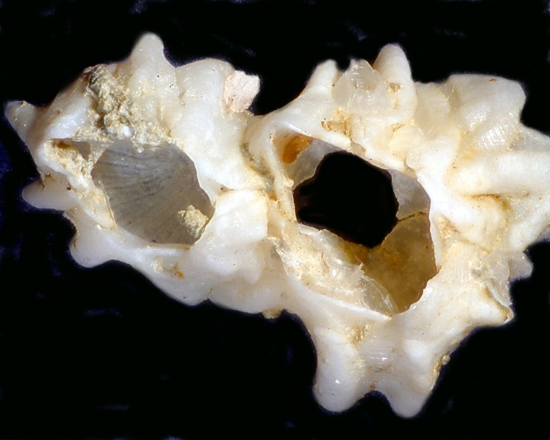
The barnacles are the “curl feet” or Cirripedia and they live, from our point of view, upside down. The “tentacles” by means of which they feed are actually their feet. They are possessed of strong calcareous plates and below is an image which shows you 2 rock barnacles that have been hollowed out so that just the “shell” remains.
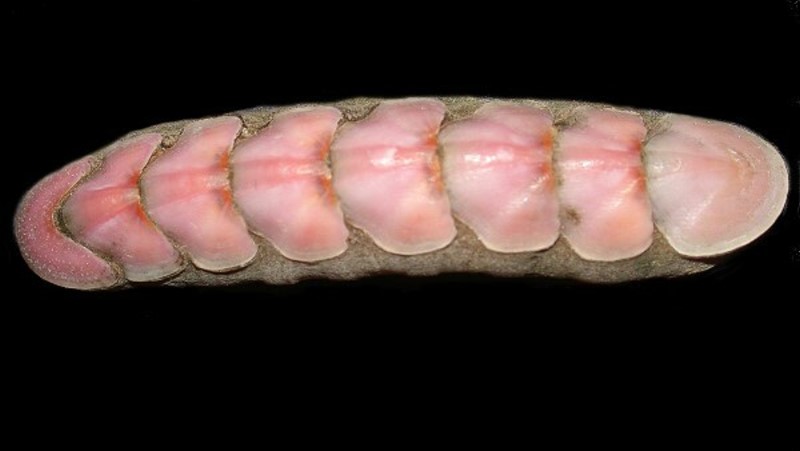
Enormous numbers of marine organisms have developed protective armor of one sort or another. Chitons too have a series of calcareous plates that help protect them. Incredibly some species also have develop “eyes” (ocelli) and may have thousands of them. If you want to read further about chitons and their eyes, I have written several articles for Micscape on these intriguing creatures.
Below is a colorful example of a chiton.
I have teased my wife that we could take a couple of these and remove 10 of the plates and then she could glue them onto her fingernails. She was not impressed.
Some animals camouflage themselves by using bits of detritus from their habitat; there are, for example, the decorator crabs, some mollusks that cover their shells with other shells, crabs that place anemones on the carapace or carry them around in their claws to use as defense. I’m going to show you a bristle worm that covers itself with bits of detritus and creates a very effective camouflage.
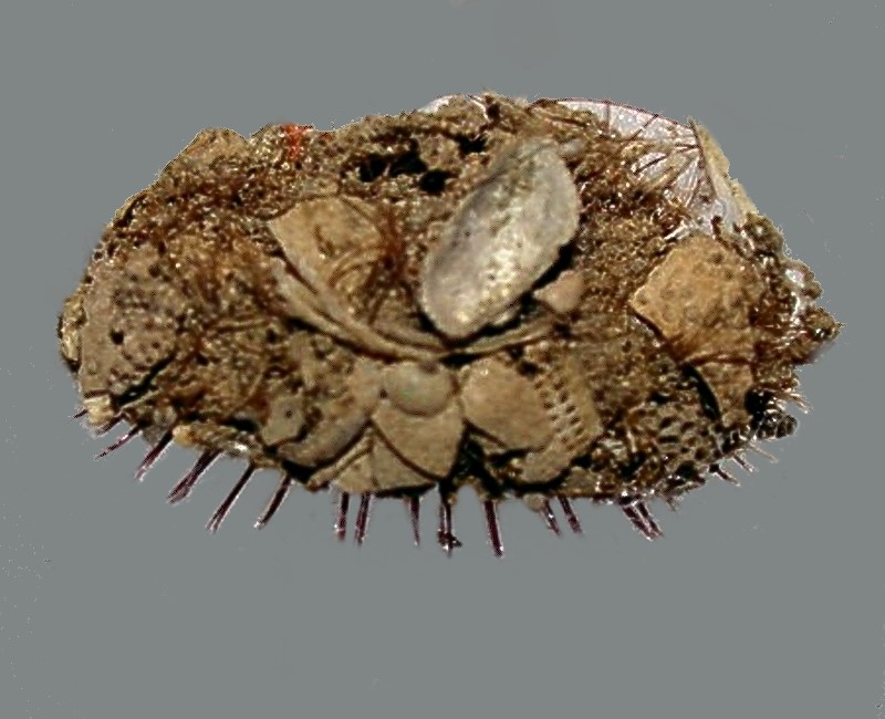
As you can see, there are fragments of bivalve mollusk shells, a small gastropod shell, and a number of piece of sea urchin test.
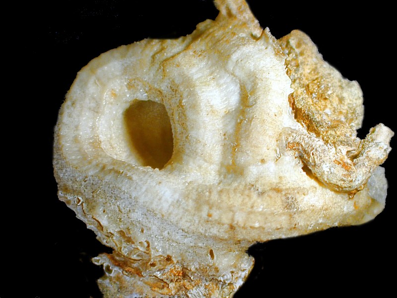
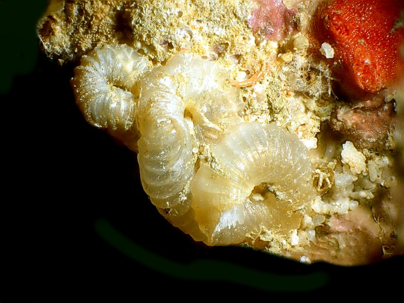
Finally, let us look at a couple of worm tubes. Usually these are calcareous and often spiral and on this first one, you can see worm tubes on the worm tube.
In this second image, you can see 3 coiled tubes which have a translucent character and may well be a complex mixture of organic and calcareous material. You will also notice some bright red and pink material which looks porous and one might think that his is a bit of red coral; however, amazingly it is a very unusual foraminiferan called Miniacina miniacea.
I was first introduced to these remarkable unusual creatures by means of a sample from Brian Darnton which he collected in Malta. I think he was having a bit of fun by asking me what I thought these red bits were. I, of course, jumped at the conclusion of red coral and then he kindly informed me as to their true nature.
I hope you have enjoyed this whirlwind tour of some Philippine marine specimens.
All comments to the author Richard Howey are welcomed.
Editor's note: Visit Richard Howey's new website at http://rhowey.googlepages.com/home where he plans to share aspects of his wide interests.
Microscopy UK Front
Page
Micscape
Magazine
Article
Library
Published in the August 2017 edition of Micscape Magazine.
Please report any Web problems or offer general comments to the Micscape Editor .
Micscape is the on-line monthly magazine of the Microscopy UK website at Microscopy-UK .
©
Onview.net Ltd, Microscopy-UK, and all contributors 1995
onwards. All rights reserved.
Main site is at
www.microscopy-uk.org.uk .