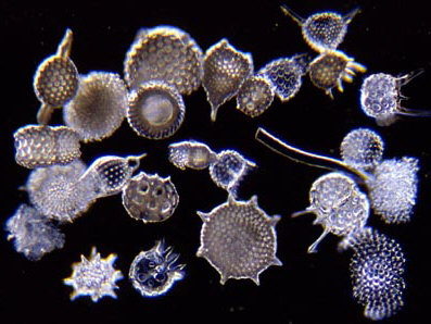
Radiolaria shells. Darkfield illumination.
Image by Wim van Egmond.
Mounting Radiolaria Shells |
There is a long tradition in microscopy of a blending of scientific and aesthetic perspectives. Almost every microscopist has had a particular group or type of specimen to which he or she has a special partiality. For some, it is the marvel of micro-crystals under polarization; for others, it is diatoms or desmids, or foraminifera or radiolaria. It's not really necessary to create a hierarchical ranking; each of these types of specimens has its own unique charm and interest. Certainly, no one can deny that radiolaria are among the most beautiful and intricate denizens of the microworld.
Radiolaria shells. Darkfield illumination.
Image by Wim van Egmond.
From a cursory examination of bibliographies, I am under the
impression that the literature on radiolaria is rather limited
compared to the literature on forams and diatoms. There certainly
doesn't seem to be any convenient reference that provides a good
survey for the amateur, which means that getting a sense of the
taxonomy of this group is very difficult. There is an excellent
technical book simply called Radiolaria by O. Roger
Anderson published by Springer Verlag, but its current price is
$205.00!! If they sold it for a quarter of that price, they
probably would sell ten times as many. Springer is notorious for
charging outrageous prices for technical books and having them
show up a few years later remaindered for a tenth or even a
twentieth of their original prices. Seems to me a funny way to
run a business. Hey, is anybody from Springer out there
listening?
All of this is further complicated by the fact that many of the
forms are known only by their shells and this is true of both
fossil and extant species. To make matters even more interesting,
the shells of some species show some morphological variation and
furthermore most samples are processed in such a manner that a
significant proportion of the specimens are chipped or broken and
very often this involves the loss of spines and other structures
which may be crucial in identification.
In this discussion, I will limit myself to dealing with shells
from "radiolarian ooze." Dealing with living,
planktonic forms is entirely another matter and one which,
unfortunately, I know nothing about.
So, where do we start? The first thing is to purchase a small
vial of cleaned shells from "radiolarian ooze." This
ooze can be several hundred feet thick at certain spots on the
ocean floor. It is not so easy to dredge up and not easy to
clean, so the samples are moderately expensive. When you get your
vial from a biological supply house, your initial reaction on
examining it may be shock—What? I just paid $20 for a half
inch of sand in the bottom of this little glass tube? But rest
assured, you have made a wonderful investment; that tiny sample
will contain thousands and thousands of elegant glass (silica)
shells.
The sample may be stored in distilled water, water and glycerin,
or alcohol. If you wish to examine the shells in a liquid medium,
the distilled water is preferable. Don't use tap water or
artesian water as these contain salts which can form deposits on
the shells if you decide to let them dry for dry mounting or they
can cause annoying precipitates in mounting media.
A classical problem in mounting radiolaria (and diatoms) is their
transparency as a consequence of their siliceous composition. If
you use a standard mounting medium, then you are looking through
glass lenses, glass prisms, a glass coverslip, a glass slide and
more glass in the condenser which is providing the illumination
for these tiny glass shells mounted in a substance that has a
refractive index very close to that of glass—and so, the
radiolaria become virtually invisible. The traditional solution
has been to mount such specimens in a medium with a considerably
higher refractive index than glass. This approach has several
disadvantages. Some of the best media are now virtually
unobtainable. Most available media are very expensive and
frequently present a greater problem with air bubbles forming
around the specimen than do standard media. Worst of all, some
media that produce the highest refractive indices, such as
realgar, involve extremely dangerous techniques that should never
be attempted by the amateur.
Almost every type of specimen presents special challenges in
order to view it under optimal conditions, but radiolaria are
among the most difficult. As we have already noted, the fact that
they are glass presents special obstacles and even though diatoms
are similarly constituted, they are generally thinner and, as a
consequence, do not present the same degree of difficulty in
terms of depth of field. My recommendation is that if you want
mounts of radiolaria in a medium with a high refractive index,
purchase them from a biological supply house which produces high
quality preparations. These are relatively inexpensive and a good
addition to one's collection of permanent slides.
For making my own preparations of radiolaria, I decided on a
quite different approach. It occurred to me that if I could find
a means of coating them, then I could use oblique illumination
and also not have to worry about having a mounting medium with a
high refractive index So, the first task was to find some kind of
stain or coating which would provide the right sort of contrast.
Attempting to stain silica using biological stains would, I knew,
be hopeless; however, I thought that perhaps a thick, but not too
thick solution of stain, might deposit a thin film and provide
the right sort of contrast. I tried Malachite Green, Alizarin Red
S, Orange G, Nigrosin, and Neutral Red—and the results were
either no staining or a sort of sticky mess that obscured the
structural details of the shells. Finally I decided to try out
the silver staining method that I had developed for forams. I
have already described this technique in another article titled: Silverizing Forams; however,
I will give a brief summary here as it applies to radiolaria.
It is imperative that the specimens be clean and free from salt
deposits. Put a sample in the bottom of a small vial and
vigorously add distilled water using a pipet or wash bottle. This
will agitate the tiny shells in the water helping to remove any
deposits. Allow the sample to settle for about an hour and then
using a micro-pipet, carefully remove most of the water. Repeat
this process several times always being sure to allow sufficient
time for the shells to settle back to the bottom. Transfer a
minute sample of the shells to a clean slide and, when dry,
examine them under the microscope. If the shells are till not
clean, it may be that there is an alkaline deposit. This can
generally be removed by treating the sample with dilute acetic
acid in the manner in which the washing with distilled water was
done. After the treatment with acid, it is then necessary to
rinse the sample several times with distilled water again.
Once the shells are clean, we can begin the staining procedure.
Use a small, shallow glass or plastic dish and transfer a small
sample of the shells to one edge of the dish in as little water
as possible. Add 2 or 3 drops of 1% silver nitrate solution (This
can usually be made up by a local pharmacy) and allow the
specimens to sit for 5 to 10 minutes or even longer. Time is not
crucial to this part of the procedure.
Prepare a Vitamin C solution by dissolving a 1000 mg. tablet in
60 cc. of distilled water. Add 2 or 3 drops of this to the shells
in the dish. At this point, timing becomes important. The
specimens will begin to turn brown as the silver begins to
precipitate and then a gray film will start to form on the
surface of the fluid. About 2 minutes after the gray film forms,
remove as much liquid as possible using a micro-pipet. Next
squirt 90% alcohol over the radiolaria. This helps remove
undesirable clumps of silver particles from the specimens, but
without removing the tiny layer which we want, since silver
nitrate while readily soluble in water, is only very slightly
soluble in alcohol.
Transfer a small drop of radiolaria to a clean slide and allow
the alcohol to evaporate. Add a drop of xylene, position the
radiolaria and then add the mounting medium. (Caution: Xylene is
poisonous and highly flammable.) The silver stain produces very
good contrast and specimens stained in this manner are best
observed with oblique top illumination.
A Note Regarding Silver Nitrate
All metal salts should be handled with caution and silver
nitrate is no exception. Both the crystals and the solutions are
poisonous and caustic. Silver nitrate is also highly reactive and
should not be mixed with chemicals other than those specified.
Comments to the author Richard
Howey welcomed.
Editor's notes:
The author's other articles on-line can be found by typing in
'Howey' in the search engine of the Article Library, link below.
Related articles. Also see Brian Darnton's and Mike Samworth's articles on aspects of the radiolaria.
Published in the August 1999 edition of Micscape Magazine.
Please report any Web problems
or offer general comments to the Micscape Editor,
via the contact on current Micscape Index.
Micscape is the on-line monthly
magazine of the Microscopy UK web
site at Microscopy-UK