|
|
Experiments in attaching a video
camera by Tom Korolev (aged 12), Netherlands |
The images you see in this article look like average photomicrographs, but the techniques used to make them are something other than average. The photomicrographs were made using a Hitachi camera and an Olympus UV student microscope most commonly found in student laboratories of universities and high schools from all over the world. Because the microscope was not equipped with a camera adapter, we were forced to think hard before we could make the first photomicrograph.
|
|
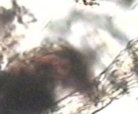
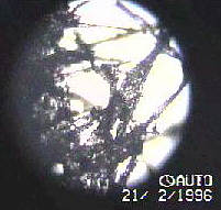
| Because we were more interested in video microscopy than in making still images, we had to stand in awkward positions clutching the video camera in one hand while focusing the microscope with the other. This poor quality image (right) was taken during the first period of microscopy. This is an image of a human wound. |
|
The images of this period were worthless as images but valuable from the point of view that we found that we could at least see the samples we were looking at, through the camera. We figured that we needed something to keep the camera stable so I tried to obtain a tripod of some sort. Unable to find a cheap camera stand we decided to make a camera stand ourselves. This was a hard task for we had little materials.
In the end I produced some 'THING' that had the right height for the microscope, and also had the "sophisticated" bolt that allowed the camera to be inclined.
The images we made afterwards were stable but poorly focused due to the fact that the camera gave only black and white images on the preview screen. This inaccurate prediction of the outcome of the photomicrograph coupled with the fact that the condenser couldn't dim low contrast objects well enough created low quality images.
Then after a short period of time we discovered a very interesting device called the "ALARIS" video transport connector. It enabled us to connect the ordinary video camera to the computer where we could preview photomicrographs in color and digitally process them to create nice looking pictures. This also enabled me create still images and make video clips. The images I made on the computer have fairly good quality compared to the quality obtainable digital cameras of today produce.
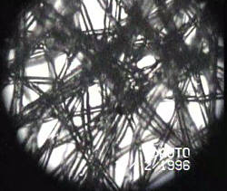
These are two reasonably high quality images I produced when I perfected my techniques on the computer. The first image (below) is an image of synthetic fibres. And the second image shows its natural counter part, fibres of paper. You can see the difference between organic and inorganic fibres by noticing that inorganic fibres are much straighter than organic fibres.
|
|
We also solved the problem of dimming low contrast objects by placing thin paper on the filter holder. Later we made further changes and developments to our techniques and equipment, and started using a tripod instead of my home-made camera stand. The tripod enabled us to have much greater control over the camera's position.
I hope this article interests you in the field of photomicrography even if you do not have the perfect equipment.
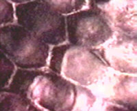
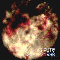
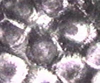
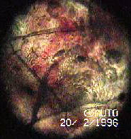
These are some of the finally produced images, the first one is an image of some onion cells the second is an image of a human wound it contains skin cells dirt and blood. Human wounds were some of the most popular objects of Victorian microscopists.
|
|
These are two microorganisms both of the Rotifer Class. The first is a Philodina it like all rotifers has a wheel organ that it uses both to suck in food, digest it and throw the waste out of its hind end. The wheel organ also acts as a jet engine to propel it through the water. The sessile rotifer also has a wheel organ and the rotifer is attached to some microflora.
If you have any questions or comments or want to obtain a video clip or all of my images contact me by email: Tom Korolev.
Editor's note: many thanks to Tom for submitting this to Micscape and great to hear from a young enthusiast. Tom writes that he has a particular interest in rotifers and photomicrography.
Published in the August 1999 edition of Micscape Magazine.
Please report any
Web problems or offer general comments to the Micscape Editor,
via the contact on current Micscape Index.
Micscape is the
on-line monthly magazine of the Microscopy UK web
site at Microscopy-UK