Steve Durr recently gave me an excellent photomicrograph of a desmid of the genus Closterium, I hope it has not deteriorated too much by scanning and saving in jpeg format. This photo' had been taken on a microscope using Normarski Differential Interference Contrast (DIC). Looking at Fig.1 you should see a row of circles that appear to be in cameo (sculptured in relief). Now look at Fig.2 which has been turned through 180°; the circles should now appear to be in intaglio (incised) where all the sculpturing appears to be on the surface of the cell. In fact the nucleus between the two halves of the chloroplast is in the middle of the cell, the other circles which are pyrenoids are within the chloroplast.
It is often difficult to tell even with normal transmitted light if you are looking at projections, depressions or even holes; showing how important it is to view an organism from more than one aspect. DIC gives wonderful photomicrographs but unless you are familiar with the organism pictured you might get a false impression of its structure.
Thanks to Steve Durr who took the photo' using a x40 Normarski Plan Fluorite objective on a Leitz Orthoplan microscope fitted with a Leitz Orthomat camera. The photo' is published with his permission.
Comments by e-mail are welcomed to
Bill Ells.
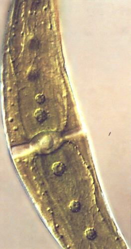
Figure 1 |
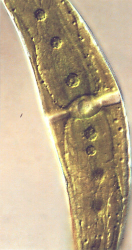
Figure 2 |