 |
|
Part 2 : Salps and doliolids |
|
||||
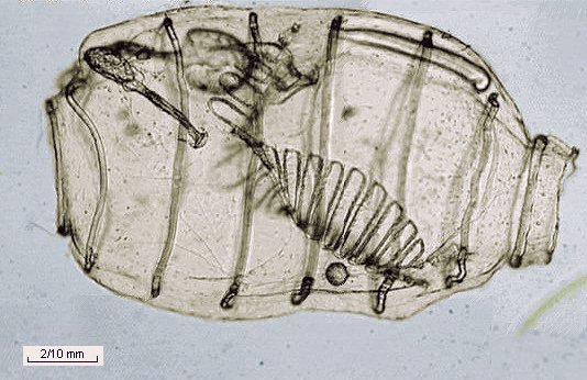
| A salp's body has muscular
annular strips. By successive and quick contractions of these muscles,
beginning near the anterior siphon, water is ejected by the posterior siphon
and the animal moves forward by a process similar to jet propulsion.
Generally salps live in colonies and have delicate internal structures. Unfortunately I have no pictures of them. I thought that the picture below was a salp, but it's a closely related species called a doliolid. The main difference is that salps don't have internal cilia to catch food, and they must move continuously to let seawater circulate inside them. Doliolids bodies are very transparent, as you can see in the picture below, and delicate structures are evident. These are covered with cilia and create currents to make the water circulate and help the organism catch food particles. (The author thanks Larry Madin of the Wood Hole Oceanographic Institution - for identification of this creature). |
|
|
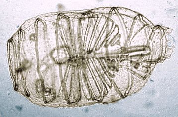 |
'Top' view showing
the delicate pattern of ciliate slits.
Anterior siphon is on the right hand side. Note: This is not the same individual as that in the picture above. |
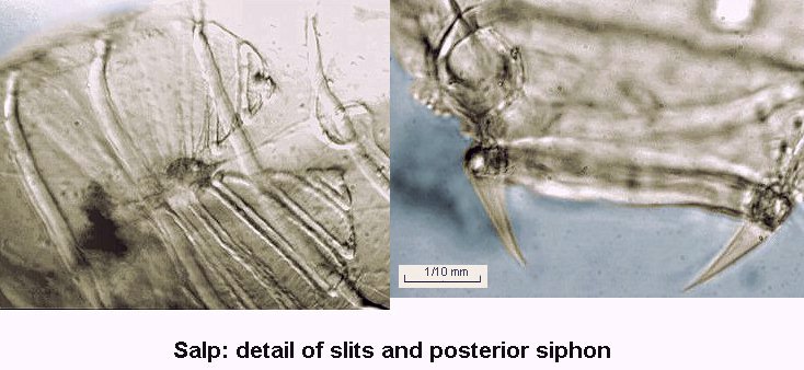
| Detailed pictures below show internal gill slits and aperture of the posterior siphon, which is very different to the anterior; the posterior siphon seems to be surrounded by sorts of deflectors. The heart is also easily visible but mainly in darkfield. (See video clip). |
 |
|
|
|
All photographs © Jean-Marie Cavanihac 2000
Published in the December 2000 edition of Micscape Magazine.
Please report any Web problems or offer general comments
to the Micscape
Editor,
via the contact on current Micscape Index.
Micscape is the on-line monthly magazine of the Microscopy
UK web
site at Microscopy-UK
WIDTH=1