
by Rosemarie Arbur, Oregon, US |

by Rosemarie Arbur, Oregon, US |
| If protists had common names, members of the genus Tillina might be "comma creatures" because of their odd shape. Their macronucleus is in the round part of the comma (their anterior end), and their water-expelling vacuole is in its tail. At 125-250 µm, they're easily twice the size of their relatives the Colpoda, and they're comparable in toughness. Colpoda cysts remain viable for years, and the Tillina I'm writing of had probably been encysted for at least ten months. I took a tablespoon of dried soil from the high-water mark on an irrigation canal (which got very little water last summer), and in fewer than 24 hours after I added boiled well-water to it, there were some big comma-shaped organisms swimming around. | 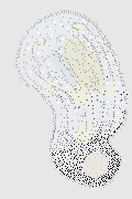 |
| I waited too long before I looked at them carefully—the Tillina population in the water had peaked and was declining. Possibly as a result of my procrastination, I ended up viewing something I certainly had not expected. It had looked like a Tillina under my stereomicroscope before I pipetted it out onto a slide. But under a cover glass, magnified 400X, it was a pretty questionable dab of protoplasm. |
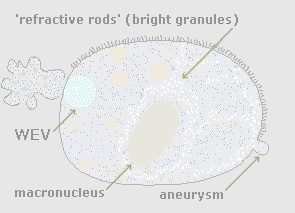 |
"Is this a Tillina?" I asked myself. When I had put it on the slide, I'd been hoping that the organism I'd pipetted wasn't just a big Colpoda, for I hadn't yet seen any Tillina that I knew were Tillina. I was in the mood to find and identify a new-to-me critter, and the big "comma creature" looked promising—at least it had before I got a good look at it. After I realized that this organism had fallen on hard times (later I learned that it had become a "flattened cell"), I became a little bit stubborn and decided that I'd just have to see what clues it offered about its identity and about anything else of relevance to what protists have and do. |
| The organism was 220 x 153 µm; checking its attributes against what Jahn and Kudo list for Tillina, I began to believe that I had found one. When I spotted 4-µm-long bright granules in its cytoplasm that corresponded to Kudo's "refractile rods 3-7 µm long" I grew hopeful that this Tillina was an esoteric-sounding T. canalifera, since Kudo mentions the rods in connection with only one species. But that was after I'd observed it awhile. Looking at the stationary organism on my slide, I noticed that it had moving cilia along just one side of its body; it also had a water-expelling vacuole at its rear end, emptying and filling as if to confirm the critter's dubious vitality. |
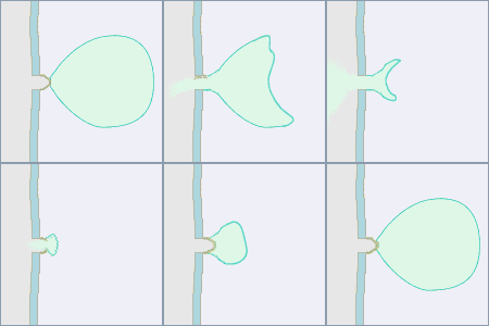
| In How to Know the Protozoa, Jahn writes of "water-expelling vesicles" or "WEVs" instead of "contractile vacuoles"; he emphasizes that they don't contract but are pressed against the pellicle by the movement of underlying cytoplasm. A WEV seems to contract when the pore through which it empties is centered between the spherical organelle and the microscope's objective. (I based the illustrations to the right on Jahn's Figure 424.) Notice how the membrane breaks and then re-grows each time the vacuole empties and refills. The organism's expenditure of energy to sustain this continuous process and the speed with which it forms the membrane amaze me. | 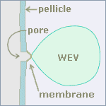 |
|
 |
| On the third emptying of the WEV that I saw, a big chunk of cytoplasm (65 µm in diameter) seemed to erupt from it. The pellicle at the posterior end gave way and the bright rods in the cytoplasm moved toward the anterior end, where a little aneurysm had developed but remained stable. For two or three minutes, things were definitely in flux: more cytoplasm leaked out, and I couldn't locate the WEV. Then I saw it again, three or four minutes later; the organism became pear-shaped and larger than before (265 x 171 µm); its cilia resumed normal-looking motion, now on all sides of its body. |
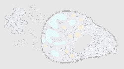 |
In fact, however, the organism had not grown, nor had I seen the original WEV. The Tillina's misadventures had flattened the remaining cellular material so that the whole organism was very nearly on the same focal plane, even with a 40X objective. The macronucleus remained intact, so the pellicle was "tented" above it and flat elsewhere, like Pooh Bear's carpet on top of the heffalump. |
| What I had thought the organism's WEV was, I'm almost certain now, one of its collecting canals. As I watched, I saw "another WEV," and then another, all pumping busily. The bright granules in the cytoplasm and then these multiple water-collecting canals —this critter was pretty surely a Tillina canalifera. But are the collecting canals supposed to pump? | 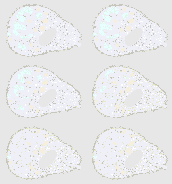 |
| Here I've re-drawn and animated the twelve panels that Jahn uses to show how Paramecium's WEV fills with and then expels water. The curved lines at the top are cilia. The opening in the pellicle is a permanent pore, sometimes sealed by a continually re-grown membrane. The WEV is fed by the collecting canals at its sides. | ||
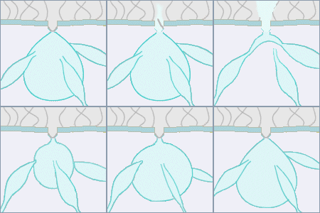 |
Jahn's drawings show that as the central vacuole fills and empties, the collecting canals change shape; and that when the vacuole is empty or nearly so, the canals are momentarily detached from it. Their varying shapes imply that they contain water that they empty into the WEV once they re-attach to it. They do seem to be pumping; it's not terribly unlikely that they'd pump even if they became unattached to the WEV. | |
|
Since both T. canalifera and T. magna have the same kind of collecting-canal system as Paramecium, it's pretty safe to conclude that the vacuoles I observed emptying and then filling in the flattened Tillina were the organism's collecting canals —or all but one of them were. If they ordinarily grew an opening into the WEV, as Jahn's drawings of Paramecium's collecting canals show, they quite plausibly could grow an opening into . . . into what? Possibly not "into" but "out of," out of the organism; given the disorganized state of this poor Tillina, it's probable that there were ruptures of the pellicle that removed the necessity of each canal's having its own exit pore. |
||
| The collecting canals of the Tillina were collecting. They may have been pumping water out of the organism, or they may have merely been pumping water out of themselves into the organism's leaking rear end. In either case, their work was futile, for within twenty minutes a much larger anterior aneurysm pushed through the pellicle and moments later burst. The pumping collecting canals may have extended the critter's life for half an hour (no mean feat), but the situation in its front end was irremediable: the Tillina canalifera died. | 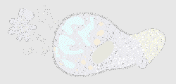 |
|
| Lest the Tillina above misrepresent the genus, below are two normal, healthy ones, recently excysted from an infusion made from a second collection of irrigation-canal soil. They're smaller, averaging 124 by 68 µm, because they'd excysted the day I observed them and hadn't had much opportunity to eat and grow. | ||
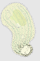 |
The organism on the left appears as it might in bright-field illumination. The dark dots toward the posterior result from the density of the granules or rods in its cytoplasm and low magnification; the longitudinal lines are the WEV's collecting canals. I show seven, since I counted seven and eight repeatedly; I never saw the entire water-collecting and -expelling system in one glance, nor do I think I could have even if the organisms had been inactive. The other illustration shows the organism as it might look in dark-field illumination. | 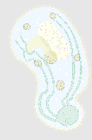 |
| The granules or rods in the cytoplasm appear very bright and are here somewhat difficult to see, though with the 40X objective and a switch to bright-field they are plain and silver-white. I've left in only four of the seven to nine collecting canals that Tillina canalifera has, the better to show the macronucleus and oral vestibule that are side-by-side in the center of the body, the forming food vacuole just beneath the latter, and the circulating food vacuoles. | ||
| The indentation just forward of the WEV is the most visible part of a groove that spirals around the Tillina's body, its other end slightly in front of the opening of the oral vestibule. |
| Most ciliates with grooves have them forward of their cytostomes where they guide food particles toward the rear and into their mouths. Tillina's oral opening is at the wrong end of the groove for that. Tillina's is not an oral groove. |  |
It's possible that the groove is locomotive. When the organism swims for some distance it rotates around its long axis, and the groove seems to stabilize its body, making it look hydrodynamic instead of ungainly. |
| Being hydrodynamic means
using less energy, and swimming without wobbling too much does keep the
oral opening near the front, where food particles drift by and might
be pulled in by perioral cilia (my underlinings express my doubt). But
Tillina don't always rotate when they swim; as often as not, their
bodies move forward and back while maintaining the same orientation, and
sometimes they wobble, too, and look like pale Paramecium bursaria.
It's far more likely that any advantage conferred by the groove has to
do with Tillina's being able to "process" chemical information about
what's ahead of it as it swims in its streamlined and relatively faster
mode (i.e., its chemoreception of things that signal ingestibles, warmer
or cooler water, maybe even predators). It would be wonderful to know the
evolutionary origin of Tillina's not-oral groove and its complex
system of water-collecting canals. Other members of the family Colpodidae
have neither, though T. magna and T. canalifera have both.
What began as my stubbornness about trying to identify what I'd correctly guessed was an organism too big to be a Colpoda became a kind of detective game: the "flattened cell" didn't reveal its characteristic shape, but it did have those bright granules in its cytoplasm, and its multiple "WEV"s had to have been remnants of water-collecting canals. So, despite its being flattened and moribund, the unfortunate Tillina canalifera was my introduction to a "new" species of ciliate. Just as exciting was its demand that I find out more about collecting-canal systems and grooves around organisms' bodies. Pretty soon I'm going to get a third sample of that irrigation-canal soil, find bigger but still healthy Tillina, and then see if I can somehow get my illumination just right. I may be able to watch the water-collecting canals actually emptying into the WEV; I may also be able to discover how far forward of Tillina's oral-vestibular opening its spiral groove extends, whether or not the ciliation in the groove is different from the other body cilia, and other things much too small for the rest of the world to notice. |
Comments to the author Rosemarie Arbur welcome.
Please report any Web problems or
offer general comments to the
Micscape
Editor,
via the contact on current Micscape
Index.
Micscape is the on-line monthly magazine
of the Microscopy UK web
site at Microscopy-UK
WIDTH=1