| Regular readers will be
getting to know that I like to study microscopic marine life! Many times,
when I've observed creatures contained in my seawater samples, I've encountered
some biological structures which seem to be shared between various phyla,
which were often widely separated in the evolutionary tree. These structures
are implicated in spatial location for these critters, and these structures
also play a role in humans.
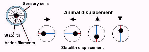
For equilibrium control in man, the inner ear contains some tiny 'stones' called otoliths (from the Greek: otos=ear and lithos=stone). In fact these stones are crystals of minerals like calcium carbonate. The stones are inside three semi-circular canals, each one working in one of the three dimensions. As these stones are mineral, they are heavier than surrounding biological tissues: when they move they push on ciliated cells which produce a nervous stimulation, which is used by the brain to compute our body position. Another ear part called the vestibule, is used to detect rotation and acceleration movements by using a similar principle. This process is responsible for seasickness: if you are on a boat deck which is pitching and rolling and you look at an object on the boat, the object is stationary to your eyes but the inner ear is detecting body motion according to the boat movements, thus altering brain perception. To attenuate this indisposition, look at a horizon line, clouds, sea waves.. or any object outside the boat. It's surprising to find such a sophisticated process in other creatures, but particularly in marine zooplankton, including the larval stages of primitive animals. Some examples are described below. Equilibrium organs are called statocysts. They contain statoliths (stones) made with dense material such as calcium or magnesium salts crystals which are in contact with specialized cells. Many research studies are in progress on this topic and a hypothesis would be that actin fibers connect the statoliths to the cells. How do statoliths work? The simple drawing I've drawn below shows how. When an animal moves, a statolith remains stationary for a brief instant because of inertial resistance, so it pushes more against the fibers located on the side opposite to the direction of motion (red lines). In the same way statoliths acting under the force of gravity allows cells to recognize up and down displacement.
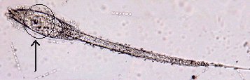
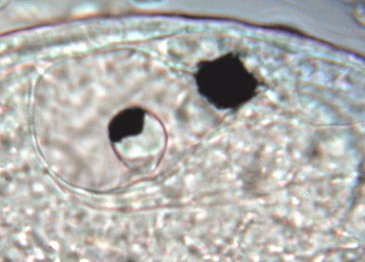
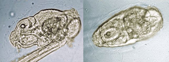
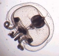
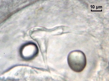
TUNICATE LARVA: The first picture below shows a tunicate swimming larva (tadpole) which metamorphoses in a few days into the very different form of a sessile adult (for example ascidian). (See this Micscape article on tunicates.) The statolith which is used for equilibrium is clearly visible as an 'eye-like' feature containing an irregular shaped stone. The other black dot above is an ocelli which is probably light sensitive. These two organs disappear in the adult stage.
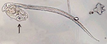
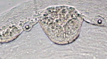
Lower image: closer view with x40 objective. APPENDICULARIANS or LARVACEANS: These are tunicate species which remain in a larval like stage. (See article on tunicates.) As shown in the image below (arrowed) the statoliths have a pearl-like spherical shape and seem to be contained within an oval chamber centered over the pharynx.
Below are closer views: side view and top view of larvacean body, x15 objective.
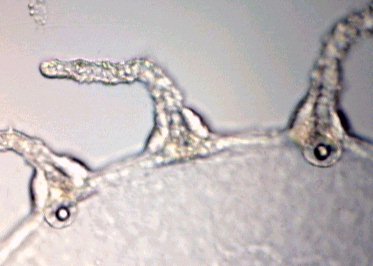
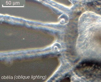
OBELIA JELLYFISH FORM: Cnidarians form another phylum which include the well known jellyfish. In a sexual mode of reproduction in cnidaria like the Obelia shown below, specialised individuals in Obelia colonies (gonozoids) have this swimming jellyfish-like form which possesses gonads. (See Micscape article on cnidarians/bryozoans.) If you look at the delicate tentacles suspended around the 'umbrella', you can see near their base, ampulla containing a round shaped statolith: but not for each tentacle, more often for every other three or four. 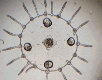
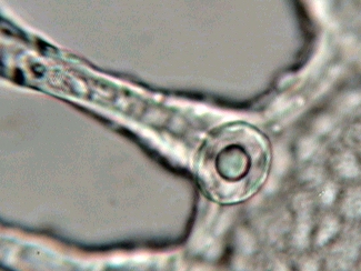
No ciliated structures can be seen in the images above of the statoliths (i.e. as proposed in their mode of action), even at high magnification. (A x40 objective was used but the linear video enlargement is near to x1400 here!) The statoliths are just seen as centered in a round sac. TRUE JELLYFISH: Zooplankton often contains true tiny jellyfish with four long tentacles. The statoliths aren't located near the tentacles but at the margin of the 'umbrella' as shown below.
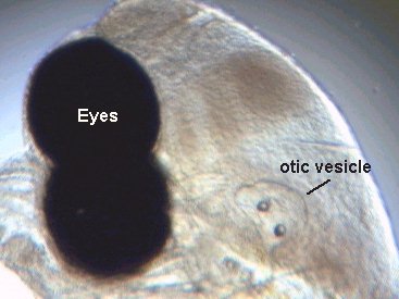
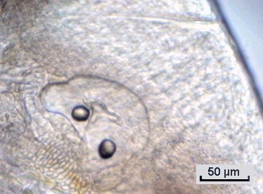
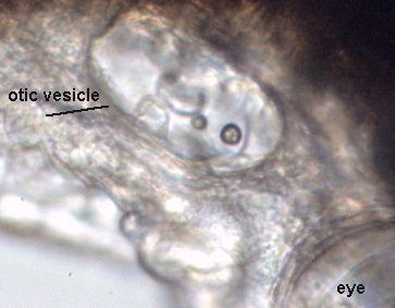
FISHES: It is particularly easy to observe otoliths in young fishes or even in fish embryos still in their eggs. All the structrures are still transparent and the two otic vesicles can be seen behind the eyes. In the pictures below taken of a fish just after hatching, the two otoliths are clearly visible, and also the internal structure of the ear (maybe one semi-circular canal too). In some species a third otolith appears some days after hatching. Unfortunately, it's not possible for me to identify the fish the species in the pictures. The lower right picture is taken of another species of fish. You can extract more large otoliths from the head of an adult fish, but they are not transparent and often have irregular shapes. (Other statolith pictures in a goby embryo can be seen in this Micscape article.) In squid, the statolith is also present. It has a laminated structure and exhibits the peculiarity of adding a layer for each day of its life, like the yearly growth rings in the wood of trees.
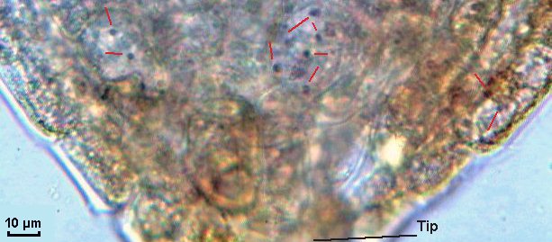
It is astonishing to find the same principle used in plants. When seeds are under the ground, how do the seed leaves grow toward the surface while the roots grow in the opposite direction? Root cells and particularly apical cells (at the root tip) contains some dense starch grains which act exactly like statolith calcium crystals under the force of gravity. It's possible to see them within cells of a young root stained with alcohol iodine solution. Starch grains become purple (red arrows) and are only found in the cells near the root tip, as shown in the picture below. It's not a slice but the tip of the root crushed under the coverslip! Many experiments of seed growing in micro-gravity have been made during space flights of the American Shuttle and in this case roots are observed to grow in any direction. (Sorry, I have been unable to rent the Shuttle to try this experiment myself!)
|