 |
 |
The Eye and Illumination with the Brightfield Microscope Some thoughts about the suitability of visual imaging with various illumination techniques By Paul James (UK) |
| This article concerns itself with Visual aspects of microscopy with various illumination techniques and not Photography. Therefore the reader should treat the illustrations below as a guide only. |
Imaging by eye the emerging rays issuing from the eyepiece rarely raises any questions in microscopy, which to some extent is understandable as we tend to assume everything has been attended to in the lengthy history of microscope technology. In truth most facets of applied microscopy have been sorted out, but there will always remain the fact that the light that bathes the retina from the eyepiece is not always as evolution intended it to be. The eyepiece itself is not instrumental in this less than ideal situation because it is the suitability of a number of substage illumination techniques which is at the heart of this article.
We are so used to imaging our surroundings in the normal manner, as indeed our very ancient forebears evolved to do, that many, if not all beginners in the art might subconsciously assume the imaging light emerging from the eyepiece is like any other which the eye accepts from a normal earthly scene. Depending on the illumination used, the simple fact is that this assumed premise might not be the case, and indeed for a large number of microscopists using certain illumination techniques it surely isn't.
Brightfield is undoubtedly the first transmission lighting method in the long history of microscopy. In its basic form it is a technique which can be compared with the projection of say a 35mm colour slide, but not onto a screen but directly into the eyes! The natural earth bound reflective scenes which our eyes have been perfected to embrace does not issue forth from the eyepiece in brightfield microscopy. The BF condition is at its worst when we observe isolated specimens which allow unhindered light around the peripheral field to pass to the eye. Of course this is unavoidable, and any discomfort can be averted by simple means such as diminishing the intensity either by crossed polars, neutral density filters, rheostat control over the light source, and incorrectly, but temptingly the substage iris. However none of these precautions have any affect on the way our vision copes with the end result which can still be highly contrasty and transmissive. As specimens under the coverslip vary greatly it would be folly to castigate out of hand such an old technique which is used daily by both professional and amateur microscopists, yet the unavoidable fact is that our eyes have to cope with an often severely contrasty image.
Images/Tone charts
The following images and tone charts accompany respective illumination techniques to reveal the basic characteristics of each. They are average run of the mill examples which nevertheless share the same characteristic light output with other examples using the same technique.
|
|
A typical BF image where much of the light input is in the brighter zones. As long as the field intensity is legitimately tamed it is tolerable. The bulk of the light energy of the highlights comes from the background and even in this sample where it is relatively low it is still dominant. Remember that your field intensity through the 'scope will in all probability be very much higher than this representation on screen.
 |
 |
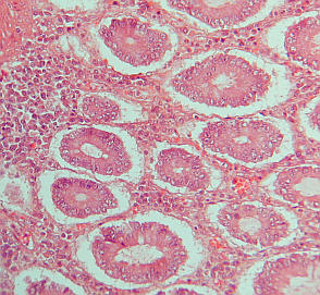
Histology section again in BF with less dynamic tonal range, but very eye friendly. There is little in an image like this that the eye cannot make sense of as the vast majority of the tones fall within the most sensitive intensity regions for the eye.
 |
|
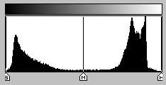
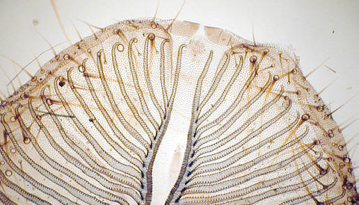
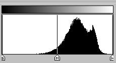
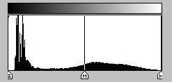
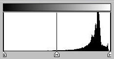
Here is an example where a dense specimen, moth tongues, shows the unsuitability of standard BF. There is little in the way of mid to upper mid tone imagery which the eye is most sensitive to issuing from the specimen. The two spikes in the tone chart are caused by the field and the opacity of the specimen. A diffusion screen cures this in visual use very effectively, though photographically it still remains a difficult subject and the cure not easily noticed in a photomicrograph, but are clearly evident by eye, and any resistance to this seemingly muddifying technique should be tamed by the reasoning that it is the eye which we are concerned with in this article and not the camera. Kohler's illumination which is possibly the most commonly implemented brightfield technique in microscope stands, was construed for the principle purpose of yielding a more evenly lit field from a point light source for the photographic plate. The eye simply does not possess the same sensitivity as the camera's film/ccd to an unevenly illuminated field of view nor does it require the intensity of output that the latter does.
 |
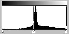 |
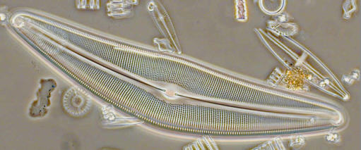
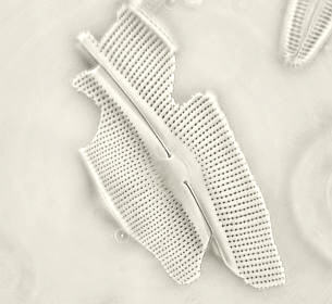
Phase Contrast usually results in an image which though contrast enhanced has less overall brightness. This image above demonstrates high midtone output and a broader range of tones at a much lower output level yielding an eye friendly yet detailed representation of the diatom. The background accounts for the peak at the centre of the tone curve.
 |
 |
Darkfield illumination as represented above shows a tone graph with modest output of mid to higher tones, yet the image has all the necessary information without being too contrasty. The darkfield surrounding the specimen does not hinder its scrutiny so long as the difference in their intensities is not too severe. Too strong an illumination in DF obliterates much detail. Use of a 'phase' or annular stop just above the na of the objective in use, seems to yield the most delicate and revealing imagery with low overall light output to the eye.
 |
 |
Above COL illumination technique using an example to demonstrate that a small bandwidth of mid to higher tone output achieves the transfer of information to the eye in the region in which it is most sensitive and perceptive. Here the subtle gradations of tone can be readily seen and interpreted, which in a much darker image would prove too difficult for the eye. The principal virtue of COL apart from the inherent contrast inducing property with fine detail, is that the overall intensity of light and the tonal expansion of the image is very evident to the eye. The main spike in the tone chart is of course the 'field', which being not too dissimilar to the subject is clearly identified by the eye because of the eye's acuity to the variation of subtle tones in this region.
CONCLUSIONS
The unison of 'eye @ brain' conjures images in a different way to that of the camera, though they both share some fundamental elements of course. It is a fact however that many microscope designs include it seems almost exclusively the provision for a light source which is overly bright as well as an illumination train devised to provided the camera plate with a bright and uniformly lit field. In principle, it would appear to be perfectly suitable for visual use, but the experience demonstrates otherwise for most specimens. It seems pointless and lacking in commonsense to soldier on with such a visually debilitating situation without trying a remedy. Thus there are two alternatives: choose a more suitable illumination technique if the specimen warrants it, such as DF, Phase Contrast, COL, Oblique etc.. and see if this helps, and if not then tame the intensity and diffuse the light by trial and error. You will soon become accustomed to manipulating the substage and importantly realise that the way light is used to illuminate a specimen is perhaps the most important operational aspect of microscope manipulation.
I think the ultimate image which reveals much about the shape and texture of the specimen, is one which is derived from an illumination technique which puts the specimen into the mid to light 'grey' scale region of the tone curve. Here the eye is at its maximum capability to scrutinise in all respects, and as a consequence is at its most relaxed too.
When both resolution and illumination are of a very high order the views are memorable indeed.
Good Lighting!
| All comments welcome by the author Paul James |
Microscopy
UK Front Page
Micscape
Magazine
Article
Library
Please report any Web problems or offer general comments to the Micscape Editor.
Micscape is the on-line monthly magazine of the Microscopy
UK web
site at Microscopy-UK
© Onview.net Ltd, Microscopy-UK, and all contributors 1995 onwards. All rights reserved. Main site is at www.microscopy-uk.org.uk with full mirror at www.microscopy-uk.net .