THE WILD HONEYSUCKLE
Fair flower, that dost so comely
grow,
Hid in this silent, dull retreat,
Untouched thy homed blossoms blow,
Unseen thy little branches greet:
No roving foot shall crush thee
here,
No busy hand provoke a tear.
..................
Philip Freneau
(1752-1832)
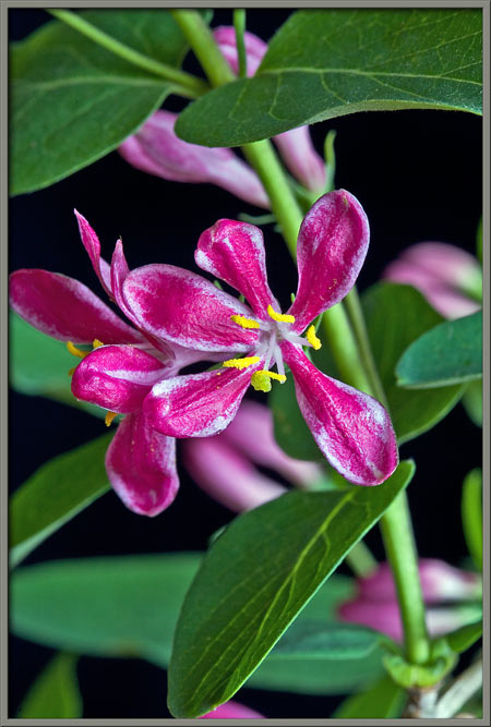
Honeysuckle bushes are a common
sight in southern Ontario where I live. Most have flowers in
shades of pink, but a few are white. About five metres from the
stream that passes through the area where I commonly search for
wildflowers, a single straggly bush, hidden from passersby by trees,
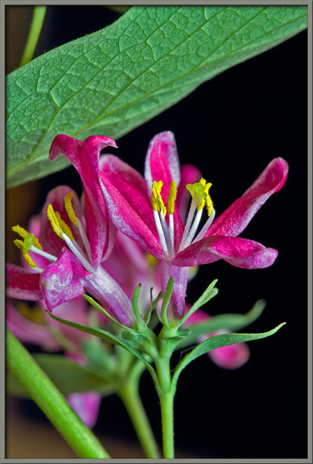
possesses the brilliant red blooms that can be seen above. I
suspect that the seed that grew into the mature plant was dropped,
(need I say more?), by a bird perched on a branch of the tree that
overhangs this particular bush. The fruit, that will be seen
later in the article, is a favourite food of birds, and small
mammals. The species is particularly cold tolerant, and thus
survives our extremely cold winters.
Tatarian honeysuckle is sometimes
referred to as bush honeysuckle, or twinsisters (due to the fact
that the blooms occur in pairs at the end of a stem.) It
originated in Southern Russia and Turkistan, and was first introduced
into North America in the late eighteenth century. Honeysuckles
belong to the Caprifoliaceae
family. The name derives from the Latin caprifolium which roughly
translates to goats leaf. (Honeysuckle leaves were a favourite
food of goats.)
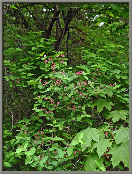
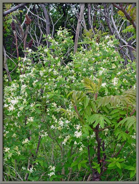
As can be seen below, the bush from
which the flowers were obtained, is almost completely surrounded by
trees and saplings.
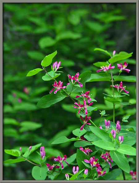
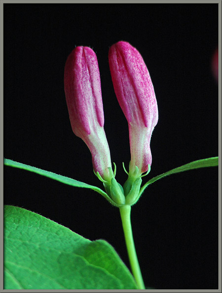
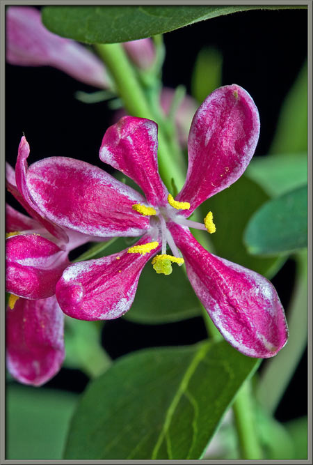
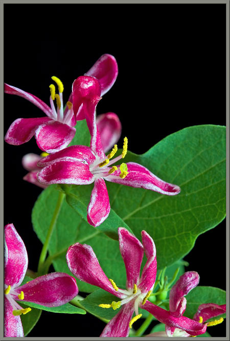
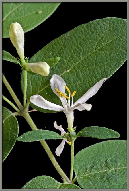
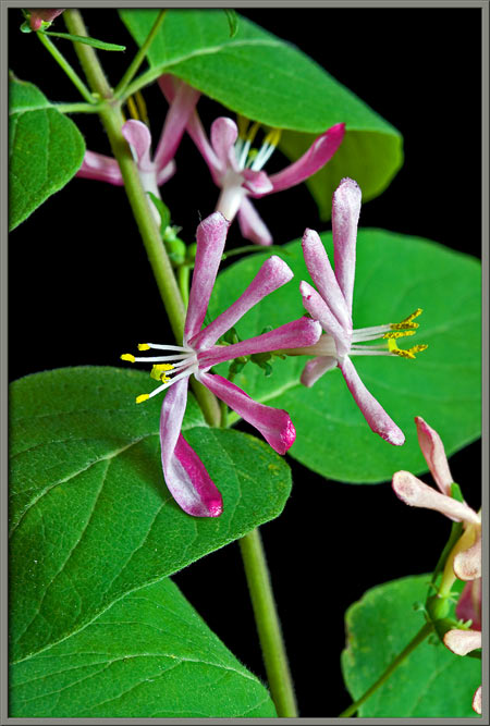
Notice in the image at right below,
that the oval leaves are arranged opposite one another, and that the
stems supporting the pairs of flowers grow from the points of
intersection of the leaves to the stem, (the axils).
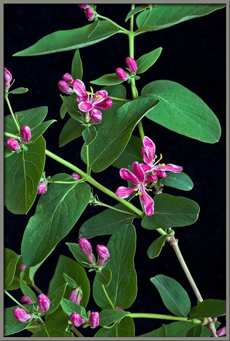
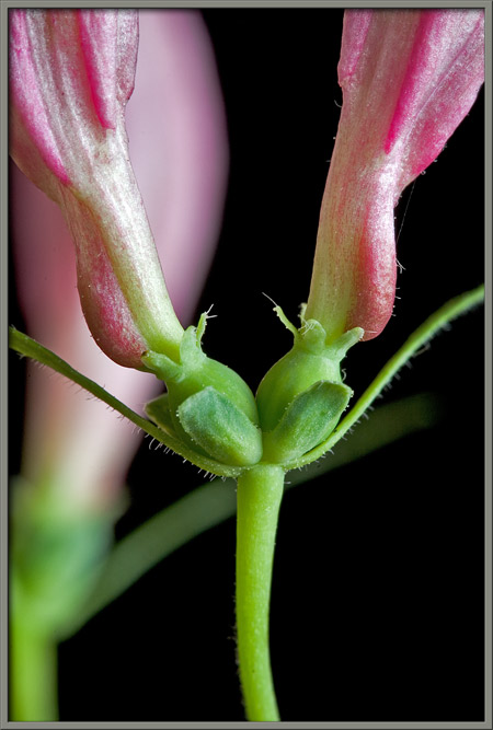
Beneath each pair of buds there are
two leaflets. Immediately above the leaflets, there are two sepals
(modified leaves) that partially cover each of the green, egg-shaped,
immature ovaries. At the top of each ovary is a ring of very tiny
pointed, green, sepal-like structures. Notice in the image at
right, that the bottom surfaces of the lower-most leaflets are covered
with small hairs.
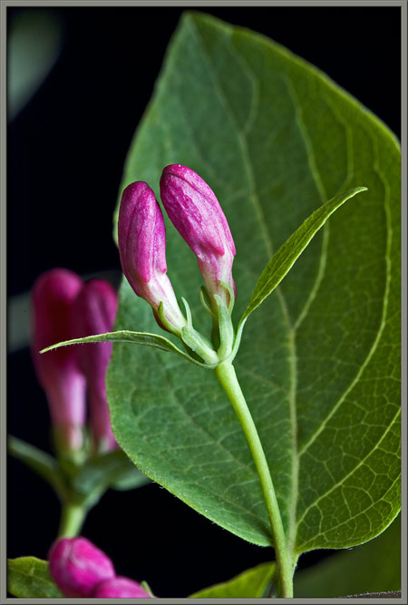
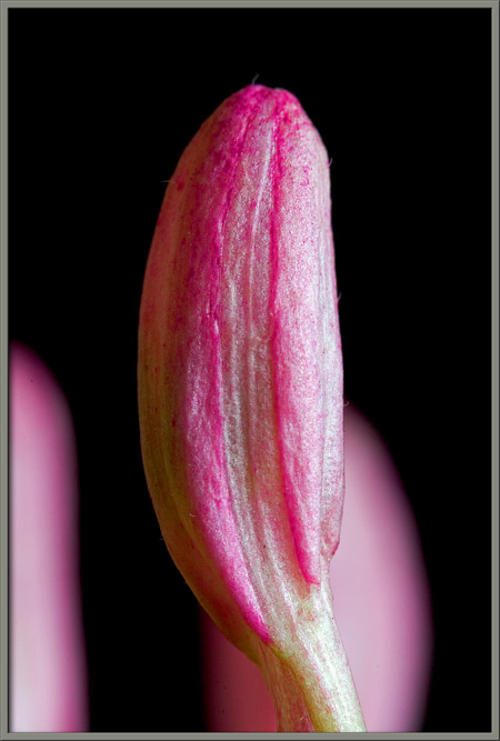
The bud itself has an elongated
red, bulbous top, and white tubular bottom. It is just possible
to discern the individual petals of the flower making up the topmost
section of a bud.
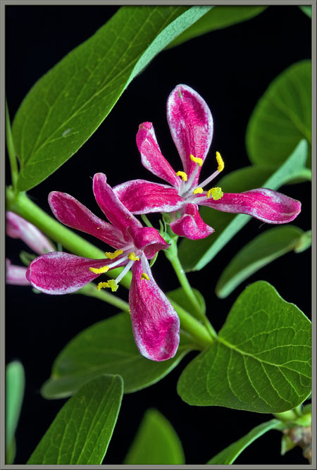
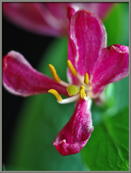
A mature flower possesses five
spoon-shaped, brilliant red petals. Notice that the edge of each
petal is white.
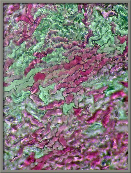
If one of the areas of intermediate
colour is examined under the microscope, its cellular structure becomes
visible.
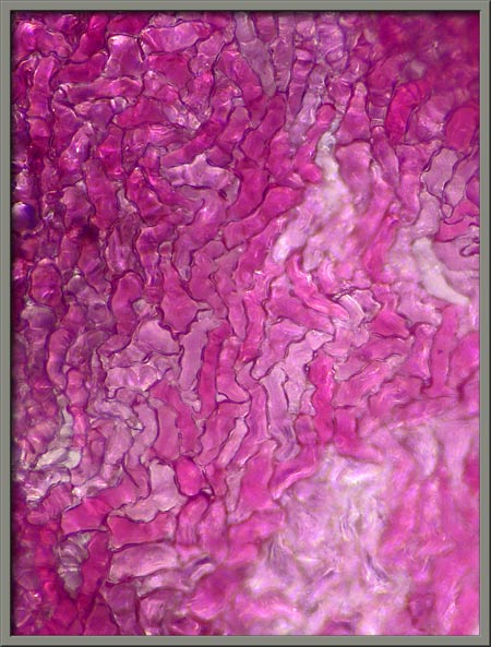
The photomicrograph on the left
below shows that the white areas of the petal are composed of cells
with no red pigment, while that on the right shows an area where the
colour is deep red due to cells containing varying amounts of red
pigment.
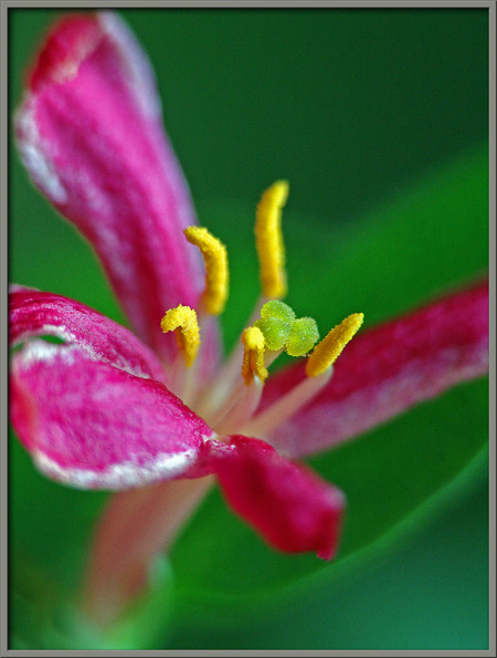
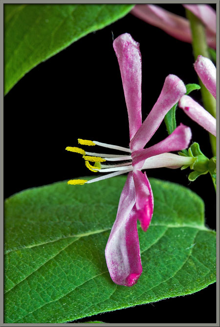
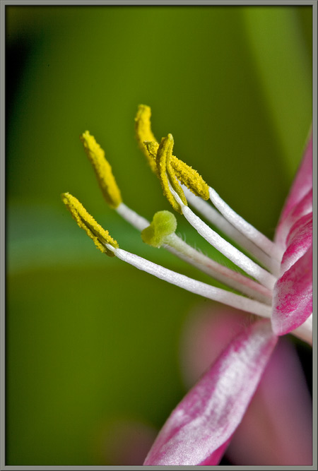
Tatarian honeysuckles reproductive
system consists of five bright yellow anthers
(male pollen producing organs) supported by white filaments, and a central, bulbous,
green stigma (female pollen
accepting organ) supported by a white style.
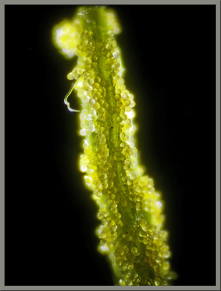
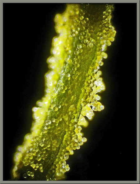
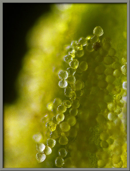
Under the microscope, an anthers
coating of spherical pollen grains is visible.
Higher magnifications reveal the
roughly spiked surface of each grain.
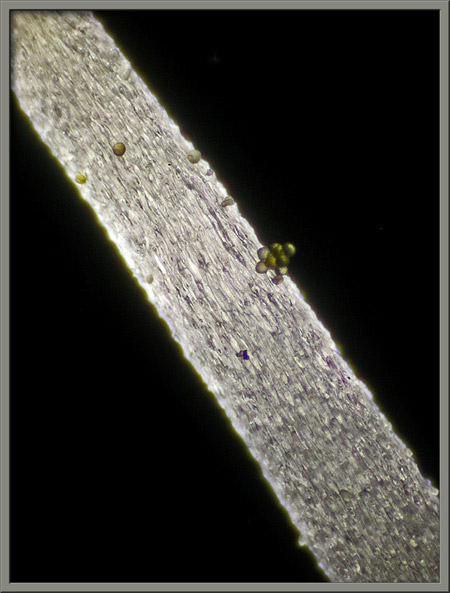
Some pollen grains adhere to a
filaments surface.
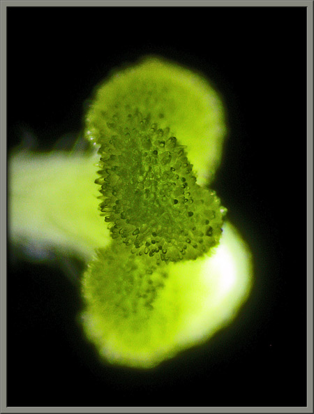
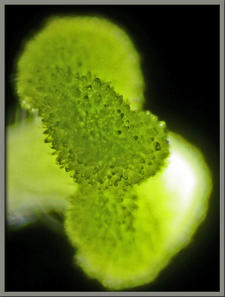
Close examination of the flowers
green stigma reveals that it is divided into three ellipsoidal
lobes. The photomicrograph on the right shows the rough surface
structure of one of the lobes.
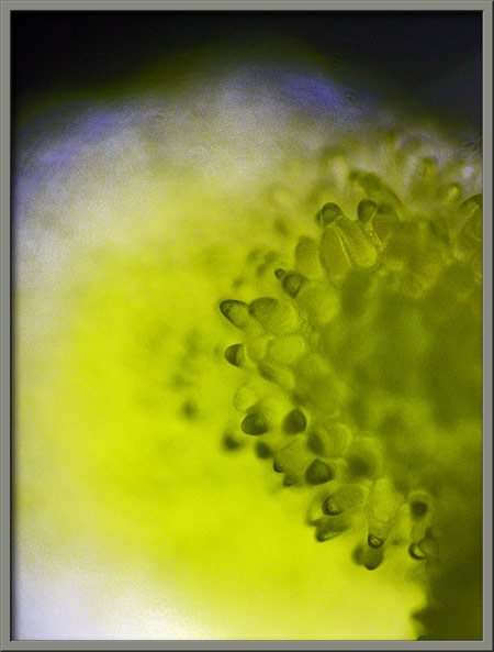
Higher magnifications show that a
lobes surface is covered with short, rounded protuberances that aid in
the collection and retention of pollen grains transferred to the flower
by insects acquiring the nectar stored at its base.
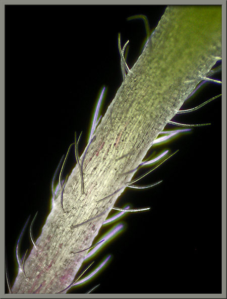
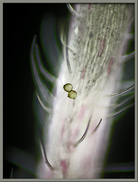
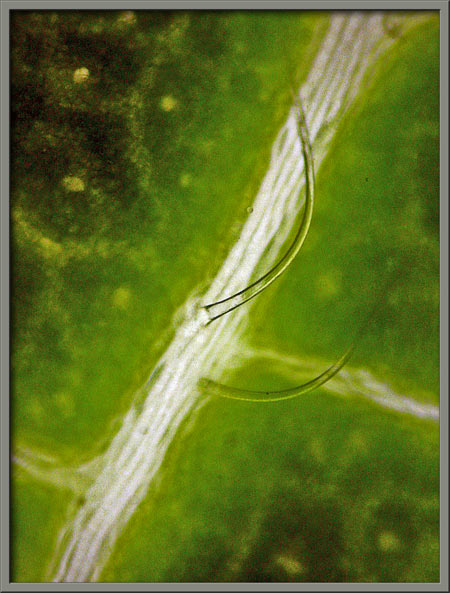
Unlike the filament, the style
supporting the stigma is covered with long hairs. The image on
the right shows a couple of pollen grains stuck to one of the hairs.
When a flower has been pollinated
(by having pollen deposited on its stigma), a pollen grain germinates
and sends a root-like tube down though the style to the flowers
ovary. The grains male sex germ travels down the tube to an
ovule in the ovary where the female sex germ is located.
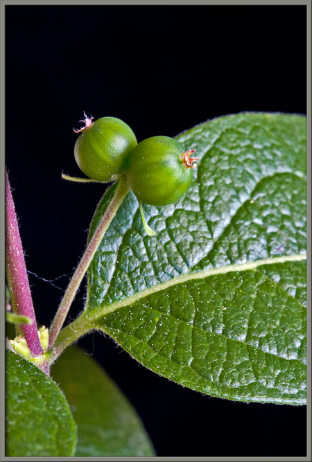
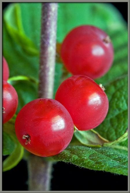
Fertilization occurs when the two sex germs combine. After
fertilization, the flowers fruit, (or in this case two fruit) begin to
develop. The image below shows two such fruit with the brown
remnants of the flowers pistils still visible.
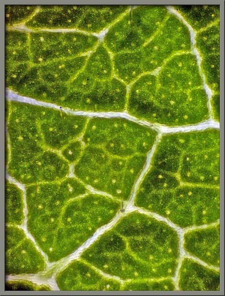
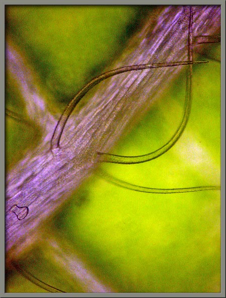
Notice the veined surface of the
leaf seen in the image above. In the lower magnification image at
left below, the intricate vein structure at the edge of a leaf can be
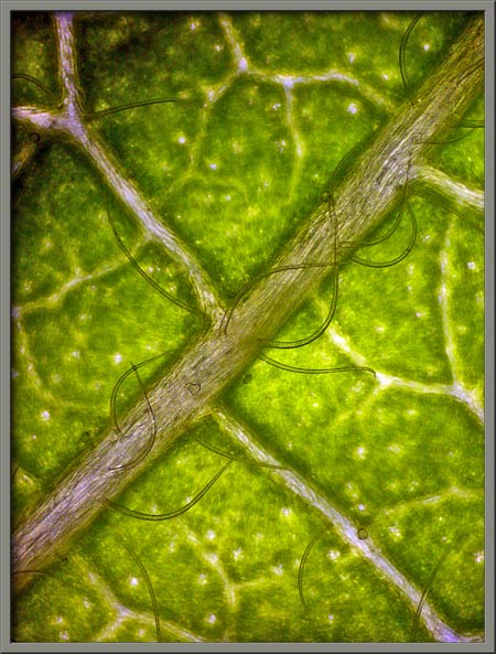
seen. The image on the right shows a higher magnification view of
one of the principal veins on the underside of the same leaf.
Note the many long hairs that grow out from this central vein.
Still higher magnifications show
the hairs more clearly.
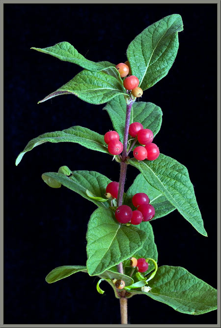
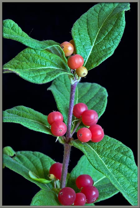
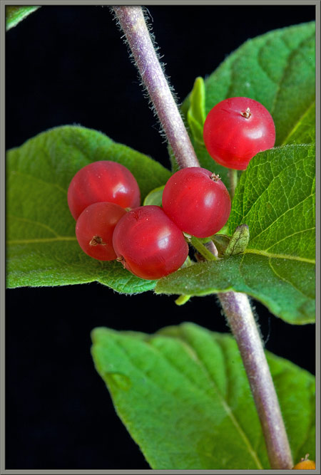
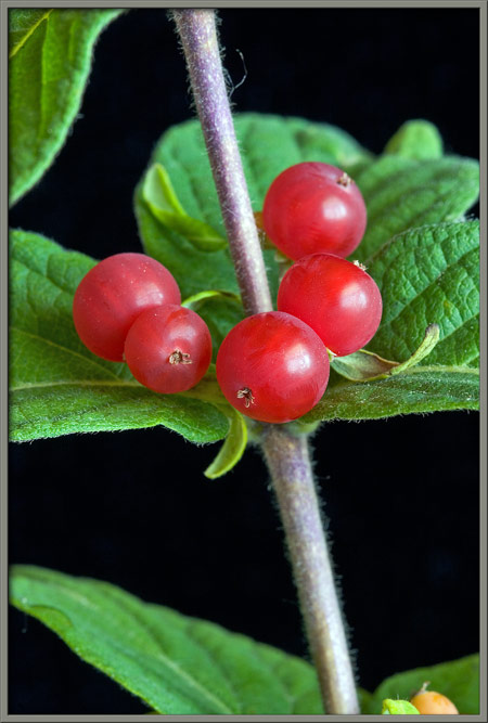
By mid July, the many fruit pairs
provide the honeysuckle bush branches with a touch of brilliant
colour. Note at the top of the two images, that a fruit
transforms from green, through yellow and orange, to its final red
colouration.
The abundant fruit of a tatarian
honeysuckle bush are visually striking, and much prized by birds as
food.
Earlier in the article I mentioned
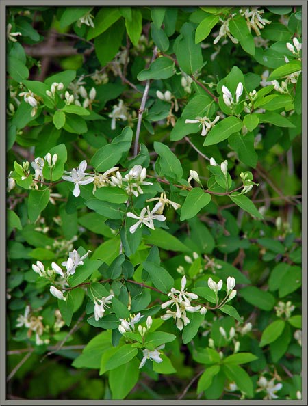
that Tatarian honeysuckle can produce flowers of other colours.
Some distance from the stream, there are several white bushes.
These flowers are identical to the
earlier ones, in all but colouration.
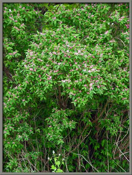
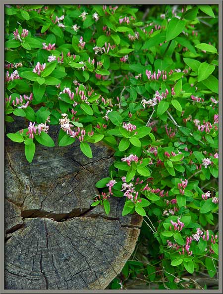
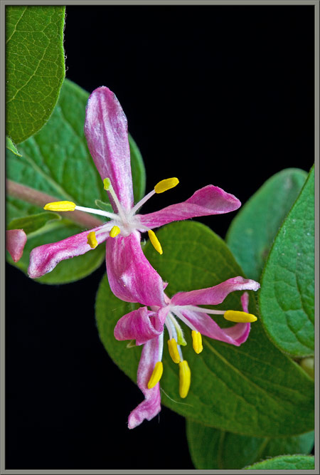
Pink honeysuckle flowers are the
most common. Two typical examples of bushes can be seen below.
Once again, only the flower colour
is different.
Notice the similarity of the
reproductive structures to those seen earlier.
Tatarian honeysuckle readily
invades old fields, open woodlands, and other disturbed sites. It
sometimes forms a dense thicket that can restrict native plant
growth. For this reason some jurisdictions consider it to
be an undesirable plant. Where I live however, this is certainly
not the case. Its colourful flowers and fruit are a welcome sight
to any passerby.
Photographic Equipment
Most of the photographs in the
article were taken with an eight megapixel Canon 20D DSLR and Canon EF
100 mm f 2.8 Macro lens. An eight megapixel Sony CyberShot DSC-F
828 equipped with achromatic close-up lenses (Canon 250D, Nikon 6T, and
Sony VCL-M3358 used singly, or in combination), was used to take a few
of the images.
The photomicrographs were taken
with a Leitz SM-Pol microscope (using a dark ground condenser), and the
Coolpix 4500.
References
The following references have been
found to be valuable in the identification of wildflowers, and they are
also a good source of information about them.
Dickinson, Timothy, et al. 2004.
The ROM Field Guide to Wildflowers of Ontario. Royal Ontario
Museum & McClelland and Stewart Ltd, Toronto, Canada.
Thieret, John W. et al. National
Audubon Society Field Guide to North American Wildflowers - Eastern
Region. 2002. Alfred A. Knopf, Inc. (Chanticleer Press, Inc. New
York)
Little, Elbert L. National Audubon
Society Field Guide to North American Trees - Eastern Region.
2004. Alfred A. Knopf, Inc. (Chanticleer Press, Inc. New York)
Kershaw, Linda. 2002. Ontario
Wildflowers. Lone Pine Publishing, Edmonton, Alberta,Canada.
Royer, France and Dickinson,
Richard. 1999. Weeds of Canada. University of Alberta
Press and Lone Pine Publishing, Edmonton, Alberta, Canada.
Crockett, Lawrence, J. 2003.
A Field Guide to Weeds (Based on Wildly Successful Plants,
1977) Sterling Publishing Company, Inc. New York, NY.
Mathews, Schuyler F.
2003. A Field Guide to Wildflowers (Adapted from Field Book
of American Wildflowers, 1902), Sterling Publishing Company, Inc.
New York, NY.
Barker, Joan. 2004. The
Encyclopedia of North American Wildflowers. Parragon Publishing,
Bath, UK.