Before the advent of digital photography most images of microscopic subjects were obtained using single lens reflex cameras or special microscope cameras. It was virtually impossible to use other cameras without the aid of special (generally expensive) reflex focusing devices because there was no way to focus the image.
When digital cameras first appeared on the scene they were, to put it charitably, not very good. However, the improvement in them has been dramatic. In fact the improvement has been so dramatic that silver halide photography is undergoing a dramatic decline with many lines of cameras and film being discontinued.
Digital single lens reflex cameras did not appear on the scene until digital imaging devices had advanced far enough to produce high quality images. In the last three or four years many new models of digital single lens reflex cameras have appeared on the market. They are significantly more expensive than other digital cameras, and they also have an enormous price range.
The sensors on most digital single lens reflex cameras have dimensions about 65% of those of 35mm film cameras. Most, if not all, can utilise lenses made for film cameras. However, because the sensors are smaller than silver halide film frames, the final image is scaled about 160% larger. The smaller image size has serious consequences when microscope photographic devices designed for film single lens reflex cameras are used for digital imaging.
With any kind of interchangeable lens single lens reflex it is senseless to attempt to shoot the images through an eyepiece using a lens on the camera. This introduces extraneous optical components that can only degrade the image.
Special projection eyepieces that are corrected for projecting images directly into 35mm single lens reflex cameras have been produced for decades. When these are used with digital cameras, however, the small size of the light sensitive device results in an higher magnification than would have been the case had film been used. (Images are sometimes less satisfactory with ordinary eyepieces because spherical aberration is introduced by this.)
A large fraction of microscope objectives exhibit chromatic difference of magnification and require special compensating eyepieces. The projection eyepieces were also produced with this compensation. When compensating eyepieces are not used with these objectives, the images away from the centre of the field of view show strong colour fringes. Most 160mm tube length objectives that are either flat field, fluorite, apochromatic, or high power require compensating eyepieces. Some manufacturers deliberately made even more of their objectives this way so that users would not have to change eyepieces all the time.
Some manufacturers made very low power projection eyepieces. These probably are the best ones for digital single lens reflex cameras because of their small sensor size.
It IS possible to use microscope objectives directly to project images directly unto photographic film or a solid state device. Significant spherical aberration is introduced by doing this, and images can be fringed with colour from chromatic differences in magnification. It is apparent that by mounting the camera as close to the top of the microscope as possible and by using a microscope with infinity corrected objectives where the tube lens corrects for chromatic differences in magnification, both of these problems could be reduced or eliminated.
Single lens reflex cameras, both for film and digital have focal plane shutters that consist of curtains that move across the light sensitive surface. The mirrors that permit viewing through the lens during focusing also need to be moved out of the light path prior to exposure. These mechanical motions can blur the image, and when these motions are transmitted to the microscope itself the blur can be dramatically magnified. Most of the special film cameras that were designed before digital photography used leaf shutters because they produced less mechanical motion to minimise this problem.
The small sensors of digital cameras makes it possible to use smaller focal plane shutters and smaller movable mirrors. This seems to reduce the motion problem significantly.
A few months ago I decided that the time had come to purchase a digital single lens reflex camera with interchangeable lenses. I carefully examined specifications of cameras from Nikon, Canon, and Olympus. My initial thoughts were to get a Nikon product, because I had a good set of Nikkor lenses and T-mount devices with Nikon fittings on them. When I discovered that most of my lenses could not be used with the Nikon model and that, oddly enough, they could be used on the Canon ones, I looked with much more favour at the Canon models. When I examined the models available it quickly became apparent to me that the model that made the most sense was the one that is sold as the Canon 400D or Canon Digital Rebel XTi. I have no idea why the identical camera is sold with more than one model name!
This camera has many positive features:
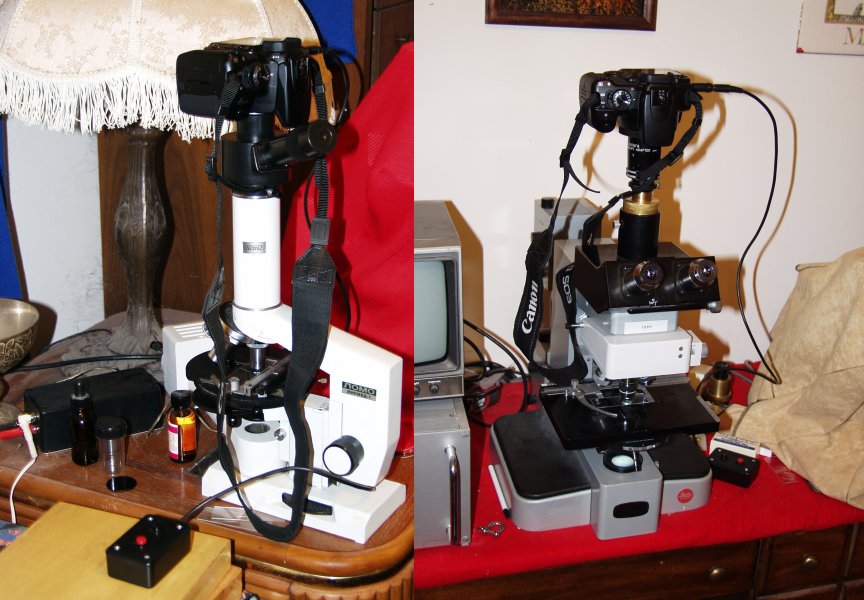
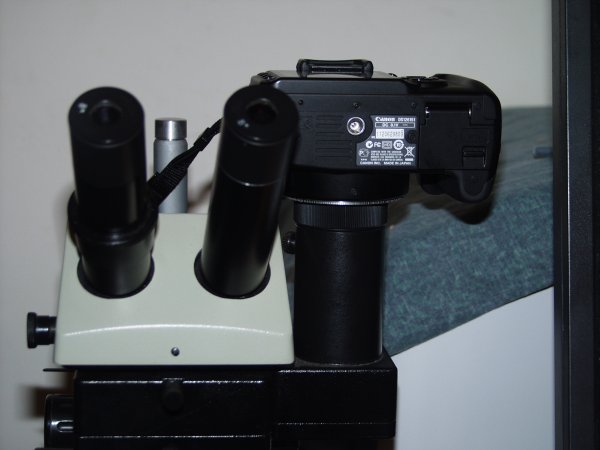
When I purchased my original LOMO microscope I also purchased the 35mm camera attachment. This device is designed for M42 cameras such as the LOMO single lens reflex. M42 is NOT the same as a T-mount, even though both are 42mm in diameter. The thread pitches are different. A very inexpensive adaptor is available that permits mounting Canon cameras onto M42 devices. The image to the left below shows this camera mounted on the LOMO camera using the LOMO 35mm camera attachment and M42 to Canon adaptor.
About 35 years ago I purchased a special single lens reflex adaptor utilising the T-mount system. The image above and to the right shows this camera mounted on a Leitz Orthoplan microscope using this device and a Canon T-mount adaptor.
The device shown mounted on the Orthoplan had a serious design defect when used with 35mm photography in that it was too short, with almost all oculars the image tended to be too small to fill the 35mm frame. It was easy, however, to correct this defect by placing a set of T-mount extension belows between the device and the camera. With the smaller image sensors of this Canon microscope no extension tubes are necessary.
Although this exact device has not been produced for decades, similar devices are available now. It would also not be especially difficult to machine a similar device using a lathe using aluminium stock.
When using either device it is best to use a remote shutter release system. The Canon 400D-Rebel XTi has two different remote shutter release systems, and infrared one, and a direct wired system. The wired system is connected to the camera by a sub miniature stereo phone jack. Although Canon sells a pair of switches to connect to the camera, it is probably better to mount two switches in a small chassis box, and connect it to the camera using one of the standard jacks. My device to do that is visible on both of the images above. I found it important to have the push button switches be different colours to mark which one is for focusing and which is for shutter release. (One does not use the focusing button when the camera is attached to a microscope, but the shutter release will not release unless the focusing button is first pressed.)
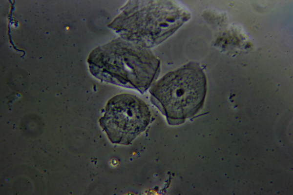
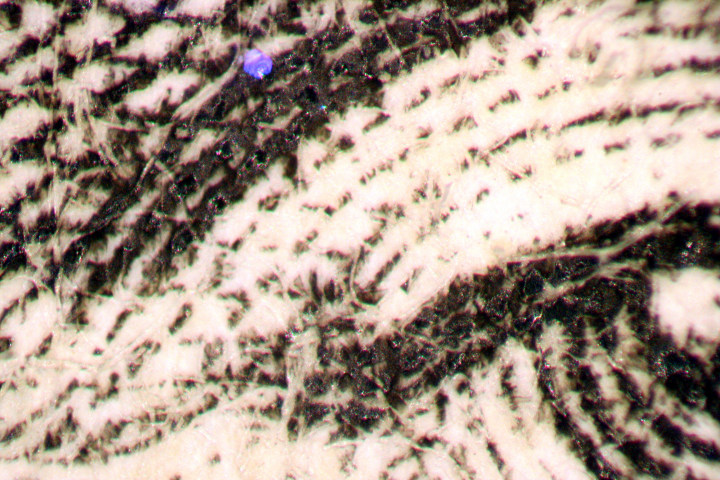
Both the LOMO mount camera adaptor and the T mount one produce similar results. The images below are all of cheek epithelial cells taken with phase contrast. These cells make extremely good test subjects.
The image below this text was obtained using the 7X LOMO projection ocular provided with the LOMO 35mm attachment kit and a 20X LOMO phase objective. (It would not be possible to get even illumination for this system without replacement of the original light bulbs in the system, here this has been replaced with a LED.)
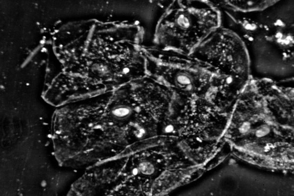
The image below this text was obtained using the same 7X lomo projection ocular, but with a HTR bright contrast 40X phase objective using green light.
The images below this text were obtained using an Olympus 2.5X projection ocular attached to the trinocular head of the Leitz Orthoplan microscope. The first image shows the results obtained with a Leitz 25X phase objective:
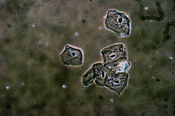
The next image was taken under similar conditions, except using a 20X Splan Olympus bright contrast objective:
The Orthoplan microscope used the standard Leitz quartz halogen illuminator.
Many people interested in microscopy have acquired one of the LOMO MBS-10 stereo microscopes. There is a trinocular attachment available for these instruments, and I obtained one with mine when I acquired it several years ago. This attachment is extraordinarily cleverly designed. The ocular holder is a slide fit, and when it is removed the top of the remaining optical tube is threaded for M42 cameras! It is a simple matter to attach the M42 to Canon adaptor described above and set the head to ΦOTO and then start snapping pictures!
The image below shows the Canon camera attached in this manner:
The MBS-10 objective turret provides 5 different magnifications. Images of flat objects seem sharp at all powers. However, the image from the 0.6X power setup are somewhat distorted and show strong field curvature, as is the case with this objective when it is used visually. The image below shows the edge of Abraham Lincoln's eyes on the US five dollar note using the 4X objective setting. Illumination is provided by the LOMO fibre optic illuminator.
The image is quite sharp, and the field is reasonably flat.
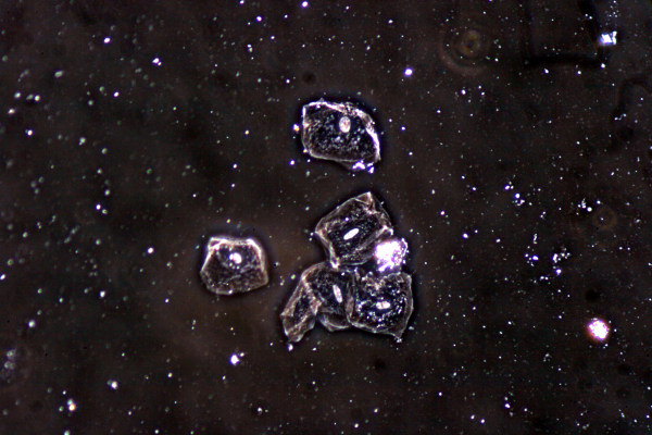
The image below shows a mineral specimen, chalcopyrite.

The image is quite sharp, but the lack of depth of field has resulted in some areas being somewhat out of focus. Note the lack of colour fringes on both of the above images.
The MBS-10 and Canon combination certainly has tremendous possibilities for obtaining images of objects that measure a few millimetres across and that are flat enough to obtain enough depth of field for an overall sharp image.
There are several software program systems that permit combining several images taken at different focus planes into a single sharp image that could certainly be used here to great advantage.
All comments to the author Robert Pavlis are welcomed.
Microscopy UK Front Page
Micscape Magazine
Article Library
Please report any Web problems or offer general comments to the Micscape Editor .
Micscape is the on-line monthly magazine of the Microscopy UK website at Microscopy-UK