|
Zeiss Mikropolychromar A sophisticated way for creating 'Rheinberg illumination' by Alain BERJONVAL, France |
Introduction
Microscope users for scientific investigations like medical, botanical or zoological, have to deal with colourless objects. Consequently, such cases are often beset with poor contrast and difficulties to see or to find details.
In 1896, a British microscopist, Julius H. Rheinberg demonstrated a form of optical staining to the Royal Microscopical Society and the Quekett Club.
This technique, called “Rheinberg illumination”, is a variation of darkfield illumination using colored glass or gelatin filters to provide colors and contrast to both the specimen and the background.
It offset the need for chemical staining which does not always provide the desired contrast, necessitates the killing of the specimen and often interferes with the live conditions and vital processes of the micro-organisms.
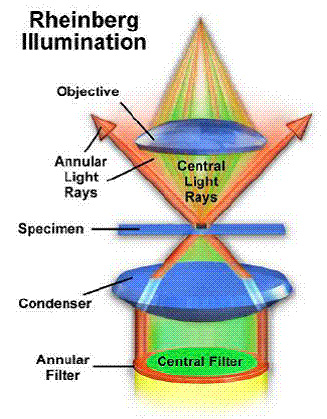
How does it work?
As said before, the Rheinberg illumination technique is a kind of darkfield illumination called optical staining. The substage condenser is arranged so that the light path from the lamp source will pass through the object at oblique angles. An objective’s numerical aperture is reduced with a light stop, iris or funnel stop. Consequently, the only light which can enter the objective is light refracted, reflected or diffracted by the object when the oblique rays strike the specimen. So, it appears bright on an black background increasing the visibility of thin or small details.
If we put colored filters (Rheinberg filters) in the condenser filter holder, we shall get a colored background corresponding to the color of the filter inserted.
Mikropolychromar description
In the 1930s, the German optical company “Carl Zeiss Jena” developed such an optical staining apparatus called the “Mikropolychromar”. The optical staining it achieved presented an advantage over ordinary darkfield illumination in that specimens are not only differentiated from the background by their relative brightness but with contrasting colours.
By means of this optical contrast staining device, the finest details and structures could be revealed with a remarkable distinctness. In fact, the “Mikropolychromar” combined the advantages of brightfield and darkfield with striking colour contrast.
In the diagram below, we can see the path of the rays through the “Mikropolychromar”.
Section through the Mikropolychromar
Path of the light rays
Controls and light path rays :
That means with a slightly eccentric position it effects a transition to a semi-bright, semi-dark field illumination (Close to oblique illumination.)
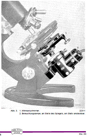
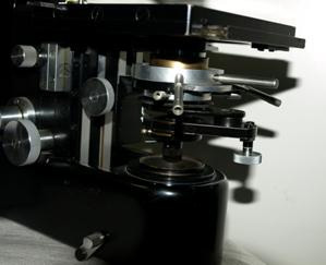
Mikropolychromar (1) fitted with its illuminating lamp (2). (Carl Zeiss Jena document ) :
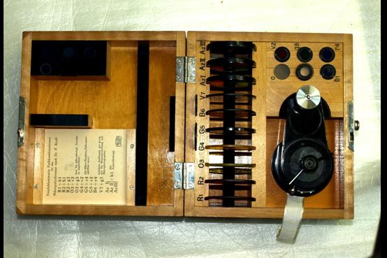
Mikropolychromar in its 1935/36 case (Document © Alain BERJONVAL)
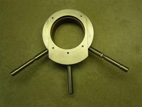
Rheinberg rings, filters and light stops
Mikropolychromar apparatus
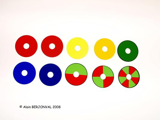
Annular Rheinberg filter rings.
(Document © Alain BERJONVAL)
How to use it?
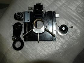
Mikropolychromar fitted on a 1933 Zeiss F or G stand. Three levers controlled the three diaphragms.
(Document © Alain BERJONVAL)
The apparatus is introduced to and screwed onto the aplanatic N.A 1.4 condenser, then observations can be started immediately once the illumination has been adjusted. Manipulation is very simple, there are no difficulties or tricky adjustments. Different kind of illumination effects can be achieved in the shortest possible time with few operations.
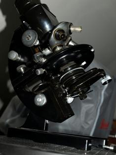
Due to ease of illumination, I fitted a “Mikropolychromar” on a Leitz Ortholux I with this self-made Leitz dovetail apparatus:
(By courtesy of Mr. Roger Bluteau)
On Ortholux I
Left. Fitted beneath Ortholux I mechanical stage.
Right. Mikropolychromar fitted on condenser holder
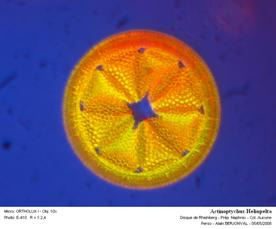
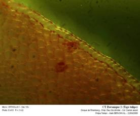
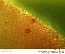
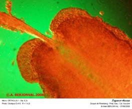
A few trials
|
|
|
|
|
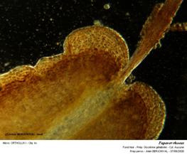
Coloured diatom and background |
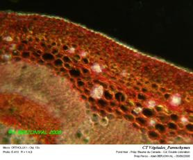
Coloured plant section and background |
Coloured plant section and background |
|
|
|
|
|
Coloured leaf and background |
Normal darfield illumination |
Normal darkfield illumination |
Conclusion
The Zeiss Mikropolychromar was developed during the 1930s, from an illumination technique demonstrated by Julius H. Rheinberg in 1896.
It combined in one apparatus all illuminating techniques known, (bright, darkfield illumination, and even oblique illumination), with optical staining.
It allowed the scientific user to deal with colourless and poor contrast specimens.
It anticipated the new illumination concepts such as phase contrast (Phaco), demonstrated by Zernike in the 1930s. (The first phase-contrast microscope prototype was made by Carl Zeiss Jena in 1936) and interference microscopy with a very simple technique based on Rheinberg illumination. Maybe, only the Leitz “Heine” condenser, can offer comparable versatility.
The purpose of this article is also to demonstrate the ability to use old illumination techniques like Rheinberg illumination or oblique illumination as a cheaper alternative to phase contrast and interference microscopy.
Comments to the author are welcome.
© A.BERJONVAL 2008

Bibliography
Carl Zeiss Jena (Zeiss documents)
Optical Microscopy by Michael W. Davidson & Mortimer Abramowitz
Leitz dovetail apparatus by : Mr Roger BLUTEAU
Photos and documents :© A. BERJONVAL
Editor's note: The English manual for the Zeiss Mikropolychromar is available to freely download as a pdf file from Gordon Couger's www.science-info.net website.
Published in the December 2008 edition of Micscape.
Please report any Web problems or offer general comments to the Micscape Editor .
Micscape is the on-line monthly magazine of the Microscopy UK web site at Microscopy-UK
©
Onview.net Ltd, Microscopy-UK, and all contributors 1995 onwards. All
rights reserved.
Main site is at
www.microscopy-uk.org.uk
with
full mirror at
www.microscopy-uk.net
.