WAR AND ITS MICROSCOPES
A review of the Spencer 60, the Tiyoda MKH, and the AO Microscope Set
Manuel del Cerro, Pittsford, NY
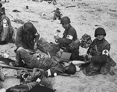
Figure 1. Attending the wounded, WWII
The admittedly loose classification of microscopes recognizes a group of lighter-than-average instruments as the “Portable”, “Field”, or “Pocket” microscopes. Portable microscopes have been produced over a span of three centuries or more. Ranging from single-lens stands to optically complex instruments they have been designed for use outside the research laboratory or the hobbyist’s living room. Portable microscopes have been the object of a comprehensive review by Mike Dingley [1] and many have been discussed in the pages of Micscape [ex. 2, 3].
Here I propose to recognize a subset of portable microscopes as the “War Microscopes.” These are instruments that were designed and built to serve under the rough conditions of the behind-the-lines hospitals. A few microscopes in my collection will help to illustrate the point.
1. PREPARING FOR WAR. THE SPENCER 60F & H
Years back I heard from a person once placed highly in the Spencer Microscope Operation, that the Spencer model 60 was designed prior to the outset of WWI primarily for use in front line hospitals. That assertion is supported by the design that is far better adapted for such use than its predecessor which was produced by the same company until 1910. The unusually heavy and robust metal shell that forms the carrying case is probably resistant to anything short a of a direct hit by a moderate-caliber bullet. The fact that the case includes, besides the microscope itself, the pipettes for differential count of white- and red-blood cells and a Neubauer hemocytometer, is consistence with use in a front-line medical facility. Some of these microscopes saw use in civilian laboratories but the weight of the kit and the nature of the accessories would make unlikely its use by say an amateur microscopists exploring the country-side. In other words, the kit is portable but not light.
Two Spencer 60 (MdC #s 240 and 006), serial numbers #26290 and #38985 will be used here as two different versions of this model. I will describe the 60H #240 (Figures 2-4), comment on the slight differences of it with 60F #006 (Figures 5-6), and refer to some original Spencer illustrations (Figures 7-8).
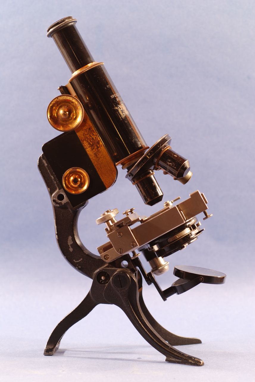
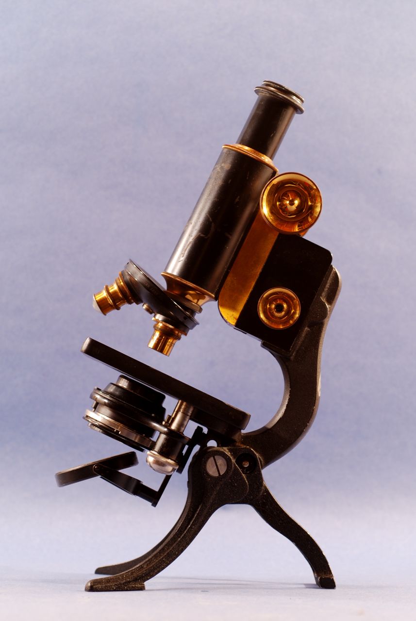
Figure 2. The Spencer model 60H in its working configuration. Notice the articulation on the metal strip that supports the fork-mounted mirror. The head of the condenser focusing screw can be seen at the left of the mirror.
The Spencer 60H microscope has a folding tripod base (Figure 2 above). A fork-mounted, plano-concave mirror is fastened to a two-piece swinging tailpiece. The condenser, iris diaphragm, and filter holder are mounted as a unit; this unit moves away the optic path if it is lowered all the way by the action of a helical screw called by Spencer the "Quick Screw” (Figures 2 and 6). There is a graduated drawtube that carries an unsigned 10x ocular. Coarse focus is by rack and pinion. Fine focus is controlled by bilateral knobs; the one on the right side has a µm scale. The stage is rectangular, 10.5 cm deep by 8.5 cm wide. One of the oculars is labeled “10x”, the other “5”. A signed, three-objective revolving nosepiece holds a 16 mm NA 0.25, a 44x NA 0.85, and 95x NA 1.30 oil immersion objective. The objectives are protected by unusual black-finished metal cups (figure 3); the objectives themselves have a brass finish.
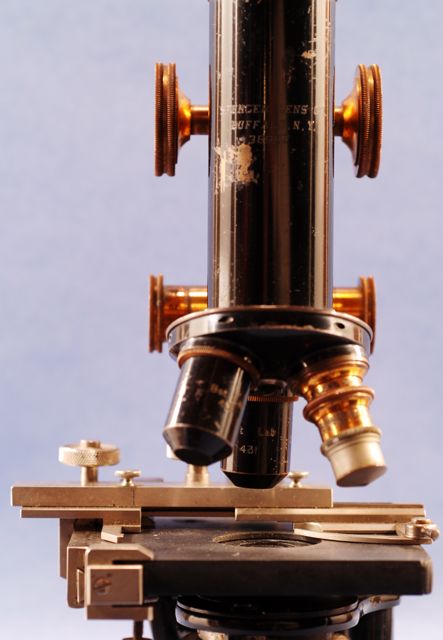
Figure 3. A close up view of the objectives in the Spencer model 60H. The two dry objectives are shown with their protective cups which has been removed in the oil-immersion objective.
The original carrying case is compact and ingeniously designed. It is made of thick metal with a black finish. The inside has a metal rack for nine slides, holders for the two oculars and the three objectives. There is also a metal container for cover glasses, an unusual chrome-finished oil immersion dispenser (Figure 4), and an optional mechanical stage, called model No. 485. The signed immersion oil dispenser has micrometric control of delivery; it is an example of precise metal machining.
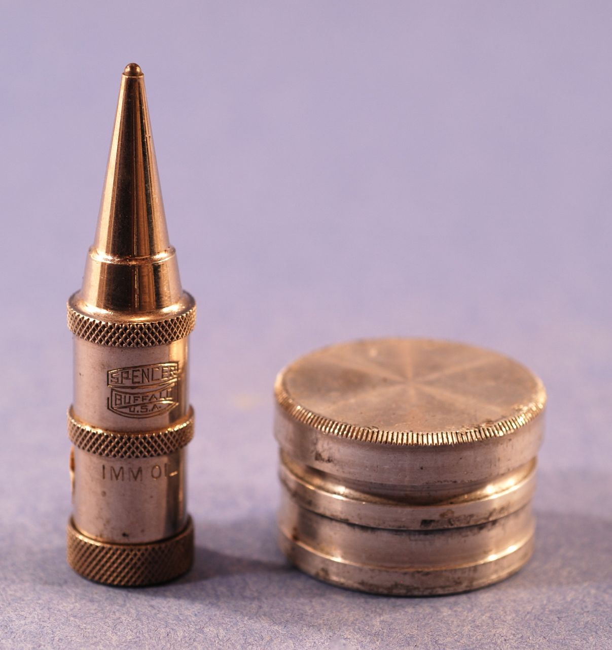
Figure 4. To the left is the metal dispenser for immersion oil included in the model 60 kit. This sturdy accessory exhibits refined workmanship and innovative design (was it the forerunner of the ball pen concept?). To the right is the cover glass holder that is also part of the kit.
Every ocular, objective, or accessory, such as the pipettes and hemocytometer, is firmly clamped inside the carrying box (Figures 5-6). The box and the microscope have the letters “ISCT” engraved on them; since the I and the S, and the C and the T overlap, it is not obvious what the exact sequence is. Circa 1912. Provenance: Lost and Found Antiques, San Francisco. Price: $400.00. Date: November 1990.
PERFORMANCE: The 16 mm objective provides a very good image with mild field curvature; the field diameter is 2,100 µm. The 44x objective provides a very good and flat image with a field diameter of 440 µm. The oil immersion objective was not tested.
COMMENTS:
1) Purtle (1974) describes a 1911 Spencer 60H, serial #25704. The design is said to be “a radical change in construction from the 1904 model.” [4]
2) The 1914 Spencer Lens Company booklet "How to use and care for the microscope" lists this microscope as the 60H Portable microscope selling for $100.00. A near identical instrument, the Model 62H had fine focus by a single knob located at the top of the pillar a design that Spencer was one of the first big makers to discard.
3) MdC Collection #006 is another example of the same design. It differs from this one in being a microscope with two dry objectives and in having a single-piece tailpiece for the mirror. It was called the 60F model [4] (Figures 7-8).
4) The 1920 catalog of the Spencer Lens Company [5] shows this microscope, its case, and accessories in excellent detail (Figures 5-6).
5) There is a microscope of this model in the MacLeay Museum, Sydney, Australia (Julian Holland: Microscopes and Microscopy. Instruments and Related Items in the Macleay Museum) an indication of the widespread use of this Buffalo, NY made instrument.
6) A similar microscope was auctioned in ebay on 2001 06 17. It was described as a “Old Rare Army Field brass and iron microscope.” The highest bid was $555.49.
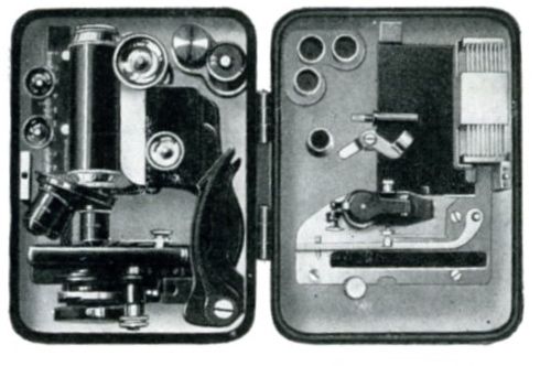
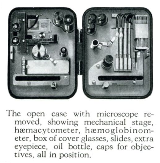
Figures 5 and 6. The metal carrying case for the Spencer 60, with and without the microscope in place.
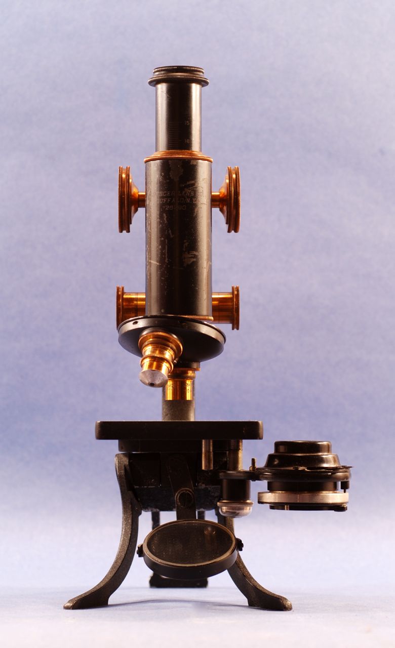
Figures 7 and 8. The Spencer 60F in side and front views. Removing the condenser from the optical path as in figure 8, and using the concave mirror for illumination provided ideal conditions for low power work.
2. IN COMBAT. THE TIYODA MKH
An article on the Tiyoda MKH was published by Jay Stanley in the August 2006 issue of MicScape [5]. Mr. Stanley notes that there is little information on this microscope, thus, the description of one other example of it seems warranted. We will then discuss here the Tiyoda MKH serial No 14050 in our collection (MdC #091) as an example of WWII military field-hospital microscope.
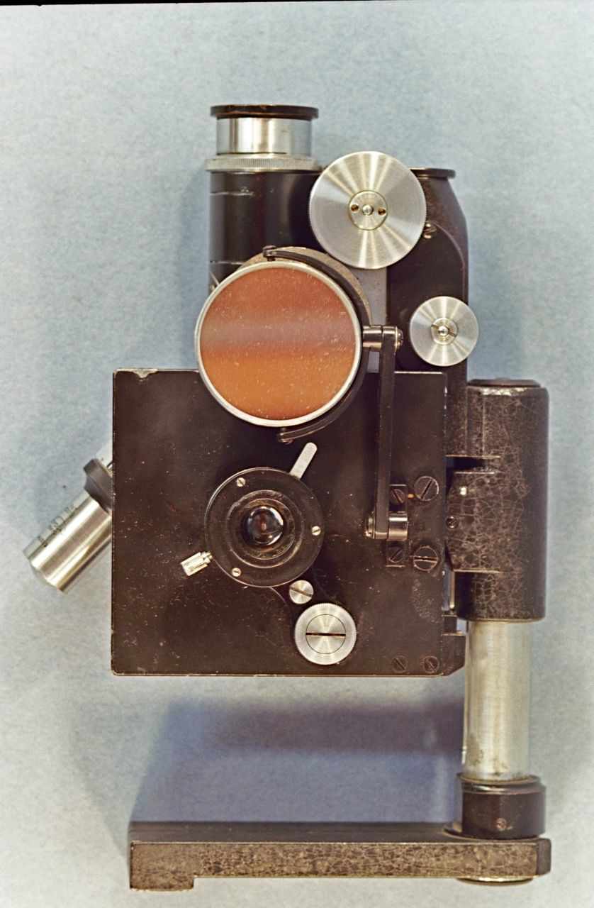
Figure 9. The Tiyoda in its folded up configuration.
This instrument is contained within a brown metal case with a hinged lid. In the closed position the folding base of this microscope is 11 cm long and 3.2 cm wide (Figure 9); in its open position the base acquires a V-shaped configuration with the arms separating 13 cm at the front (Figure 10). The vertical pillar is a metal rod 11.7 cm tall. The pillar holds an intermediate, black-finished metal piece that supports both, the stage and the arm. The fork-mounted, plano-concave mirror is set at the lower end of a swinging tailpiece, an arrangement that permits the use of oblique illumination. The illumination unit consists of filter holder, iris diaphragm, and condenser; the unit can be focused by means of a helical screw located under the right side of the stage.
The plain mechanical stage is 7.8 cm wide and 8.3 cm deep. The stage can be swiveled back to a vertical position for storage or it can be locked in its normal horizontal position by tightening a small screw located on the arm below the right side fine-focus. The arm houses the mechanisms for fine and coarse focus; these are controlled by side-knobs. A small metal plate at the top of the arm is inscribed with the Company logo, the model identification – MKH – and the abbreviation “Pat. 214050.”
The body is black-finished, 7.8 cm long, and it is engraved with the company logo, the word Tokyo and the serial number. There is a plain drawtube inscribed with a circle indicating the optimal extension required by the provided objectives. The 5x ocular is a signed. The revolving nosepiece carries two objectives, which are non-parfocal. The two objectives are a 10x NA 0.30 and a 40x NA 0.65; both are signed and carry serial numbers. A 90x oil immersion objective is stored in the carrying case; the 40x objective needs to be removed in order to store the microscope.
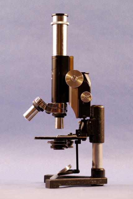
Figure 10. The Tiyoda in its working configuration.
The carrying case is made of metal finished in the “Army Brown” color that was widely used for camouflage during WWII (Figure 11). The box measures 18.3 cm wide, 6.2 cm tall, and 13.3 cm deep; it closes by a lock and hinge mechanism. Inside the box there are metal containers for the 40x and 90x objectives, an extra ocular (signed 10x). There are also two small canisters labeled “x” and “c” on their tops; each has a spring to press small bottles of xylene and Canada balsam, or immersion oil against the cap. The inside of the lid is stamped with Japanese characters encircled by an oval. Provenance: ebay auction. Price: $132.50. Date: 2005 08 13.
OPTICAL PERFORMANCE: When used with the 5x ocular both objectives provide excellent, flat images. The field diameter of the 10x objective is 2,100 µm, and that of the 40x, 520 µm.
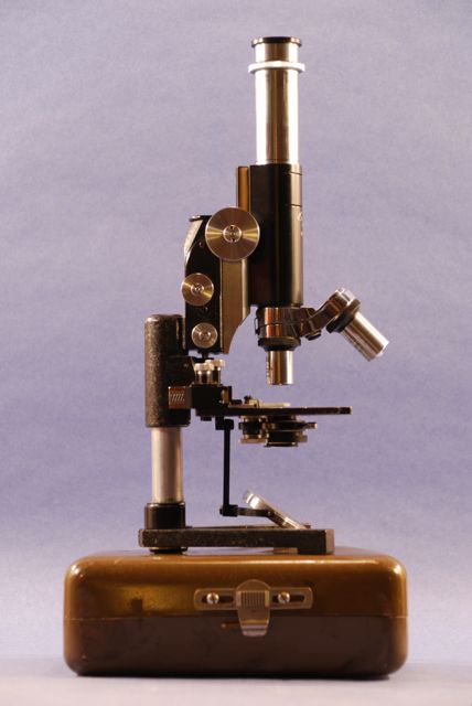
Figure 11. The Tiyoda and its carrying case.
COMMENTS:
1) The auction printout for this microscope is on file in the MdC Collection Documents box.
2) This model represents one of the several portable models made by Tiyoda.
3) According to Dingley this microscope is a copy of the Reichert “Heimdal” microscope.
4) Jay Stanley reports that this model was also called the “MKATERA Portable” and it was developed in 1929. Micscape, August 2006.
5) This is an exceptionally well-designed and constructed instrument with very good optics and stored in a very compact carrying case. The seller claims that Japanese forces left behind this microscope after the battle of Luzon, in the Philippines (15 December 1944 - 4 July 1945) and that an American serviceman recovered it.
3. SPECIAL OPERATIONS. The AO Microscope Set
American Optics Corporation, Buffalo, New York. This is a military microscope set, serial # 3050 286, made circa 1970. The microscope is 30.0 cm tall; it comes stored with its legs folded up to the sides of the arm (Figure 12). The legs are connected to each other by an articulated metal piece that when extended becomes the back leg of the tripod base (Figure 13). A metal knob allows tightening of the leg assembly for stability during use. With the legs fully extended, the instrument has a trapezoidal footprint that is 12.5 cm wide at the front, 8.6 cm wide at the back 11.5 cm in depth (Figure 14). Both, mirror and electrical illumination are provided for this microscope. The mirror is concave, 4.5 cm in diameter; it is fork-mounted at the end of a swinging tailpiece, so that it can provide oblique illumination.
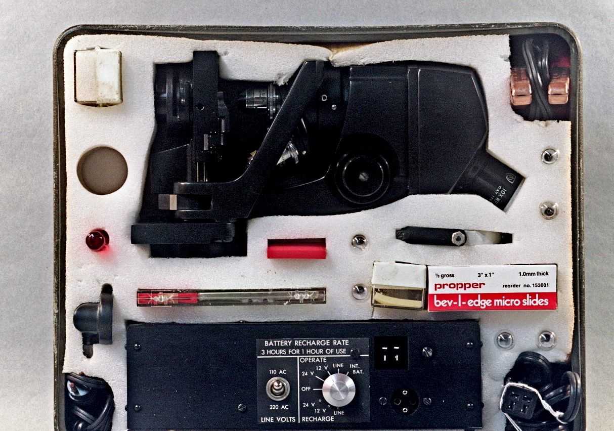
Figure 12. The AO microscope set.
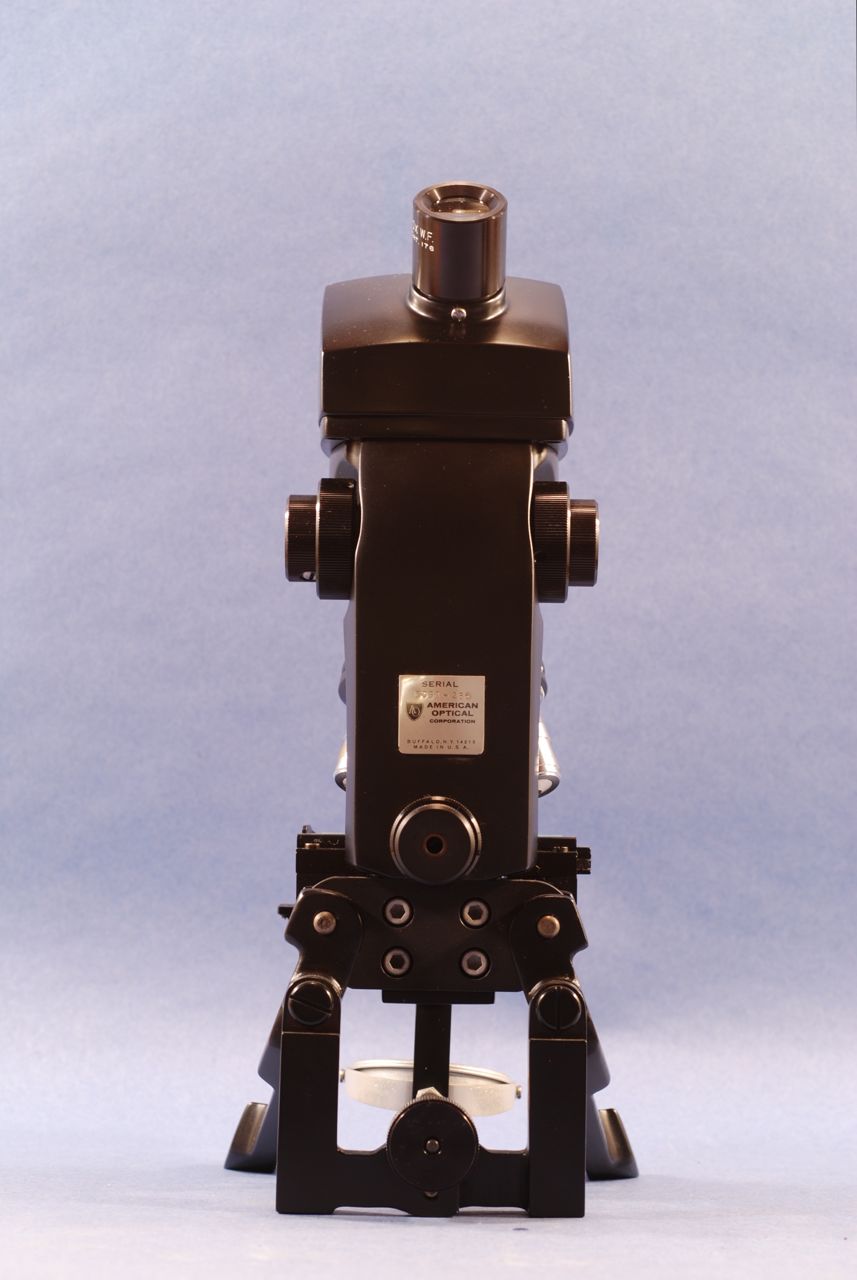
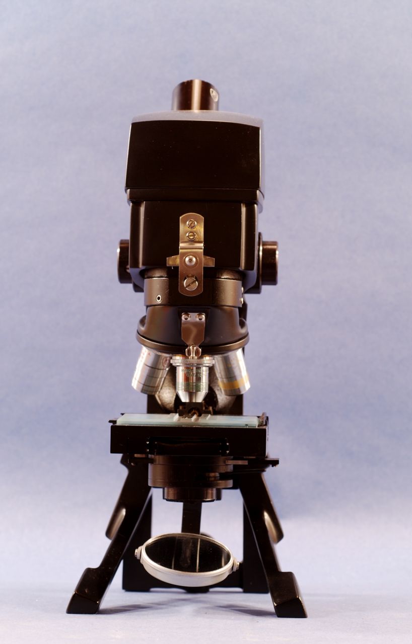
Figure 13 and 14. The AO microscope.
Electrical illumination is provided by a special device consisting of a small bulb mounted on a housing that can be attached directly to the bottom of the condenser. A rechargeable battery stored inside a black metal box powers the lamp. The battery can be energized by alternate current of either 110 or 220 volts. The output can be either 12 or 24 volts. The condenser is set at a fix position; it has an iris diaphragm and a metal slider placed between the lower and upper lenses. The slider carries two filters; one is ”daylight blue” and the other a neutral density.
The rectangular mechanical stage 18.3 cm wide and 7.0 cm deep with a spring-loaded slide holder. The movements of the mechanical stage are controlled by coaxial knobs located in the back of the column, protruding 3.0 cm towards the observer - another unique detail in the design of this instrument. The column is rectangular, 12.5 cm tall and 5.0 cm wide. A small metal plate attached to the surface of the column carries the serial number, the company logo, and the legend “American Optical Corporation Buffalo, N.Y. 215 Made in U. S. A.” The coaxial knobs for coarse and fine focus are located high at either side of the column.
The body is composed of three segments (Figure 14). The upper one houses the prisms that deflect the image towards the inclined monocular tube. The ocular is labeled 10x W.F. cat. 176. The intermediate segment of the body houses a focusing mechanism the acts on the nosepiece. The microscope is essentially pre-focused. The lower segment is continuous with the nosepiece. The revolving nosepiece carries infinite corrected objectives. These are a 10 NA 0.25, a 45x NA 0.66, and a 100x NA 1.25 with internal diaphragm. All the objectives are signed Spencer AO, and labeled with their catalog numbers and the inscription “Made in U.S.A.”
OPTICAL PERFORMANCE: The 10x objective provides a flat image of exceptional quality with a field diameter of 1,550 µm. The 45x objective also provides an image of exceptional quality with light field curvature; the field diameter is 350 µm.
The microscope is housed in a box made of heavy plastic with an olive green color suggestive of military use. Four sides of the box carry the identification: “Microscope Set 6545-926-8961.” The box is entirely filled by a Styrofoam block with cutouts for the microscope and for each component of the set. These include a box of glass slides by the Propper Company, two boxes of cover glasses by Hausser Scientific, a plastic box containing two Spencer Bright Line Hemacytometers [sic] with two boxes of hemocytometer cover glasses. There is another separate box of hemocytometer cover glasses, a screwdriver, and a fork-mounted mirror. Other cutouts hold the components of the electrical illumination system. These are a small lamp housing with its light bulb, and six extra light bulbs.
A power box contains a rechargeable battery that can be recharged from a line or from 12 or 24 v sources; it can operate the microscope either from its own internal battery, directly from a power line, or from 12 or 24 v sources. There are cables to connect the microscope to the power box and the power box to either a 120-v outlet or to a motor vehicle battery.
REFERENCES:
1) Auction printout on file in the MdC Collection Document box.
2) Information on this microscope is limited and some may still classified. The official designation of the set is “Microscope Set, Medical Laboratory Equipment Set, Lightweight, Field. FSN 6545-926-8961”
Comments made by contributors to a Yahoo Microscope forum (2007) indicate that this microscope was designed for blood work (malaria detection?) and made to focus on the chamber of a 2 mm-thick Neubauer hemocytometer (Figure 15). Accordingly it will not focus at high power on the surface of standard 1 mm-thick slides unless the objectives are shimmed or the slide is mounted on top of a blank slide. Another contributor to the Yahoo forum notes that fewer than one thousand units of this microscope were made, and still another reports from personal knowledge that this model was used in operations by (USA) Special Forces in the 1960s and 1970s.
CONCLUSION
We have identified a type of microscopes as “The War Microscopes.” Although made decades apart, the microscopes discussed in this article and their brethren show several features that are common to the group.
1- Portability. All are portable, although of course what is understood under that term varies greatly. For a person to carry the Spencer 60 in its heavy protective case for any considerable distance would be arduous. To do the same with the relatively large AO Microscope Set would be cumbersome. Both instruments and their boxes are apparently designed to be carried by transport vehicles together with whatever other medical equipment the circumstances required.
2- Sturdy construction. The Spencer 60 and the AO Microscope are solidly built. The Spencer is protected by a rigid shell of heavy metal. The AO is protected by a flexible plastic shell and abundant Styrofoam packing. The Tiyoda, although light and nimble, is sturdy in its construction; it has as a column a rod of solid metal. Its carrying case, although thin and light protects well to the instrument. The case of the Tiyoda in our collection shows damage indicative of rough handling and yet the microscope is in perfect functioning condition.
3- Specialized use. All these microscopes were designed to perform well one or a few functions. These are not “Universal” stages. The Spencer 60, particularly in its version 60H, comes close to a microscope able to perform most tasks that microscopes of that vintage were able to perform. However, any investigator or clinician of the times would have preferred to use the more conventional contemporary models that the Spencer Company was offering at substantially lower prices. Similarly, in the case of the Tiyoda MKH, investigators and clinicians in the laboratory would likely have preferred the more ergonomic models the company was offering at the time, and have little use for the protective metal shell of the MKH. The AO Microscope goes further in specialization. This is an instrument designed to study blood samples using a hemocytometer. Its ability to work either with ambient light reflected by a mirror, or using an electrical illuminator that in turn can be powered by line, rechargeable battery, or by the electrical outlet of a motor vehicle is admirable, but unnecessary in a standard laboratory environment.
One microscope model that fulfills all of the criteria but has not been discussed here is the so-called trichinae microscope. We have chosen not to include it because, although it was used by the Health Departments of many armies, specially the German in WW I, it was used as much if not more in peace time for the purpose of monitoring the safety of the pork meat consumed by the civilian populations.
Now that we have defined a group of microscopes as “War Microscopes,” let’s wish that them and their successors forever rest in peace!
Comments to the author are welcome.
REFERENCES
[1] Dingley, Mike (1997) Portable Microscopes. Micscape, October1997.
[2] Mach, Martin (2000) 30 g of microscope please! Micscape, August 2000.
[3] Stanley, Jay (2006) The Tiyoda MKH Field Microscope. Micscape, August 2006.
[4] Purtle, H. R. (Ed.) (1974). (The) Billings Microscope Collection of the Medical Museum Armed Forces Institute of Pathology. Armed Forces Institute of Pathology, Washington, DC, 2nd Edit. From the Government Printing Office, p. 127.
[5] Spencer Lens Company (1920). Catalog of Spencer Microscopes, Microtomes, and Accessories. Spencer Portable Microscope No. 60. pp. 50-51. Spencer Lens Company, Buffalo, NY, USA.
Published in the December 2008 edition of Micscape.
Please report any Web problems or offer general comments to theMicscape Editor .
Micscape is the on-line monthly magazine of the Microscopy UK web site atMicroscopy-UK
© Onview.net Ltd, Microscopy-UK, and all contributors 1995 onwards. All rights reserved.
Main site is at www.microscopy-uk.org.uk
with full mirror at www.microscopy-uk.net
.