When you take a
flower in your hand and really look at it,
it's your world
for the moment. I want to give that world to
someone else.
Most people in the city rush around so,
they have no
time to look at a flower. I want them to see
it whether they
want to or not.
- Georgia O'Keeffe (Artist)
In an earlier Micscape article, I
investigated two members of the Passifloraceae
family Passiflora caerulea,
and Passiflora coccinea x incarnata.
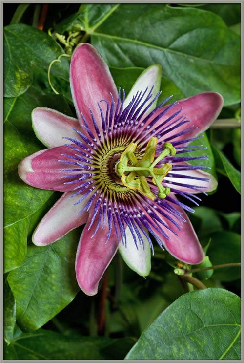
In
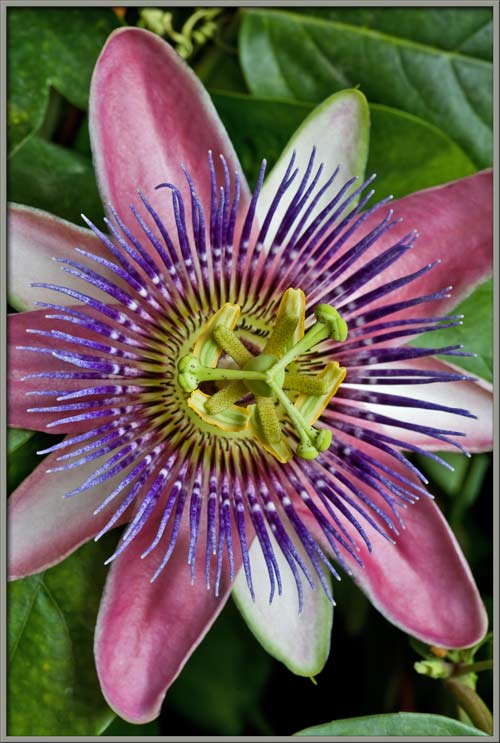
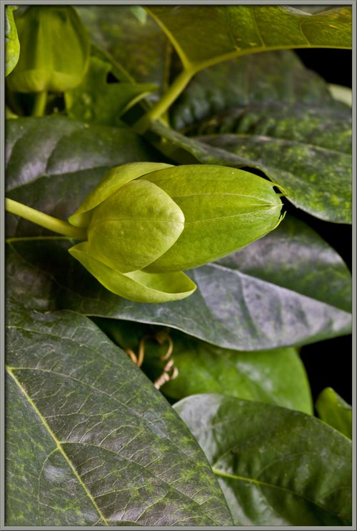
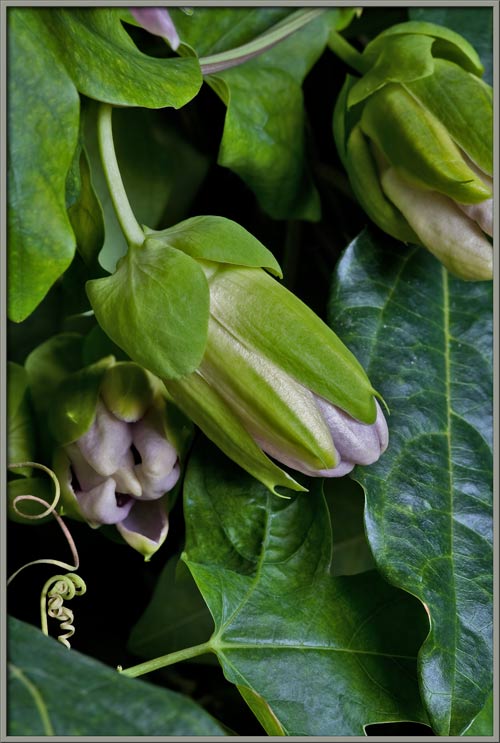
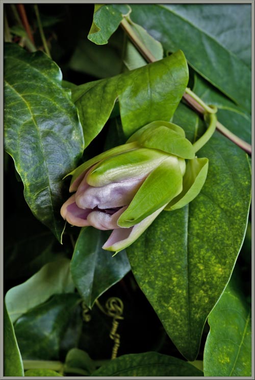
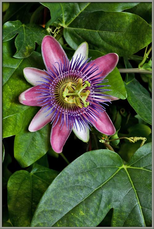
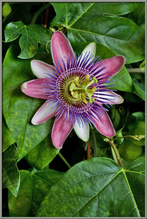
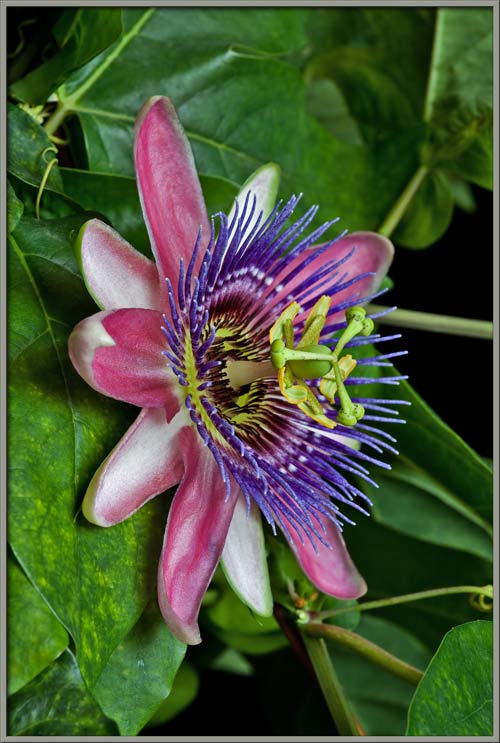
this article, I will describe the extremely beautiful hybrid Passiflora x belotti, which was
produced by crossing P. alata
with P. caerulea.
Unlike the other two species, this one has an absolutely wonderful
scent.
In 1824, Dr. Lindley named this
hybrid Passiflora alato-caerulea,
and
over the years it has been given other names: Passiflora munroi, Passiflora pfortii, Passiflora x belotii, etc.
Today, one of the commonest names is Passion Flower Imperatrice Eugenie. The
dedication is to the wife of Emperor Napoleon III.
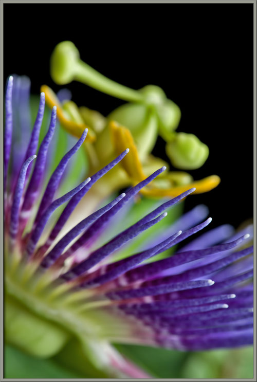
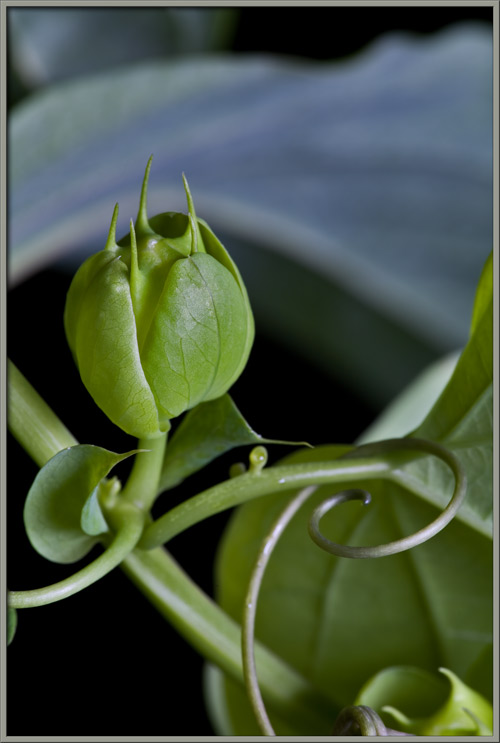
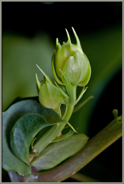
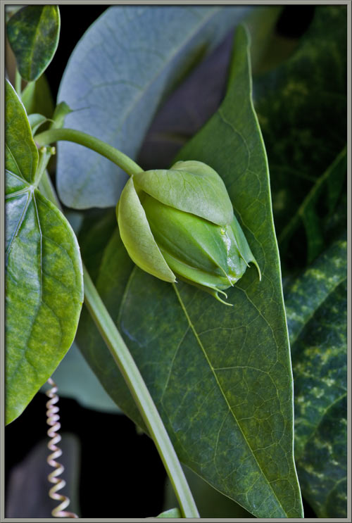
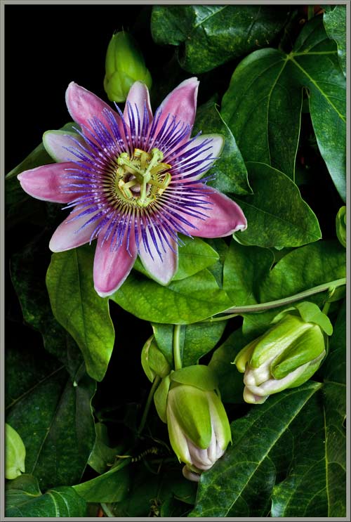
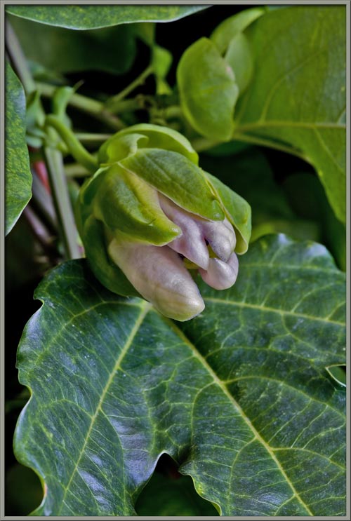
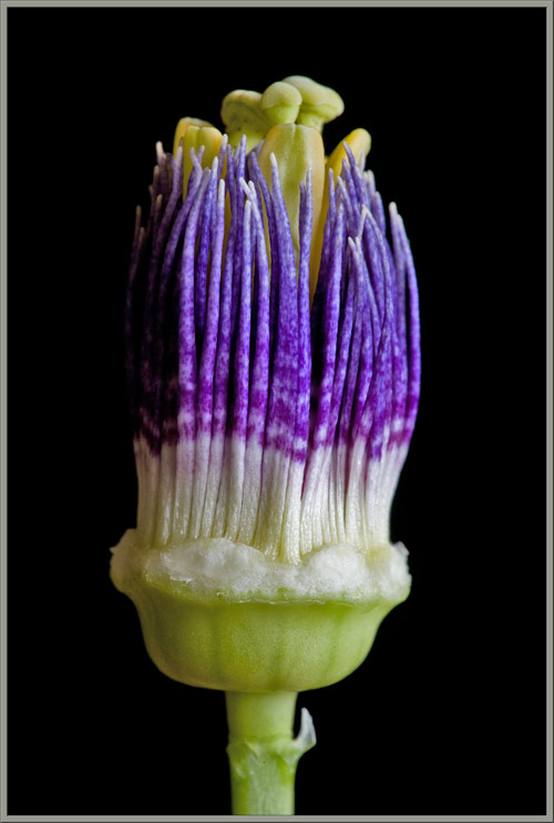
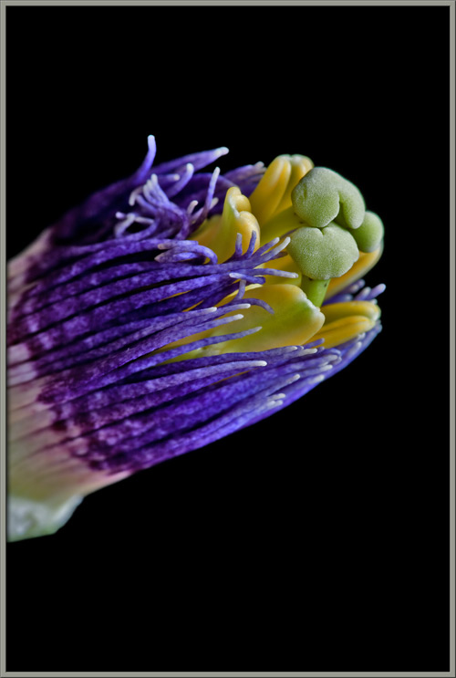
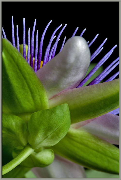
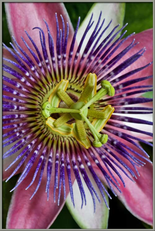
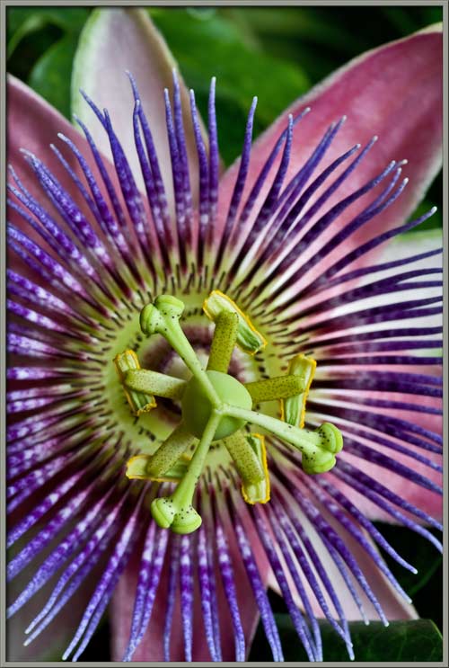
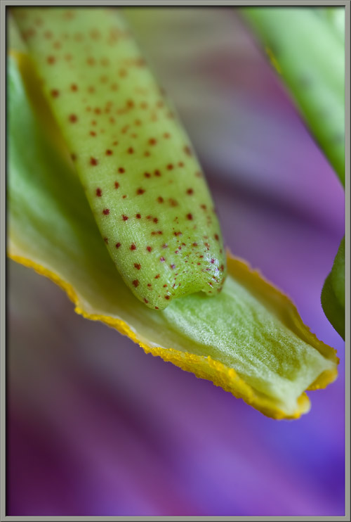
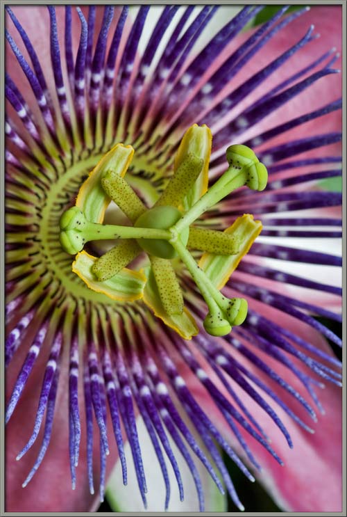
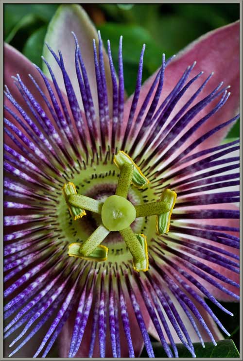
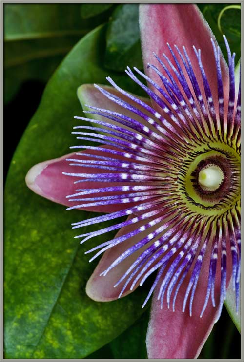
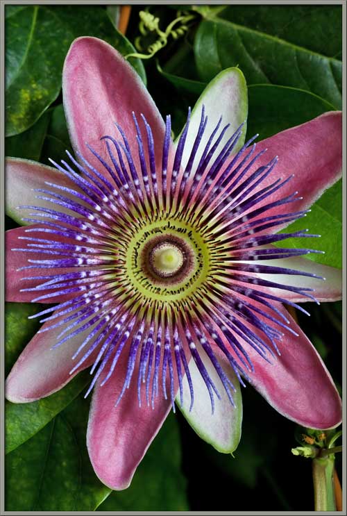
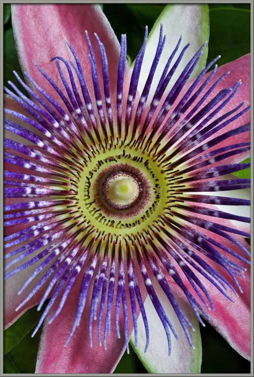
All Passion Flower blooms are
strikingly complex, and seem almost alien when compared with simpler
flowers. Sepals, petals, coronal filaments, and reproductive
structures all combine to form an amazing botanical spectacle.
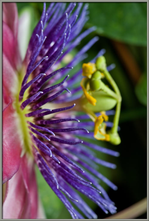
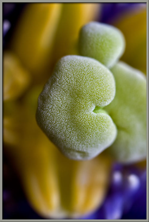
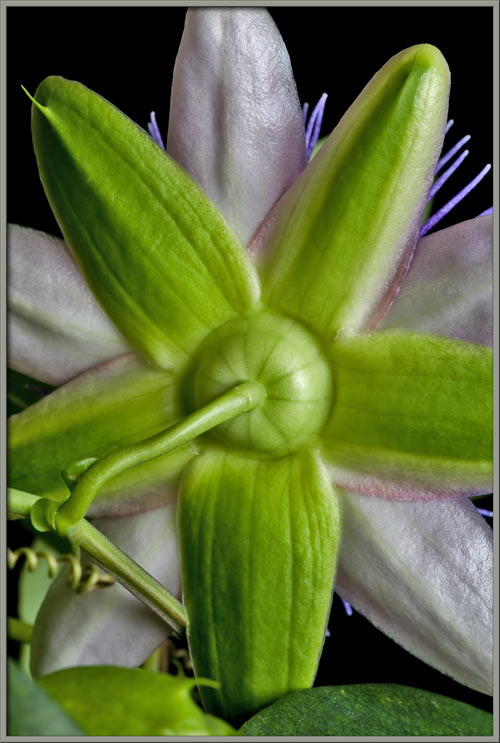
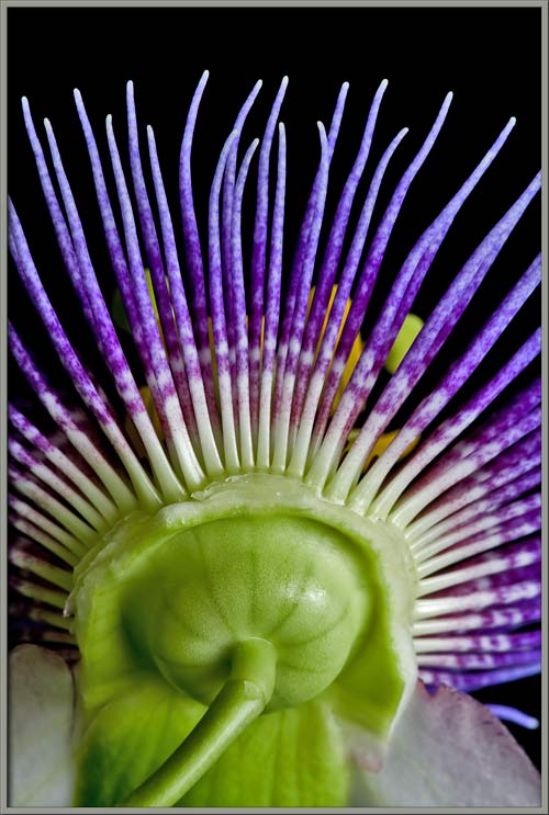
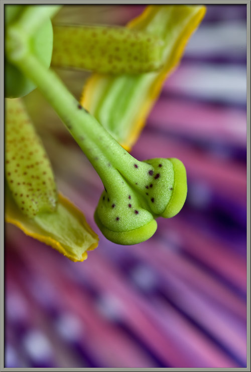
In order to obtain the
following
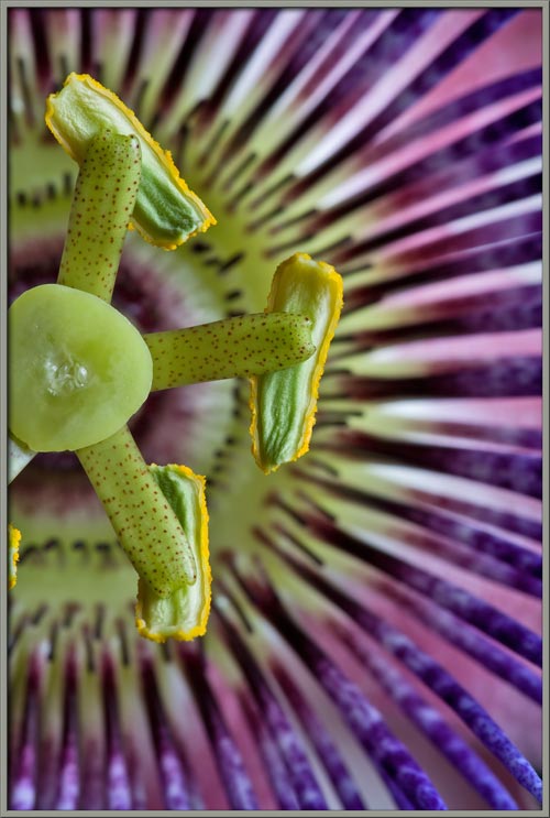
images, I carefully cut transversely through the flowers ovary to
remove the top-most section of the androgynophore containing the styles
and stigmas.
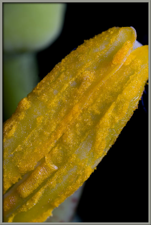
This allows the filaments and
anthers to be more easily seen.
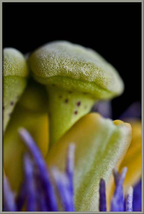
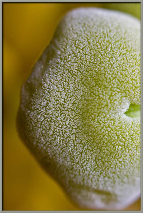
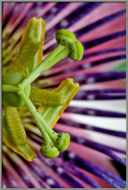
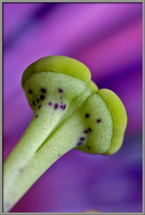
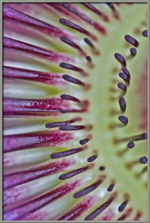
Next, I cut transversely through
the base of the androgynophore to remove the male reproductive
organs. This allows the multiple layers of coronal filaments to
be completely visible. The long
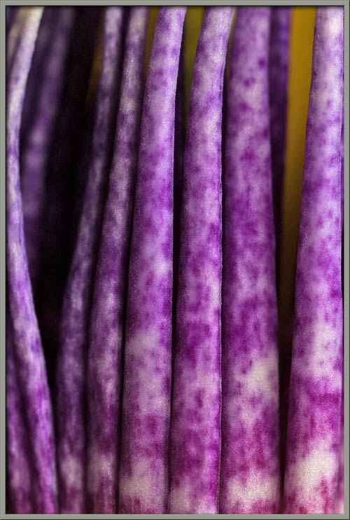
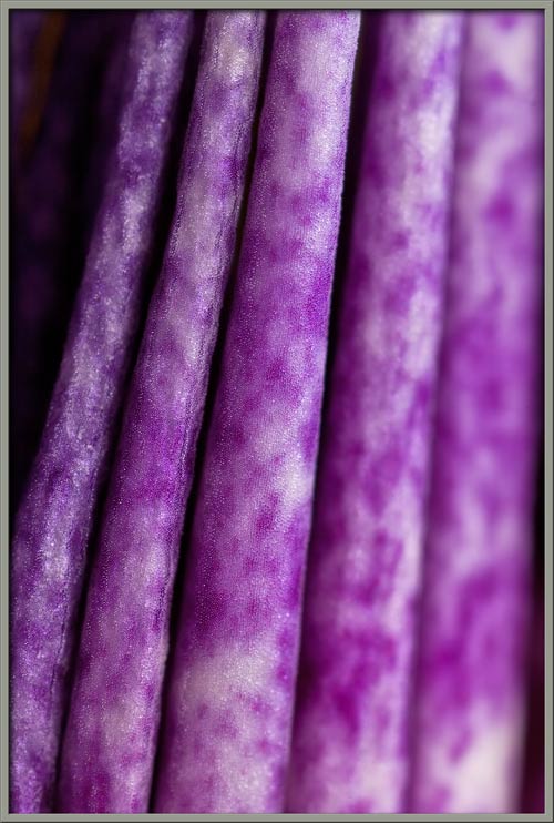
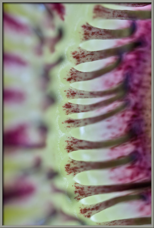
filaments form the flowers outer
corona. If you look closely at the right-hand image, you
will see that near the centre of the flower, there are several rings of
much shorter filaments.
These form the inner corona of
the flower.
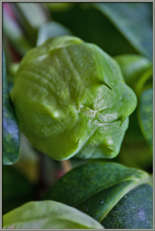
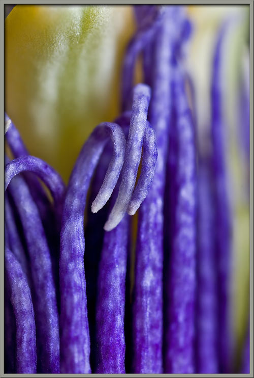
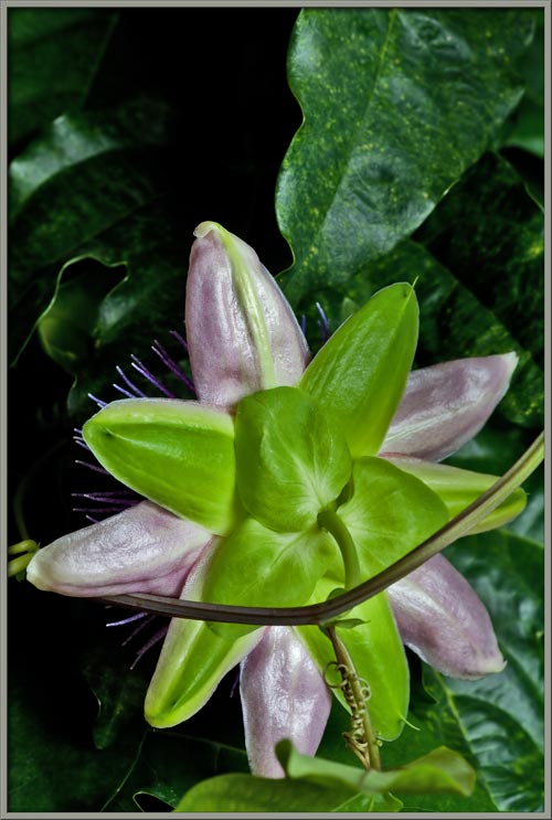
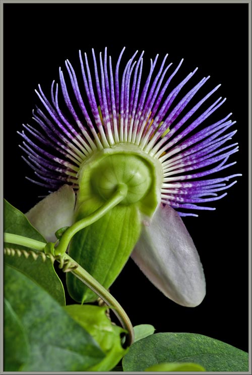
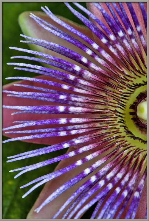
Passiflora
x
belotii plants sometimes have slight variations in
colour. One of these variations is the number of white bands that
occur on the coronal filaments.
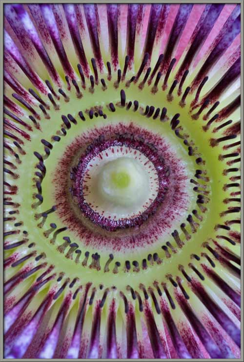
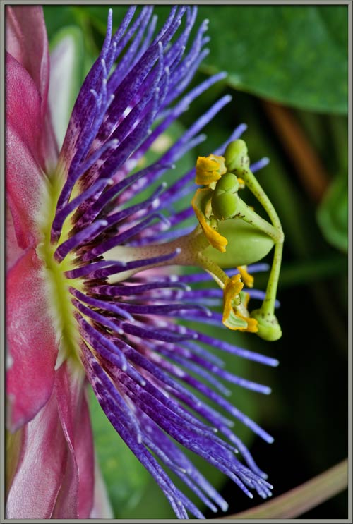
Some botanists believe that the
concentric coloured rings, or bands, visible in the flower are a
mechanism to guide insects towards the centre of the bloom where the
nectar is to be found. Of course, on the way to this sugary
treat, the insects may come in contact with anthers and stigma, and
therefore promote fertilization.
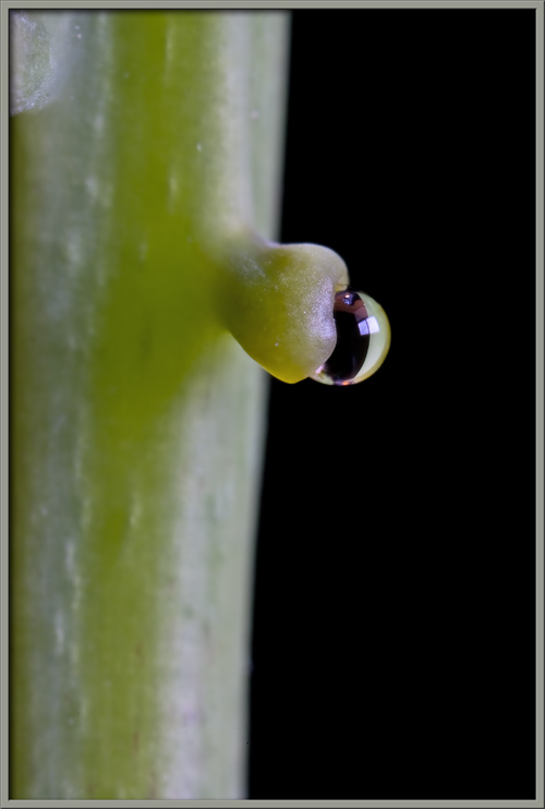
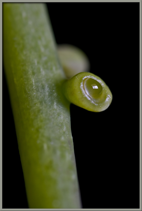
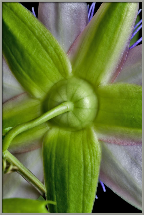
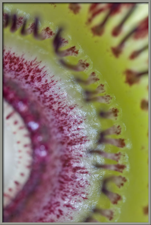
Where exactly is this nectar to be
found? It is held in a shallow, donut-shaped reservoir around the
(stub of the) androgynophore. The circular outer wall of this
reservoir is within the wide purple band that can be seen in the images
below.
The shallow reservoir containing
the flowers nectar is the light (almost white) ring around the
androgynophore stub. The purple ridge holding the nectar in place
is called the limen. The
ring of very short coronal filaments just beyond the ridge is called
the operculum.
The two images that follow show the
tops (left image), and bases (right image) of the inner corona
filaments.
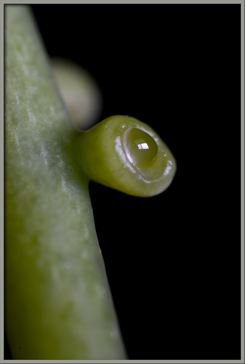
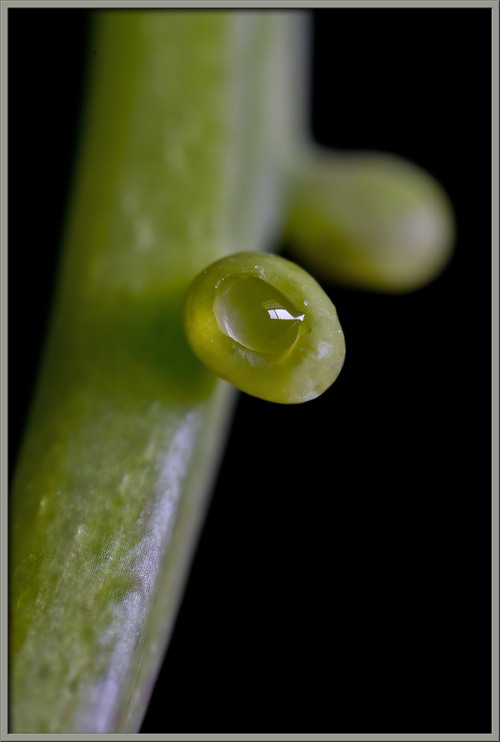
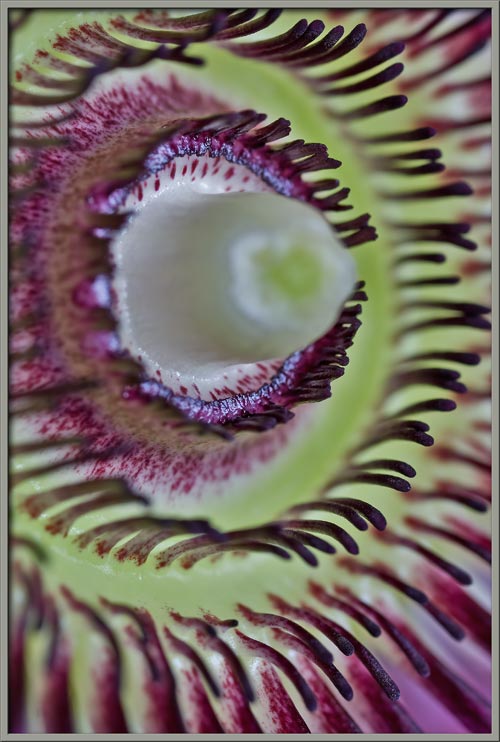
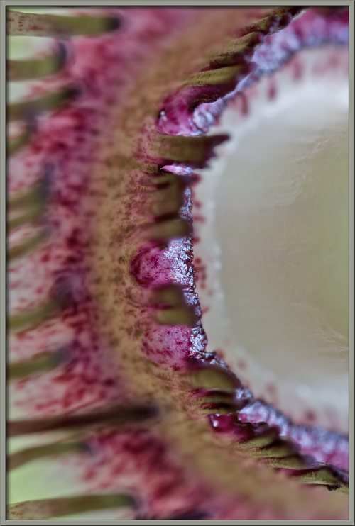
In the image below, the ring
between the limen and operculum can be seen to have a rough,
purple-spotted surface.
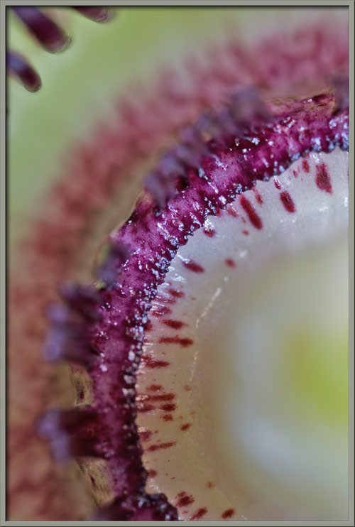
The two images that follow show the
wall-like limen that contains nectar. In both images, it is just
possible to see the shiny surface of the sticky liquid.
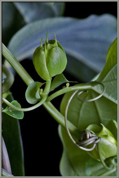
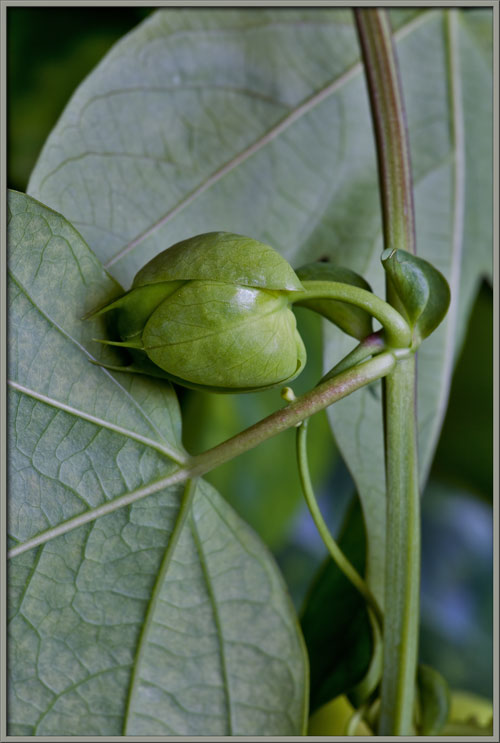
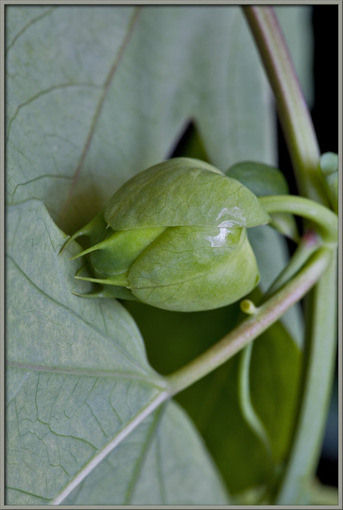
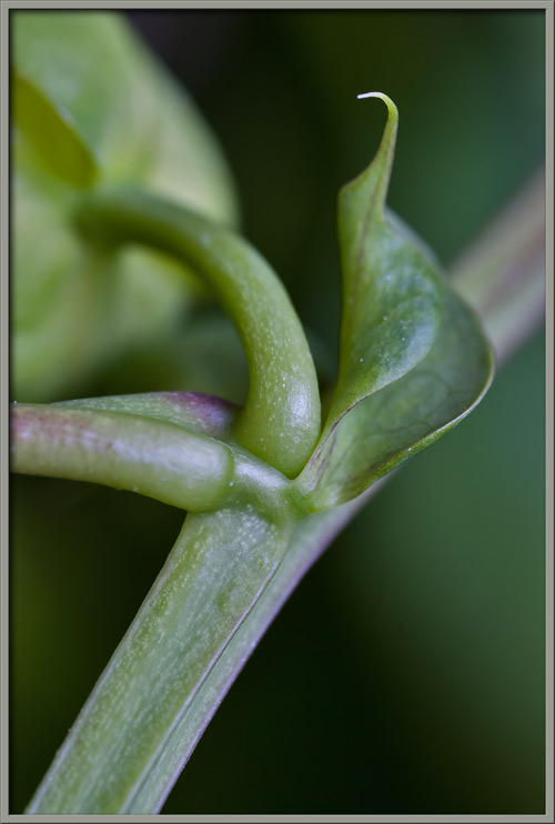
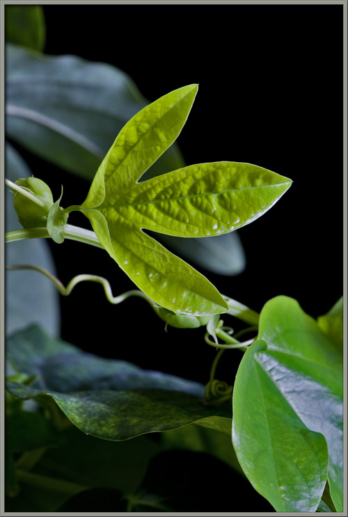
Finally, a brief look at this
plants leaves. All are three-lobed, with mature leaves being
dark green, and young ones a lighter shade of green.
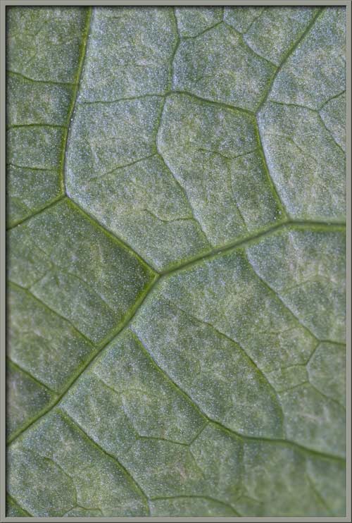
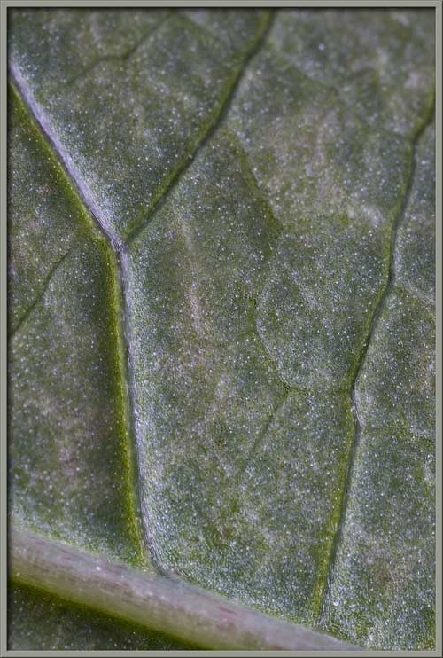
Images of the upper (left), and
lower (right) surfaces of a leaf reveal the irregular vein pattern on
its surface.
Passiflora
flowers look almost good enough to eat. This would however, be a very bad idea, since the plant
contains poisonous alkaloids (e.g. passiflorine), and cyanogenic
glucosides (e.g. hydrocyanic acid). Tissues of the plant may also
contain calcium oxalate crystals, which are also poisonous.
As a macro-photographer, I
particularly enjoy capturing unusual, complex flowers and their
structures. Passiflora
hybrids are just about as good as it gets!
Photographic Equipment
The low magnification, (to 1:1),
macro-photographs were taken using a 13 megapixel Canon 5D full frame
DSLR, using a Canon EF 180 mm 1:3.5 L Macro lens.
An 8 megapixel Canon 20D DSLR,
equipped with a specialized high magnification (1x to 5x) Canon macro
lens, the MP-E 65 mm 1:2.8, was used to take the remainder of the
images.
Reference
Zomlefer, Wendy R. 1994. Guide to
Flowering Plant Families. The University of North Carolina Press,
Chapel Hill & London.
A Flower Garden of
Macroscopic Delights
A complete graphical index of all
of my flower articles can be found here.
The Colourful World of
Chemical Crystals
A complete graphical index of all
of my crystal articles can be found here.