|
A Glass “Cup” Sponge: Part 1 by Richard L. Howey, Wyoming, USA |
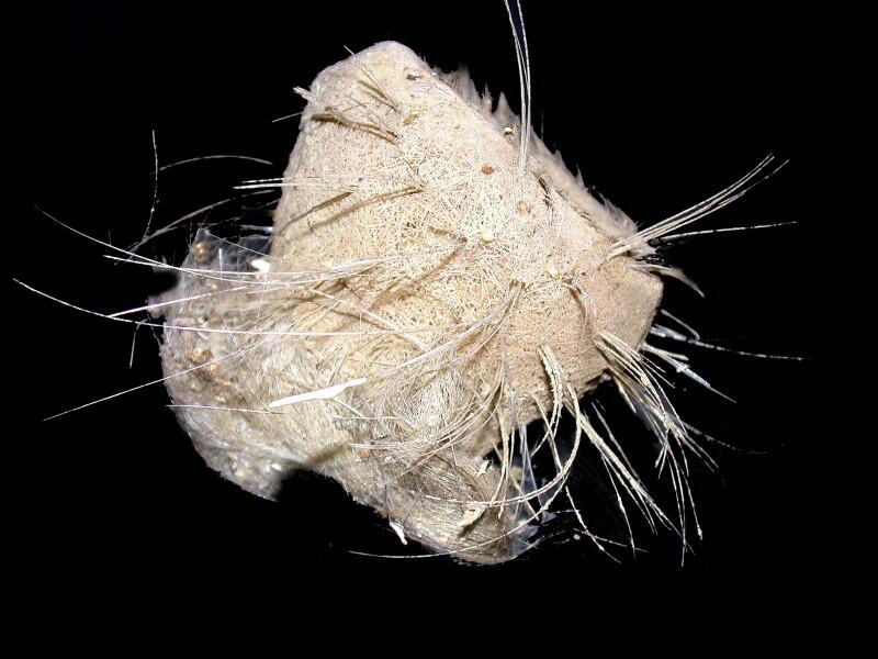
For some time now, I have been occupied with observing the spicules and other skeletal structures of selected invertebrates and of sponges in particular. In this essay, I want to look at a type of “cup” sponge from the Philippines. When it comes to spicules, this species (which I haven’t yet identified) is full of marvelous minor wonders and surprises. I have 4 specimens which vary in size from 1 inch to 4 inches in diameter not including the glass “feathers” or fibers which extend out from the central mass. Some of these are nearly the length of the diameter of the sponge but others are actually longer and 3 that I removed from the largest of these sponges were over 5 inches long.. I am going to show you 3 views of the largest specimen. This first view is an image from the side and shows the bowl or cup-like form, the projecting fibers, and the suggestion of a basal plate.
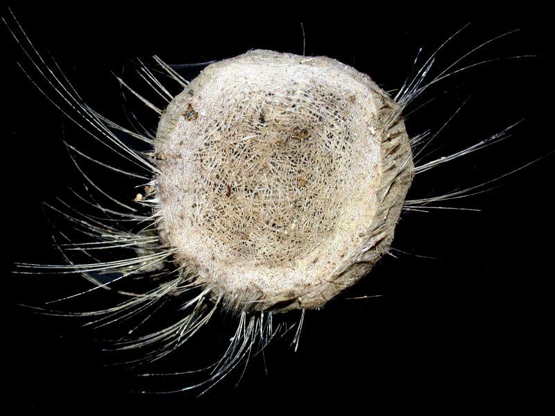
The second view is from the top looking down into the cup but, unfortunately, the depth is difficult to show in an image. The depth is a bit over an inch. Also, in this image, you can see how the long, slender fibers extend out around the main “body”
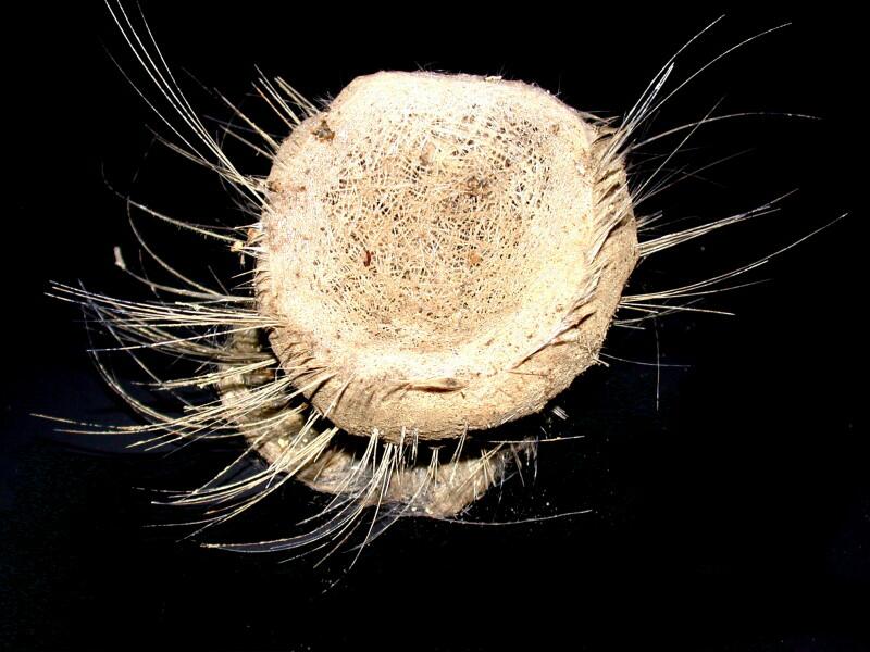
The third image is very much like the previous one, except that it is taken from a different angle which gives you another view of the basal plate which is likely an anchoring structure.
I took some forceps and pulled loose (with some effort) some small samples from the lower part of the cup and put them in a Petri dish.
---------------------------------------------------------------------------------------
A Word Of Caution: These spicules are very fine, extremely sharp, made of glass, and are highly irritating if you get any of them embedded in your skin and very dangerous if you happen to have some of the material on your hands and rub your eyes. Also, a slight draft, deep breath, or especially a sneeze can easily cause this material to float into the air which is also not good, because you don’t want glass particles in your lungs. So, if you decide to work with glass sponges, please take proper precautions. Use forceps and dissecting needles when extracting or sorting the material and if you need to handle the sponge directly wear disposable rubber or nitrile (if you are allergic to latex) gloves. For best safety, also wear goggles and a small surgical mask over your mouth and nose. A procedure which can help reduce risk, if you have a fairly small specimen, is to immerse it in water and let it sit until it has absorbed sufficient fluid to be completely covered. Then, you can do an underwater dissection without the glass and dust powder floating up into the air.
------------------------------------------------------------------------------------------
The extraction process produces considerable damage and there is really no way around this except to take multiple samples and keep cross-checking your results. One of the first things I noticed, unsurprisingly, was a plentitude of very thin, long glass fibers. However, there was, early on, a major surprise.
In a group of sea cucumbers called synaptids, there are some unusual spicules–perforated plates and anchors and it is the anchors that I’m interested in here. These spicules are calcareous and will dissolve in acids. Here is what they look like.
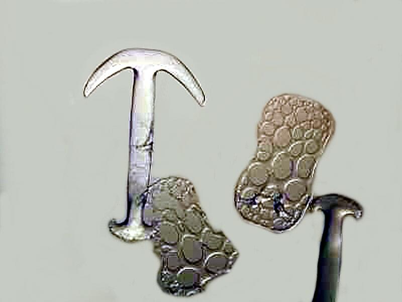
You will notice that the anchor is stout and that the shaft is relatively short and if you had a micro-boat, this is the sort of anchor you would want.
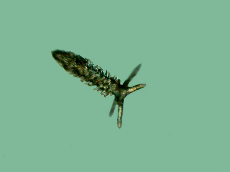
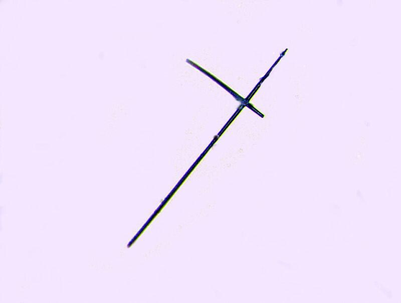
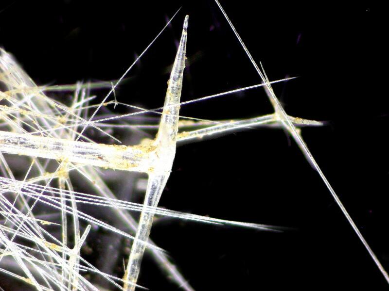
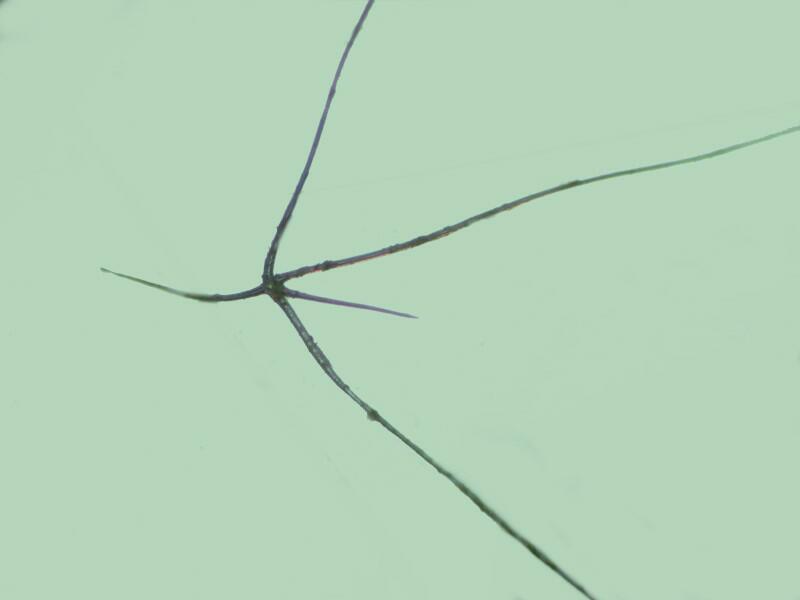
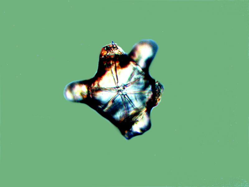
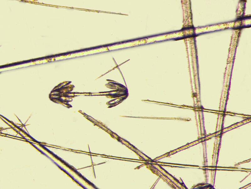
Now, I know that Mother Nature (a.k.a Big Mama) often repeats structures that solve a certain kind of problem. Just think about cilia; they are found scattered across a wide variety of organisms from protozoa to human beings. However, when I found anchor spicules in this cup sponge, I was perplexed. Here is a view of the anchor tip of one and remember that these are siliceous, not calcareous, which accounts for their vitreous appearance. Also they will not dissolve in the usual acids although, the deadly dangerous hydrofluoric acid would certainly make short work of them.
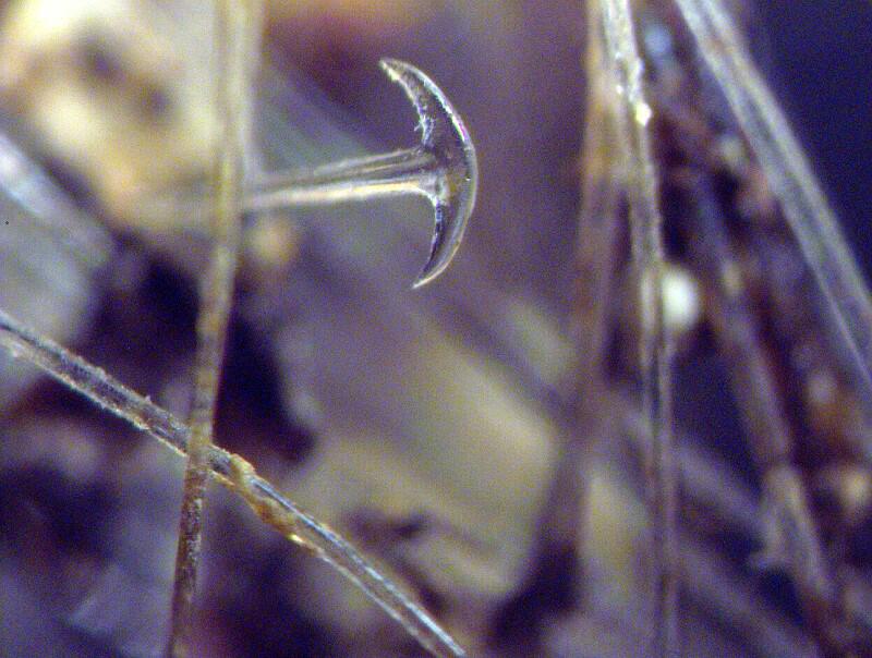
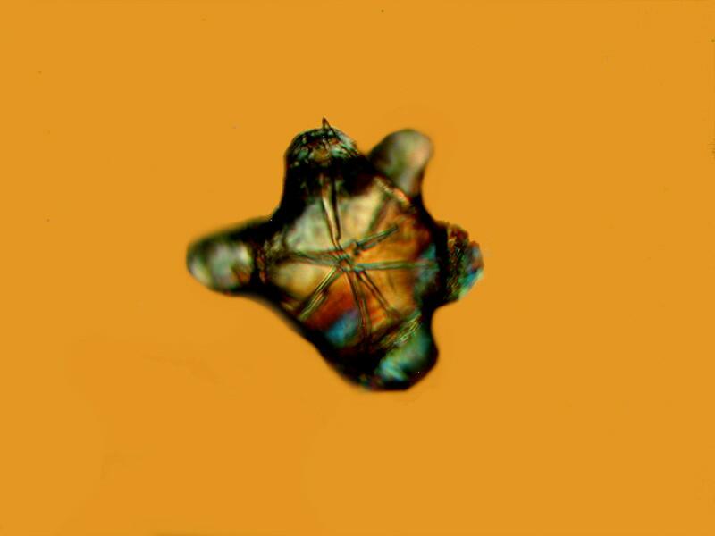
However, let me give you 2 more views. This anchor doesn’t have a nice short shaft.
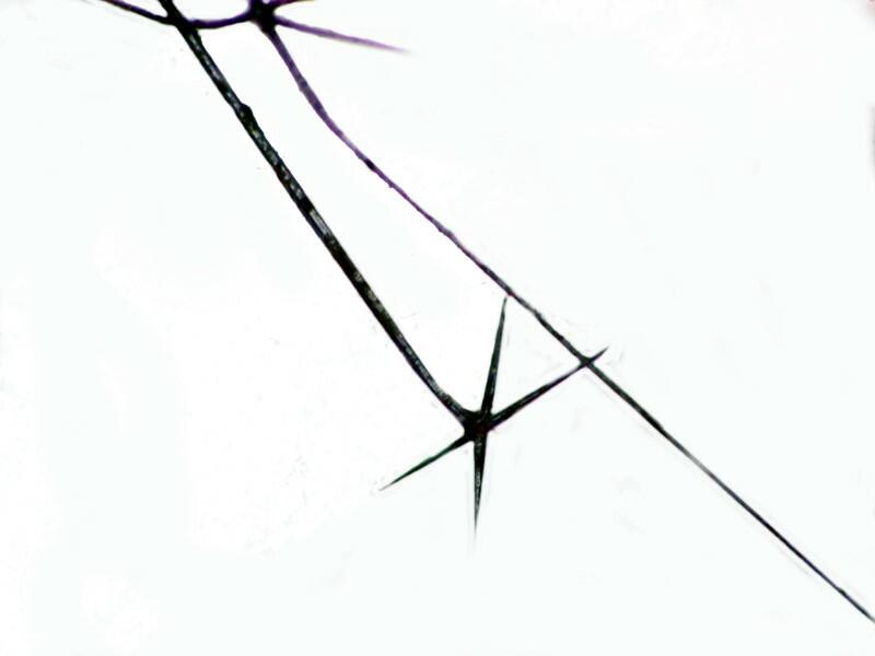
Also, note, because this a point I want to come back to–if you look closely, the long fibers in this image look as though they might be serrated in Part 2.
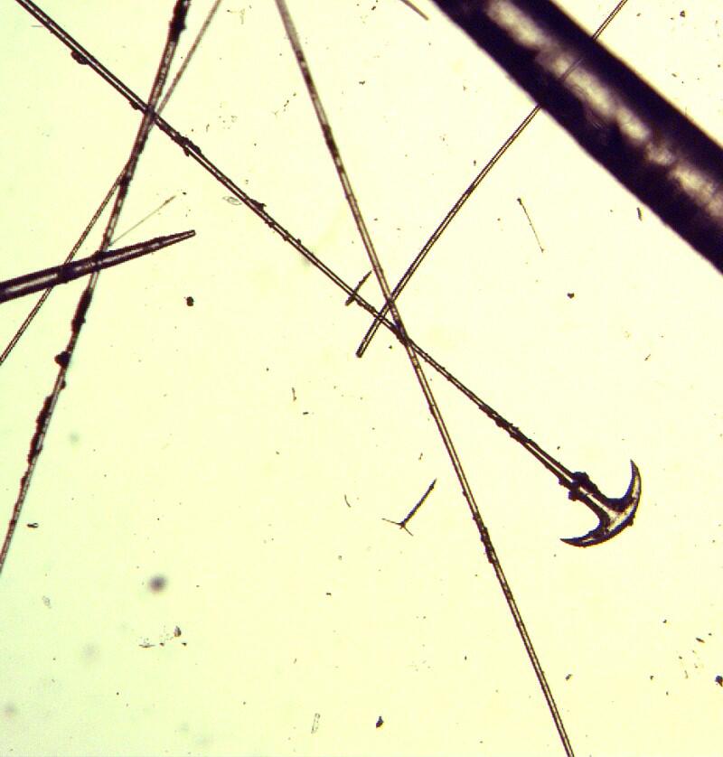
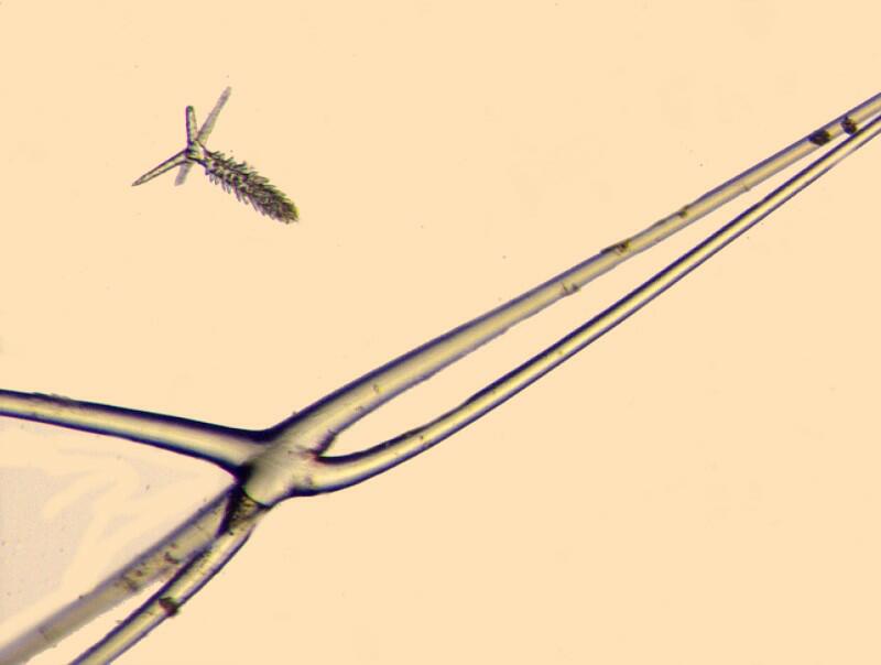
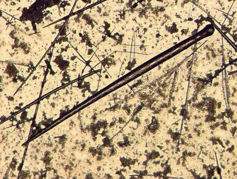
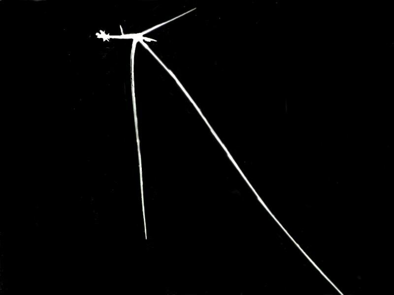
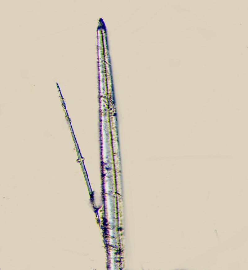
In the next photo, you can see that the shaft of the anchor is so long that it goes out of the field of view.
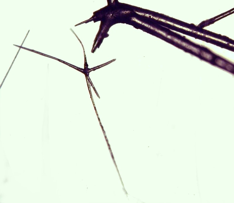
Now, this, combined with the sea cucumber anchors causes me deep puzzlement for several reasons: 1) As I mentioned above the sea cucumber anchor spicules are calcareous; the anchor spicules of this sponge are siliceous. 2) There is the question of why two (at least, superficially) strikingly similar structures should evolve in a siliceous form in a “primitive” life form (the sponge) and in a calcareous form in a quite advanced form high up on the invertebrate phylogenetic tree. 3) Then there are the fascinating and complex questions regarding function. Here one has to tread especially carefully in order not to get lured into teleological issues; namely, issues regarding purpose, intent, or meaning in a sense which extends into universalizations and ultimately absolutes. In other words, we need to approach this in terms of teleonomy, not teleology. Within a given context or set of contexts, a particular structure may have a very definite function but, this is not an entitlement to then extend this into a claim for some evidence for a cosmic or divine design or order. Some biologists, have suggested that anchors and plates of the synaptid sea cucumbers are devices which help in their locomotion, since these are basically burrowing organisms and are constantly moving through detritus and sand. I have 2 problems with this notion: 1) The spicules are rather randomly distributed and contraction could not ensure sufficient leverage to aid in locomotion unless the anchors occurred in enormous numbers. 2) An even more serious difficulty arises from the fact that the spicules are not epidermal but, dermal, and even a very thin layer of tissue would certainly diminish their efficiency in aiding in locomotion. So, from my point of view, the function of these dermal ossicles remains a puzzle.
So what about the anchor spicules in the glass cup sponge? For me, they seem equally, if not even more, problematic. They seem to occur primarily in the bottom part of the sponge and so, one might conjecture that they are used as holdfasts to help secure the sponge to its substrate. However, as we have seen, the anchor “head” is very small and attached to fibers that are very long. This afternoon, I was observing one which prompted me to do a bit of measuring. The fiber was incomplete, that is, broken off and yet measured slightly over 6,000 microns! This may prompt me to go on a quest to try to trace out an anchor with its full length in an unbroken fiber just to see if I can do it and to satisfy my curiosity. (This will probably entail wearing full surgical gear or better yet a Hazmat suit.) A bit of an exaggeration but, gloves, goggles, and forceps are certainly in order. This will almost surely involve making a number of incursions and taking several samples.
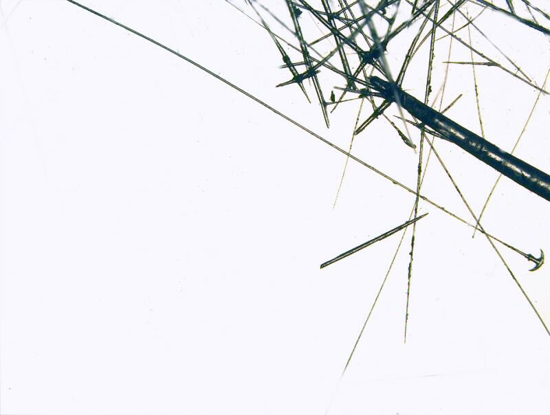
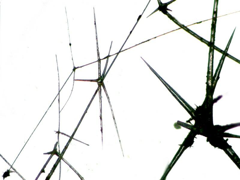
The more I study this sponge, the more amazed and puzzled I become by its spicules and its overall construction. so far, all the large spicules I have encountered are pentacts and I think that the only hexacts which I have seen are very small, but, I will have to double-check that by examining some further samples. Below is an image of some of the larger pentacts.
You will notice that the axons are quite thick and substantial. I found a short piece of one which was fractured at each end and here you can see not only its glassy character but a hint of layering as well suggesting that the deposition of the silica is indeed an ongoing process. So, perhaps, the very thin fragile-looking pentacts eventually become thick, robust pentacts.
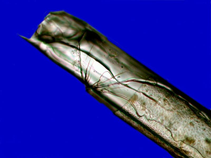
However, even more exciting for me was finding a fractured end which looked like the frayed end of a bundle of optical fibers.
This raises a couple of intriguing questions: 1) Whether or not there are spicules in this sponge that are constructed in distinctly different fashion from other spicules in the same sponge and 2) if that is the case, are there cells with distinctly different sorts of programming instructing them to construct a specific type of spicule? Or perhaps, there is an analog to stem cells and particular circumstances dictate and trigger the activity that produces a specific type of spicule. So, this might explain the variety, radical differences in shape and size, and the difference in construction of spicules in this sponge (and, of course, in other species as well.)
Admittedly, the range of my study of sponges is extremely limited (however, donations of weird sponges, provided they are not of the human variety, are always welcome). Nonetheless, this sponge continues to offer me surprise after surprise and concomitant mysteries.
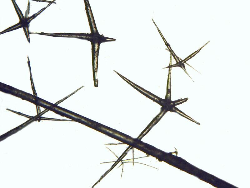
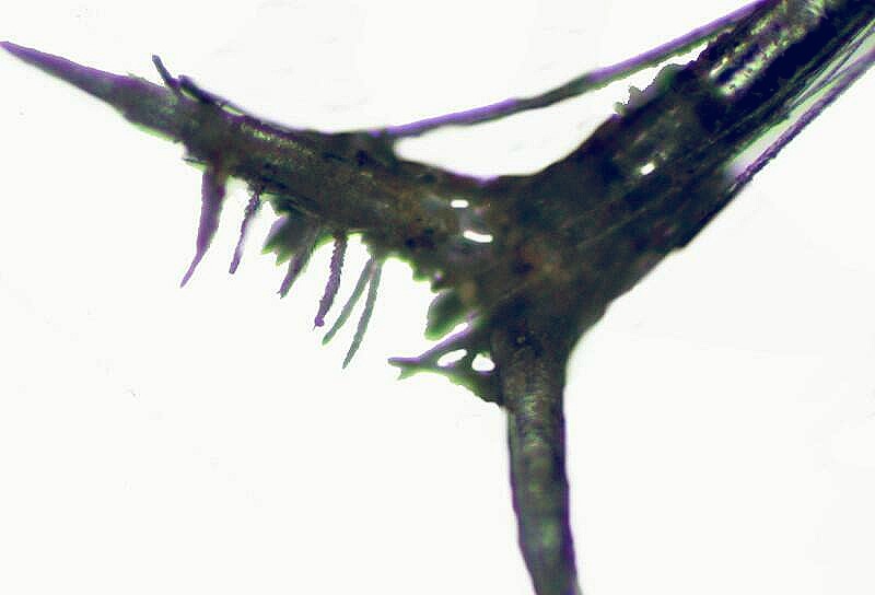
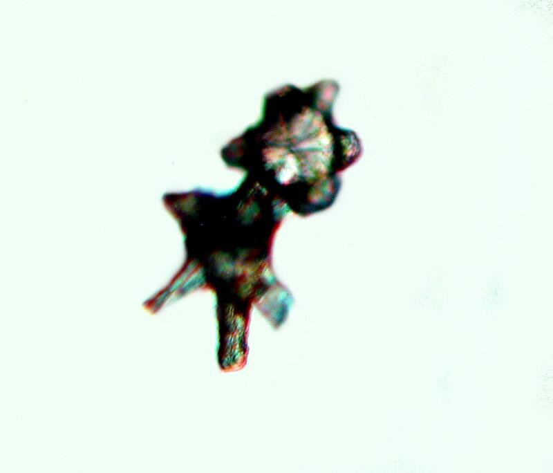
On of those surprises was finding a relatively large spicule with a series of small spines extending off of it.
I have seen such forms in sea urchin spines but, never before in sponge spicules. However, perhaps I should have expected it since this species also has some quite small spicules which are “bushy”. I call them Christmas tree spicules and below are 2 images of one.
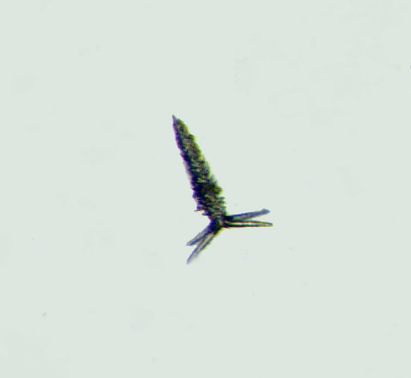
These are fairly common in the samples which I extracted but, I have no idea how they fit into the overall architecture of the sponge skeleton.
I also managed to capture an image of one along with one of the more bizarre large spicules of this organism.
For some reason, the form of this large one I found quite captivating, in large part, because again I can’t image how it integrates into the overall pattern of this sponge’s architecture.
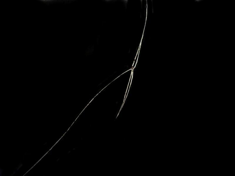
Equally puzzling, in this respect, are long, wispy pentacts which look as though they would float away at the merest breath of air and this is indeed the case once they are separated from the main body of the sponge. First, I’ll show you a rather delicate pentact
and then a very wispy one.
If you’re an aficionado of medieval weapons, well then, you’ll find some nice examples here. For example; a large dagger with an odd cross piece for the hilt.
Or spike-like monaxons which could be used in building a micro-version of an Iron Maiden.
And then there is the broad sword:
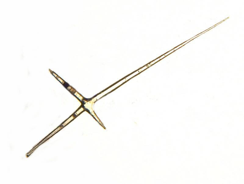
A bit more awkward to wield than you might think, because it is a pentact and there is another axon extending straight up at you from the center of the hilt.
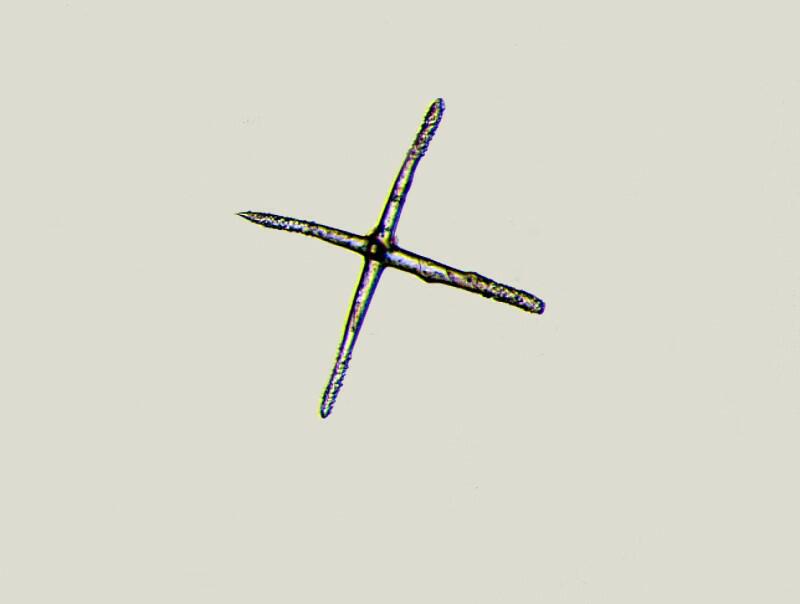
The same thing is true with this “cross” spicule below; it is also really a pentact with an axon extending up at you.
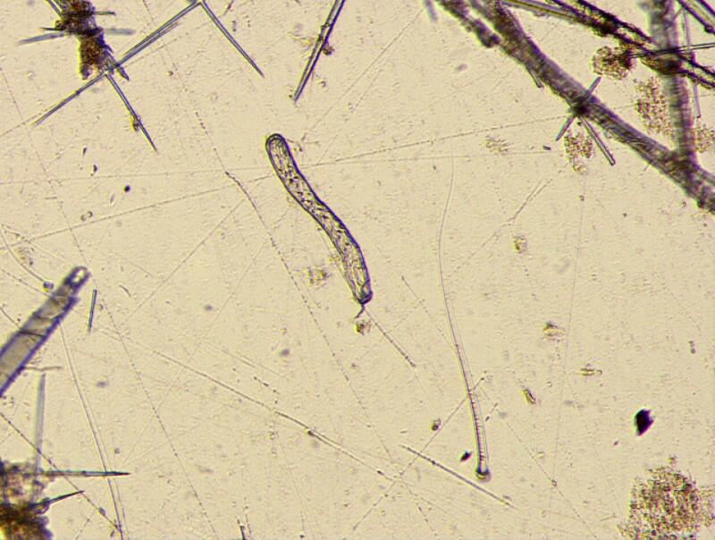
Now, let’s look at some even stranger bits and pieces. In the center of this picture is an odd little band-shaped spicule which I call (observing strict international terminological protocol) a “worm spicule”.
Here is another view of the head of an anchor spicule surrounded by slender fibrils only, in this case, the head looks less like an anchor and more like a battle-axe or pick.
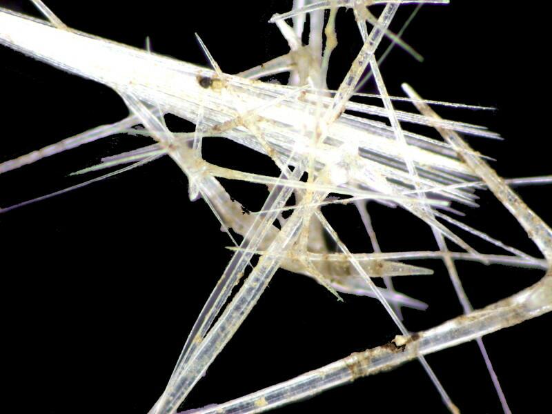
Some groups, although random, having been roughly extracted from the main body of the sponge, appear web-like.
Some spicules are so delicately wispy that they seem to be micro-aerial creatures floating through the aether.
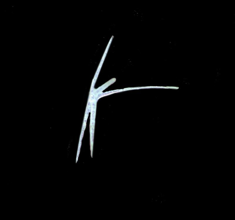

Others, however, are more like grappling hooks.
And this next one surely has to be some sort of extraordinarily long-legged Martian.
Note that at the top of the lefthand side, there is a small, odd irregular structure. This is the place where another pentact spicule was fused with the first one and has broken off. When I broke this piece to photograph it, I discovered that it had considerable depth and I was able to get only a blurred image but, enough to show where the 2 pentacts had fused.
I noticed a distinctive pattern on the upper portion and got 2 interesting images of it.
What immediately caught my attention was a series of needle-like crystals radiating out from the center to the position where the axons of the pentact had been.
I was going to go back to the original fused structure and do a series of images and stack them to get a clearer image but, by the time I prepared to do this, I had lost the piece in the jungle of spicules and pieces in the Petri dish. Nonetheless, this situation of crystals within crystals makes this all the more intriguing or, as Alice would say, “curiouser and curiouser.”
I mentioned here or in an earlier essay (I do, after all have a reputation to maintain as an absent-minded professor) that amoebocytes deposit layers of silica or calcium carbonate, depending on the kind of sponge, around an organic thread, layer after layer. I was lucky enough to find a monaxon in this specimen that quite clearly demonstrates this.
If you look closely, you can clearly see the organic “thread” or fiber running down the center of the larger spicule.
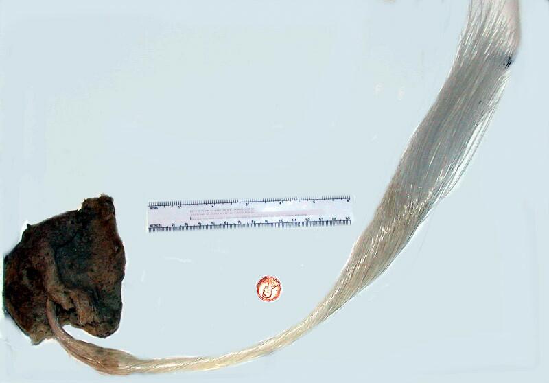
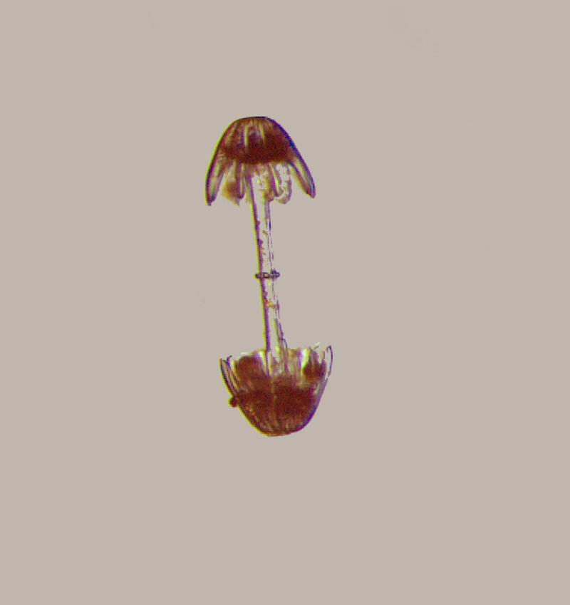
Two last big surprises. Hidden among all these rods, pentacts, and other sharp pointy things, I found spicules which I have only found in another kind of glass sponge, Hyalonema. In its general appearance and indeed its structure Hyalonema is radically different from our cup sponge, yet both types possess a nearly identical type of spicule which I call the “dumbbell” spicule.
Let me first show you just how bizarrely different Hyalonema is from our cup sponge. The penny and 6 inch ruler give you an idea of the extraordinary length of its “tail”.
And here is one of its “dumbbell” spicules
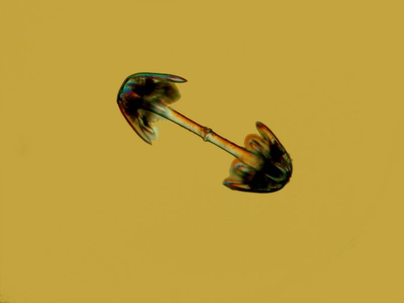
Below are 2 views of the “dumbbell” from the cup sponge.
However, perhaps the biggest surprise and the biggest puzzle come when we take a close up look at a piece of the lattice of the cup sponge.
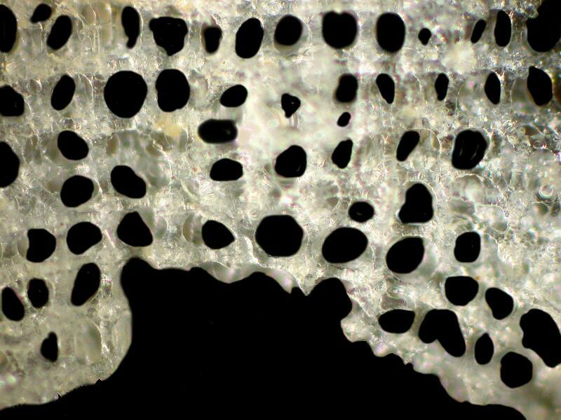
If you still need to buy a Christmas present for someone who is a fan of jigsaw puzzles, then a specimen of this cup sponge might be the ultimate gift. Look at the lattice and then go back and look at all the spicules we’ve examined and ask yourself: How by all that’s demonic, do all these bits and pieces fit together to form such a lattice?
If you think you can figure it out and can send me an answer which I find convincing, then I’ll send you an even weirder organism to wrestle with.
All comments to the author Richard Howey are welcomed.
Editor's note: Visit Richard Howey's new website at http://rhowey.googlepages.com/home where he plans to share aspects of his wide interests.
Microscopy UK Front
Page
Micscape
Magazine
Article
Library
Published in the December 2012 edition of Micscape Magazine.
Please report any Web problems or offer general comments to the Micscape Editor .
Micscape is the on-line monthly magazine of the Microscopy UK website at Microscopy-UK .
© Onview.net Ltd, Microscopy-UK, and all contributors 1995 onwards. All rights reserved. Main site is at www.microscopy-uk.org.uk .