|
The Geometry of Christmas: A Gallery by Richard L. Howey, Wyoming, USA |
There are certain patterns that recur in nature and which we often borrow for a variety of purposes–ornamentation and symbolism being common ones. Christmas over the centuries has accumulated a number of forms that are now a part of a tradition–forms which have shapes such as stars, pyramids, snowflakes, cones, spheres, circles, and cylinders as embodied in trees, tree ornaments, pine cones, and candles to mention a few. Interestingly, geography frequently gets ignored in the many places in the world which are devoid of snow and yet snowflakes and the association of a White Christmas are dominant themes.
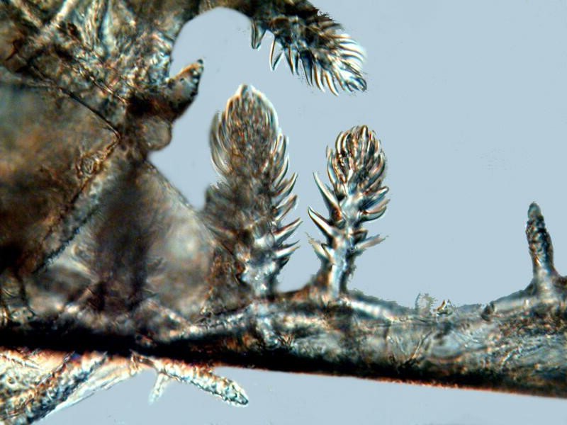
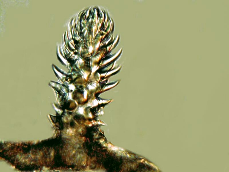
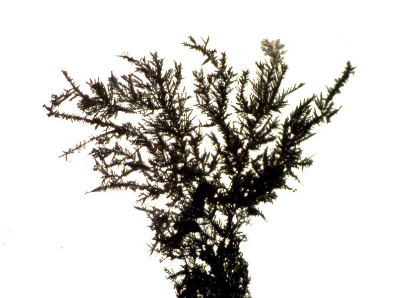
Many of these forms can be discovered in the micro-world and can exhibit a wonderful and beautiful variety. Let’s start with a couple of micro-trees which turn out to be spicules from a glass sponge (Hyalonema).
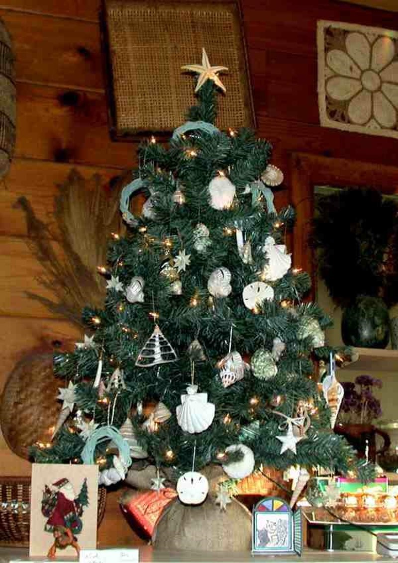
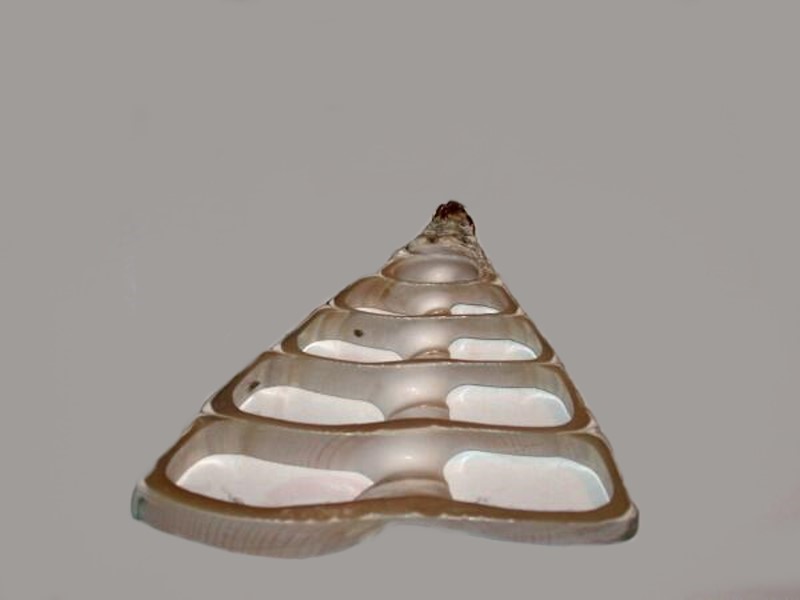
Here’s a small Christmas tree with a variety of ornaments, some types of which we’ll look at later but, for now, note that the central slide of a mollusk shell also has the appearance of a tree.
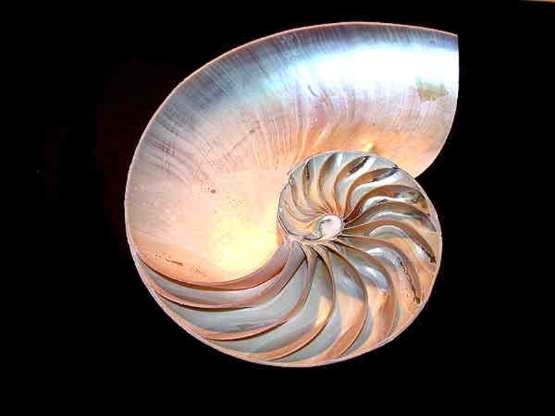
And here’s a closeup of that shell.
Many shells show wonderful patterns when sliced and, of course, a favorite of many collectors and naturalists is the Nautilus with its wonderful spirals.
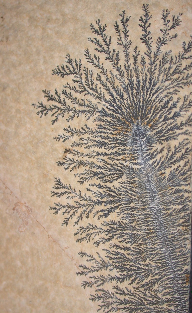
Spirals and branching or dendritic patterns are common in nature and in both organic and inorganic forms. A wonderful dendritic pattern can be found in the manganese deposits in limestone.
And at this frosty time of year, one can find dendritic patterns in ice crystals on window panes, parts of which have a certain resemblance to the “tree” spicules of the glass sponge which we looked at first.

And these in turn remind me of the silver crystals formed by letting a drop of Silver nitrate interact with a drop of Ascorbic acid.
So, now you can get out your copy of that wonderful old classic by D’Arcy Thompson On Growth and Form and find a brilliant discussion of branching. Just think, hydroids, bryozoans, certain types of algae, colonial Vorticellids, and corals all show branching and that’s only a partial list.
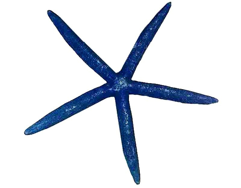
Another form that has strong associations in Christmas symbolism is, of course, the star. This is a form which throughout history has had a strong aesthetic appeal and we find it in both macro and micro states in nature. The starfish has a strong appeal to most naturalists and you will notice in the picture of the small decorated Christmas tree above, the star at the top is indeed a starfish. Generally we tend to think of starfish as pentagonal and a rather drab brown, but both of those are misleading generalizations. For example, take a look at this specimen of a blue Linckia (and, no, it’s not been dyed–this is its natural color).
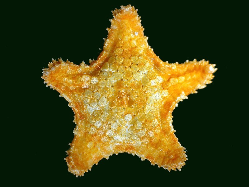
Next is a tiny specimen of an orange and yellow star with tiny circular plates throughout the dorsal surface; it is less than one inch from tip to tip of the rays.
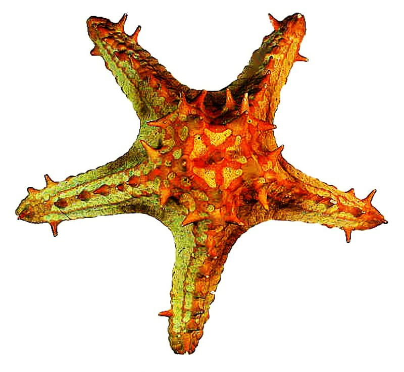
Next, let’s look at a large starfish which has brightly colored “thorns” or “knobs” on the dorsal surface. This is a specimen of the species Protoreaster.
However, to really get a sense for the brilliance and variation of color in starfish, one needs to look at living specimens, or at least images of them.
Just imagine what splendidly colorful ornamental toppings one could have for Christmas trees from these!
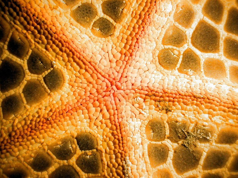
Now, let’s look at some stars within the starfish on the ventral surface. The first is from a small Philippine star.
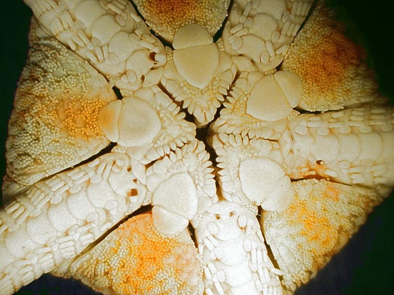
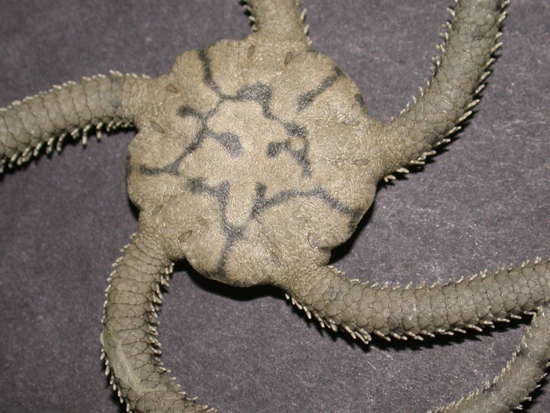
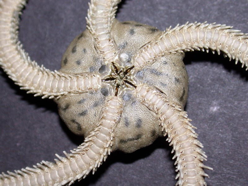
The second one is from a different group, the ophiuroids or “brittle stars”. This one shows striking pentagonal symmetry.
Here is another ophiuroid which is a relatively large one. First, the dorsal surface and then the ventral view which again reveals a “star”.
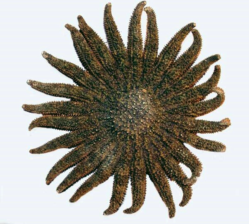
I commented earlier that not only is there a great deal of variation in color, but also in the general body shape. There are starfish with only 4 rays, some with 6, some with 8, some with 20, some with 30 or more. The Brisingida can have 6 to 20 rays. Below is an image of a Heliaster or “sun star”; this one has 25 arms.
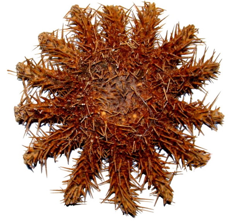
And here is a major villain, Acanthaster planci or the “Crown-of-Thorns” starfish which has been attacking the coral reefs in the South Pacific. They are extraordinarily difficult to control, since one can’t simply cut them up as they will regenerate and instead of just one, you may eventually end up with 2, 3, 4, or more new ones. Divers removing them from the reefs must exercise great care since the spines carry a painful toxin. This specimen has 14 arms.
However, I think that this would make a very nice ornament for the top of a Christmas tree. Unfortunately, however, the dried specimens can’t compete with the brilliant colors of the living organism.
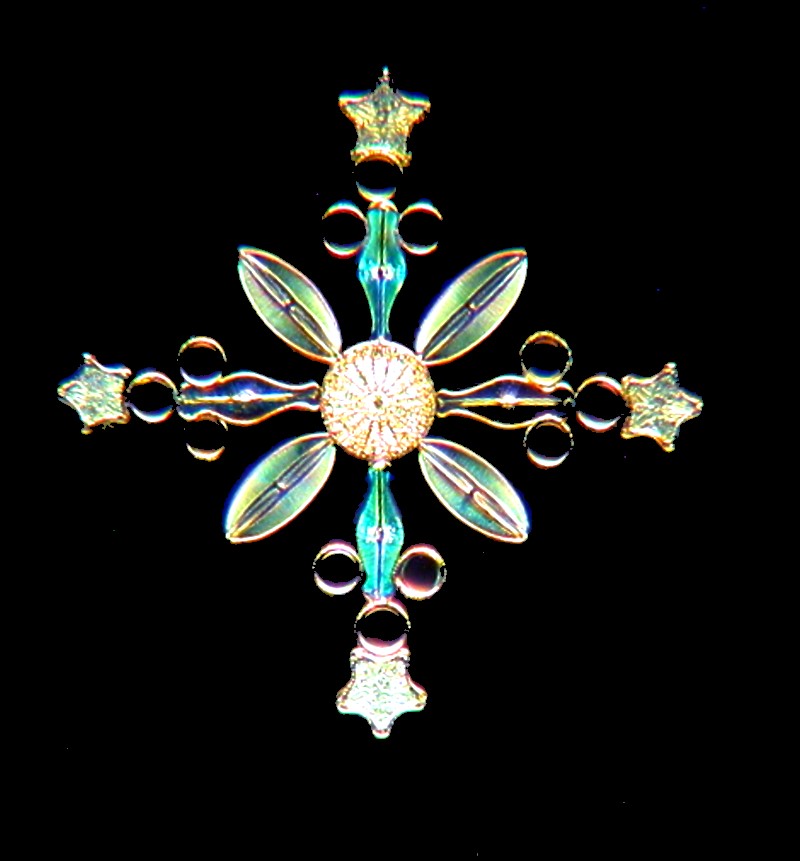
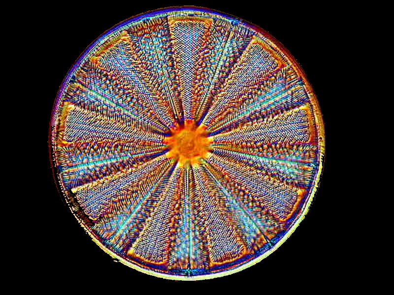
At the micro level, diatomists have made snow-flake arrangements that are both lovely and impressive. Some of these are images photographed from slides produced by Klaus Kemp and are described by him as “snowflakes”. I’ll show you a few images here along with some closeups to give you a sense of the intricacy and delicacy of these wonderful bits of nature.
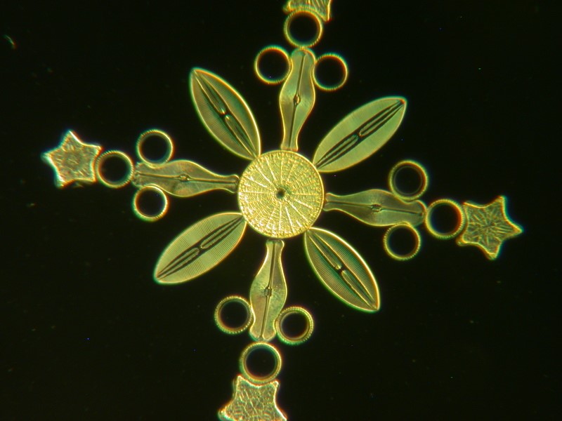
Here is the same diatom photographed with Rheinberg illumination.
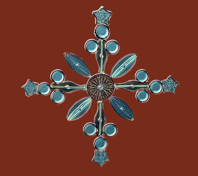
And then a diatom arrangement photographed in darkfield which provides striking contrast and brings out greater detail. Note that in addition to the general “snowflake” pattern, there are 4 stars incorporated into the arrangement.
In the one below, we again find a star, but this time it is in the center visually providing an anchor for the other diatoms.
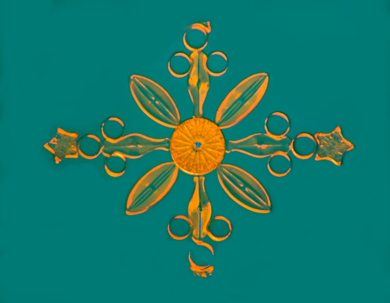
Here is another view of the snowflake with 4 stars with an alteration in the background thus significantly changing the contrast.
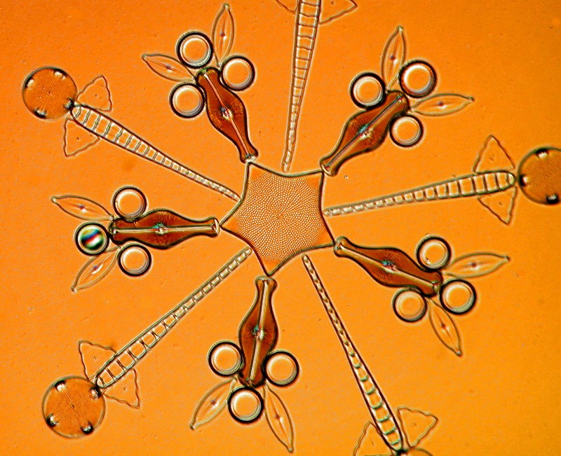
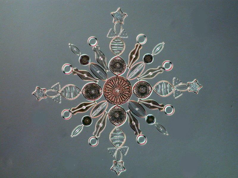
And here is a yet more intricate snowflake pattern employing additional diatoms to create a quite impressive display.
Now, if you want to compare these with photographic images of actual snowflakes, you can look at the work of Wilson A. Bentley either in a book published by Dover or you can go online.
You might well think that these snowflakes are much more complex than any diatom arrangements, but I think you might reconsider after looking at these.
In addition to snowflakes, disks and spheres are common forms that comprise the geometry of Christmas as ornaments. First off, I’ll show you a tiny disk that I think would make a splendid ornament.
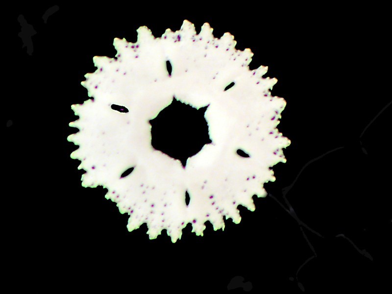
This is a calcareous plate from the tip of a tube foot of the sea urchin Strongylocentrotus purpuratus.
Interestingly, a thin cross section of the spine of many common sea urchins also appears as a disk.
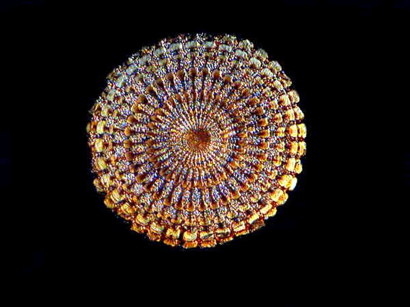
A single centric diatom when viewed from above also presents itself as a disk.
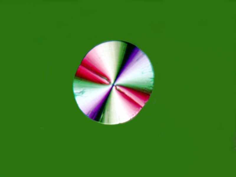
Certain chemicals will frequently form in such a way as to produce disks. Ascorbic acid both by itself and in some mixtures forms disks. The first example is just Ascorbic acid and the second is Ascorbic acid and the biological stain Indigo Carmine. Both were photographed using polarized light.
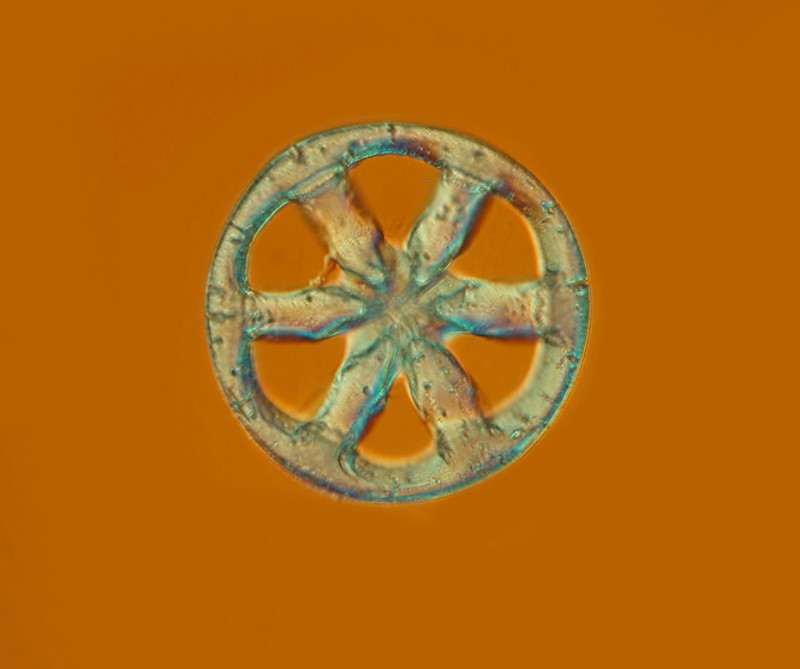
We can also find a modified disk, in this case, in the form of a wheel. This is a spicule of the sea cucumber Chirodota sp. and such spicules are found embedded just under the surface of the skin, sometimes individual, sometimes in semi-transparent packets of tissue which can contain 50 or more spicules.
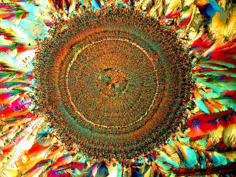
Of course, if a series of diatoms is arranged in a circle, one might contend that it also can be regarded as a disk. This handsome arrangement was photographed using polarized light and then the inverse function in the graphics software.

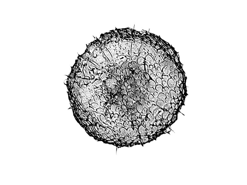
Finally, let’s look at a couple of examples of spheres and for this, we’ll turn once again to the radiolaria. The first example shown is a prickly sphere with tiny spines protruding from the surface.
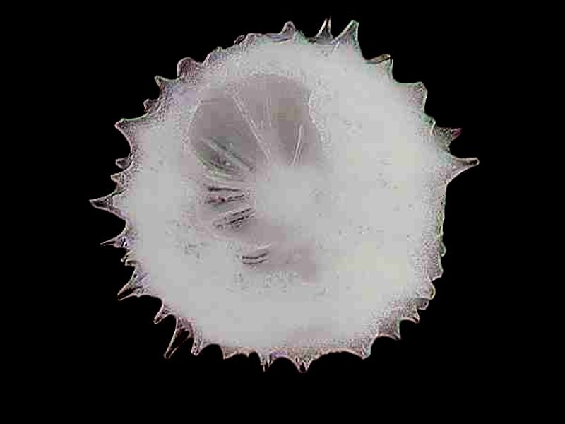
The next is intriguing; it too appears to have minute spines projecting from the surface. I found a few of these in a sample from the Andaman Sea off the coast of Burma (Myanmar).
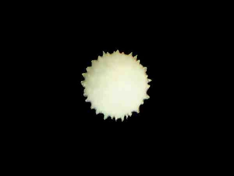
I was curious about the spines and managed to cut a section to take a peek inside and to my delight and surprise, discovered that the spines extend out from a central core and they act, in a sense, as struts for the spherical test.
When we take the trouble to look at the micro-universe which surrounds us, we can discover an abundance of geometric forms. I hope you have enjoyed this small collection of images and that you have a peaceful and wonderful holiday season.
All comments to the author Richard Howey are welcomed.
Editor's note: Visit Richard Howey's new website at http://rhowey.googlepages.com/home where he plans to share aspects of his wide interests.
Microscopy UK Front
Page
Micscape
Magazine
Article
Library
Published in the December 2016 edition of Micscape Magazine.
Please report any Web problems or offer general comments to the Micscape Editor .
Micscape is the on-line monthly magazine of the Microscopy UK website at Microscopy-UK .
©
Onview.net Ltd, Microscopy-UK, and all contributors 1995
onwards. All rights reserved.
Main site is at
www.microscopy-uk.org.uk .