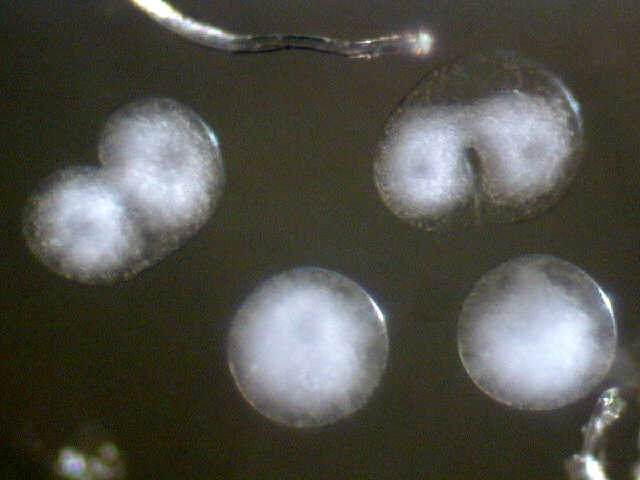
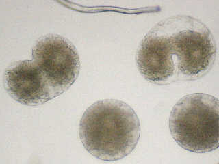
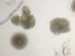
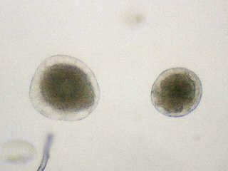
Two embryos in the two-cell stage, and two unfertilized eggs.
The Micscape Dec. 99 issue originally linked to the author's web site.
This is a mirror, with permission of the author,
of the article at http://www.ct.sakura.ne.jp/~gen-yu/sowerbyi/fwjelly2.html
Two embryos in the two-cell stage, and two unfertilized eggs.
I had reared the medusae for about one month from mid October 1999. They were all given by Miyakawa who is a veteran amateur on this species; two females were grown up from polyps in his aquarium tank, and the two wild males were collected by him in Nagoya city, central Japan.
However, as a matter of fact, female and male individuals of this species are usually not found at the same time in the same habitat. Some people think that this is due to the lack of complete investigations, but the sexual reproduction in nature may not be common.
Nevertheless, the sexual reproduction is very important. It helps to improve the genetic diversity of the population, and information about their development is valuable for the understanding of these creatures. I hope that this article makes you interested in medusae.
Pictured left are the eggs of freshwater jellyfish, C. sowerbyi. They were spawned in 20th Oct. and they are about 0.1mm in diameter. They were sunk in the bottle in which I rear medusae, and I could pick them up in a watchglass.
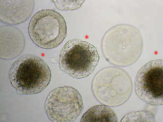
I separated females and males and I tried artificial insemination. Various eggs were observed as seen in this picture. But these all seem unfertilized because I was not able to observe any cleavage this time.
The three eggs marked with a '*' were good, whereas the other eggs were bad and cannot develop, I think. I don't know the reason why there were differences in egg viability.
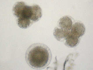
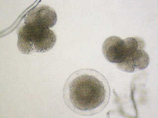
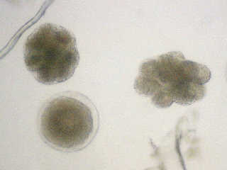
But luckily enough, near the midnight of the 27th Oct. 1999, I found
two embryos that were in the two-cell stage of cleavage! Pictures were
taken continuously until morning with intervals of about five minutes.
I show some of them here.
23:54 |
00:46 |
01:53 |
02:38 |
03:28 |
16:08 (The left is unfertilized). |
Two developing embryos can be seen in these pictures (except the last). The indicated times are when the photo was taken. Animation of the left and the right embryo are also available (each about 200KB GIF, 100x100 pixels). In these animations, disturbances in the arrangement of cells are seen especially when they divide.
 |
 |
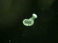
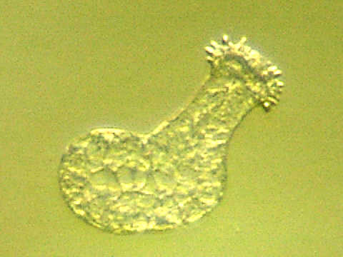
After the cleavage, the embryo began to swim. I observed a ball-shaped embryo swimming slowly in small circles. This swimming soon ceased. The embryos began to elongate and creep on the substrate. The term "planula" is applied to this swimming stage and creeping stage until they settle somewhere and become polyps.
Lefthand animation shows a creeping planula on the watchglass. The interval is ten minutes and total length is 80. The speed of the movement is variable according to the cases.
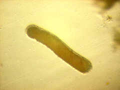
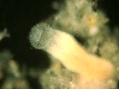
You may think that you saw this kind of animation somewhere else. Yes, I showed the movement of a "frustule", asexual larva, in the article "Freshwater Jellyfish". Frustules from polyps are very similar to planulae in their morphology and movement. Righthand picture is not a planula but a frustule.
But they are different in some points, I gather. At any rate, frustules are far larger than planulae as you see in the pictures above. Actual length of the frustule in this picture is about 0.95mm.
Some planulae became tiny polyps in several days. They are very small. Enlarged image of a tiny polyp is also available (brightfield: 20KB). The magnification on the photographs of a creeping planula, a frustule, and these two polyps are all the same.
The mouth of a tiny polyp is about 0.1mm in its diameter! What do they feed on? Protozoa? I have recently successfully fed them with rotifers, something like Rotaria. An animation is also available (ca.25min., 18frames: 59KB GIF). I hope I can grow them up to mature polyps with these kind of creatures "generated naturally" in the container.
Last update: 27-NOV-1999Photographs taken on: 21, 27, 28-OCT, 08, 22-NOV-1999
Copyright © 1999 by Gen-yu Sasaki . All rights reserved. Unauthorized reproduction prohibited.
Visit Gen-yu's home pages which illustrate an interesting selection of freshwater life and other organisms.
Please report any Web problems or
offer general comments to the Micscape
Editor,
via the contact on current Micscape
Index.
Micscape is the on-line monthly magazine
of the Microscopy UK web
site at Microscopy-UK
WIDTH=1