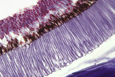
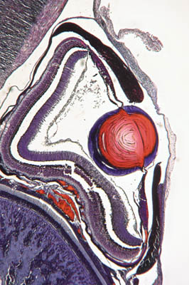
EYES under the microscope part 2

by Jan Parmentier
     

The eyes of gastropods and cephalopods exhibit a wide range of complexity. The simplest eye is a sort of 'pinhole camera' eye, consisting of a shallow pit with a retinal cell layer connected with nerve fibres. Haliotus, the abalone, is an example of this type of eye. More advanced is the eye with a deep spherical pit, filled with a crystalline lens, like the eye of Helix, the garden snail, which has its eyes at the top of each cephalic tentacle. Helix has two pairs of tentacles. In the more primitive snails you will find only one pair of tentacles, the eye is then situated near the base of each tentacle.

The eye of a cuttle fish or octopus (class Cephalopoda) is even more complex and has much in common with the eye of a vertebrate. It has a lens, consisting of two planoconvex components, and is of a fixed focal length. In the rest state the lens is focused on rather nearby objects. For distant vision, a ciliary muscle draws the lens closer to the retina.

The rod like cells of a Cephalopod eye, with high magnification

The eye of a small Cephalopod, a young Allotheutis

   
This retina contains long, rod-like sensory cells whose sensory ends point toward the front of the eye; in vertebrate eyes the orientation of the sensory cells is towards the retina. An iris diaphragm controls the amount of light entering the eye. The eyes of several Cephalopod species can be very large indeed, the largest eye ever found (in the stomach of a potfish) had a diameter of 40cm. An octopus can form a well focused picture (in colours?) of its surroundings.
There is indeed much resemblance between our eyes and those of an octopus. However, a vertebrate eye is formed as an outgrowth of the brain, while that of an octopus is formed by an invagination of the ectoderm and a closer look shows more differences.

This is a striking example of so-called convergent evolution, which means that a similar organ with a similar function is formed in quite unrelated organisms. Studying eyes is difficult enough for an amateur microscopist, educated as a chemist. I am consoled a bit by the essential photochemical part of the process of the visual process. Armed with books about the anatomy of molluscs I am trying to understand the structures I am seeing through the microscope. But standing before a large sea water aquarium with an octopus in it, clearly looking at me and following me with his eyes, I am pondering about what this octopus really perceives of me. I suspect it must be a lot!
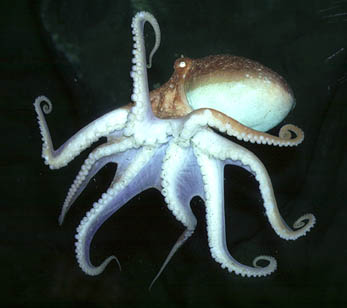
An octopus, with its large eyes, looking at me. Its eye is white, due to the reflection of the flashlight against the pigment cells behind the retina


Back to First Page

Comments to the author Jan Parmentier are welcomed.

or visit Jan's HOME PAGE

Preparation of the eye of a garden snail: John Wells
Other preparations by Johannes Lieder, Germany

All photographs © Jan Parmentier 1999

html by Wim van Egmond


Microscopy UK Front Page
Micscape Magazine
Article Library


© Microscopy UK or their contributors.

Published in the December 1999 edition of Micscape Magazine.

Please report any Web problems or offer general comments to the Micscape Editor,
via the contact on current Micscape Index.

Micscape is the on-line monthly magazine of the Microscopy UK web
site at Microscopy-UK

WIDTH=1


© Onview.net Ltd, Microscopy-UK, and all contributors 1995 onwards. All rights reserved. Main site is at www.microscopy-uk.org.uk with full mirror at www.microscopy-uk.net.