Image gallery by the author.
 |
 |
 |
 |
|
by Roland Mortimer, Rio de Janeiro, Brazil |
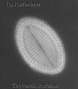
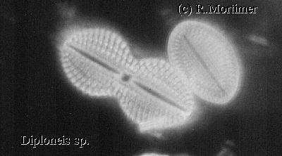
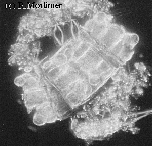
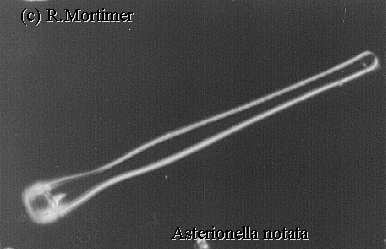
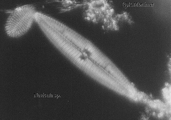
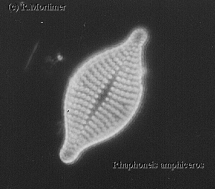
There is almost nothing as beautiful as looking at diatoms under dark-field illumination, whether they be live or just empty frustules. I recently got hold of a Heine condenser made by Ernst Leitz which fits my Leitz Ortholux, this condenser gives bright/phase and dark-field illumination by moving a mirrored lens much like the lens in a standard dark-field condenser up or down an outer tube by rack and pinion. There are intermediate positions where neither of the above types of illumination are rigidly obtained, here, many beautiful effects, some almost 3-D can be obtained.I found diatom frustules are better photographed in dark-field when they are mounted dry than mounted in high refractive index resins. The diatom material is placed on a clean slide and a clean cover added, the liquid allowed to thoroughly dry out then examined with a x20 objective. The objective I actually used for the images was a Leitz x40 n.a. 0.65 phase in combination with the Heine condenser.
Inevitably there is some extraneous matter surrounding one or two images, usually this consists of fine sand grains the same size or smaller than the diatom frustules and are very difficult to remove. I would like to thank Klaus Kemp of Microlife Services once again for his help in identifying some of the diatoms and Jan Parmentier of the Dutch Club who kindly sent me the phase objective.
Comments to the author Roland Mortimer welcomed.
Image gallery by the author.
Please report any Web problems or
offer general comments to the Micscape
Editor,
via the contact on current Micscape
Index.
Micscape is the on-line monthly magazine
of the Microscopy UK web
site at Microscopy-UK