The pictures in the image gallery below were scanned color photos. The photos in turn were taken from 35mm slides projected on to a white wall.For the original slides, I used 35mm Kodak 100 ASA Elite Chrome film. They were taken through the microscope using a 25X Leitz plan objective and 10X eyepiece. The camera was used as a film transport with no lens attached. The initial magnification was approximately 220X (on the slide film).
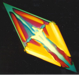
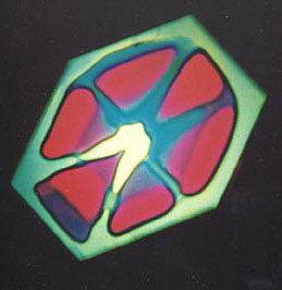
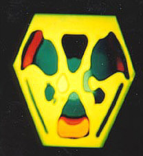
I used polarized light to bring out the colors and applied heat and pressure on the chemical compounds (hyroquinone + sodium sulfite) to form the crystals. Low heat is applied to the powdered mixture (using a bunsen burner flame 6 inches away). A small disc is put on top to apply pressure after first placing the heated compounds on a glass slide.
All the pictures are of the same chemicals and the amazing thing is that each one is unique in both its shape and colors.
Comments by e-mail are welcomed to Jim Evarts.
Images of sodium sulfite + hyroquinone between crossed polar filters
 |
 |
 |
|
Images © James Evarts.