About ten or so years ago I ordered, rather absent mindedly, several natural history books from a catalog I had received. One of the books was a definite afterthought at the time, as I had never heard of it before. The title A History of Infusoria caught my eye, as did the supposed publication date- 1869. The price was fairly low too, especially for an illustrated Victorian book.Weeks later I received the book and discovered that it was a rebound copy of A History of Infusoria, Living and Fossil: Arranged According to Die Infusionsthierchen of C. G. Ehrenberg by Andrew Pritchard, M.R.I., and that it had really been published by Whittaker and Co., London in 1845. I was at first disappointed that they had not sent exactly what I had ordered but then discovered that the book had twelve delightful hand colored plates of everything from Volvox to various rotifers, all delicately detailed as only Victorian illustrators could. The details were fantastic (although some may be a bit fanciful) and the style was enchanting.
I have ever since cherished this wonderful book with its quaint color plates. After seeing Dave Walkers gallery of Victorian microscopy illustrations I thought that I might share some of these neat plates with others interested in such things. Following are five of the color plates and a few notes on each, including a few of the names given to the illustrations in the text.
This gallery will, I hope, give the reader a flavor of this remarkable book with its especially remarkable artwork. I doubt that I would ever be able to pick up a similar volume for the small amount that I originally paid.
Comments to the compiler David Richman are welcomed.
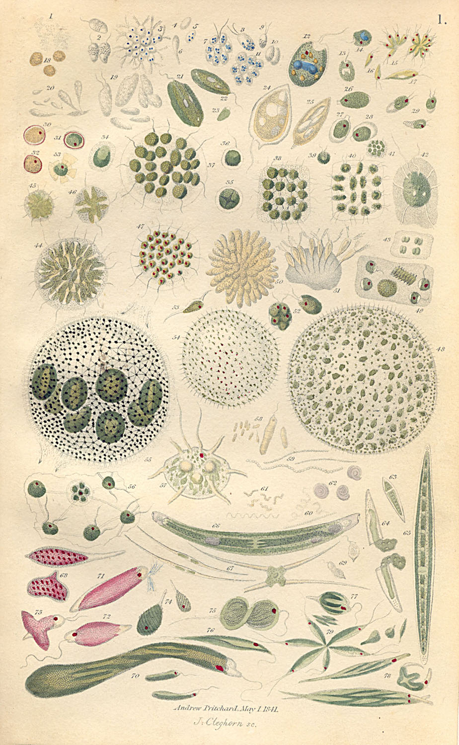
| Plate 1 is a bit discolored, but clear and has on it illustrations primarily of flagellates. Figure 55 is of Volvox globator, while 47 represents Eudorina elegans according to the text. (There are no plate or figure captions as such; all are listed within the text, which runs to over 400 pages). Figure 61 shows a group of the bacterium Spirillum undula, while 59 is of Vibrio bacillas. Figures 70 and 76 represent species of Euglena or relatives. |
 |
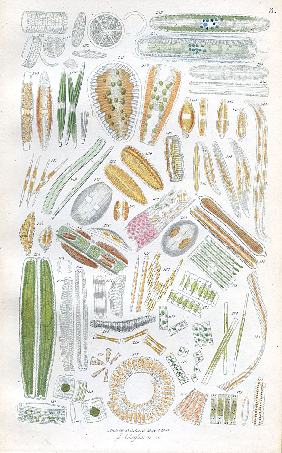
| Plate 3 appears to consist mostly of various species of diatoms (Plate 2 had amoebas and desmids), which were at this time still included within the Infusoria. Soon they would be removed to the plants and then in more modern times placed first with Protoctists and then into a kingdom of their own. The strange sliding Bacillaria paradoxa is shown in Figure 167. |
 |
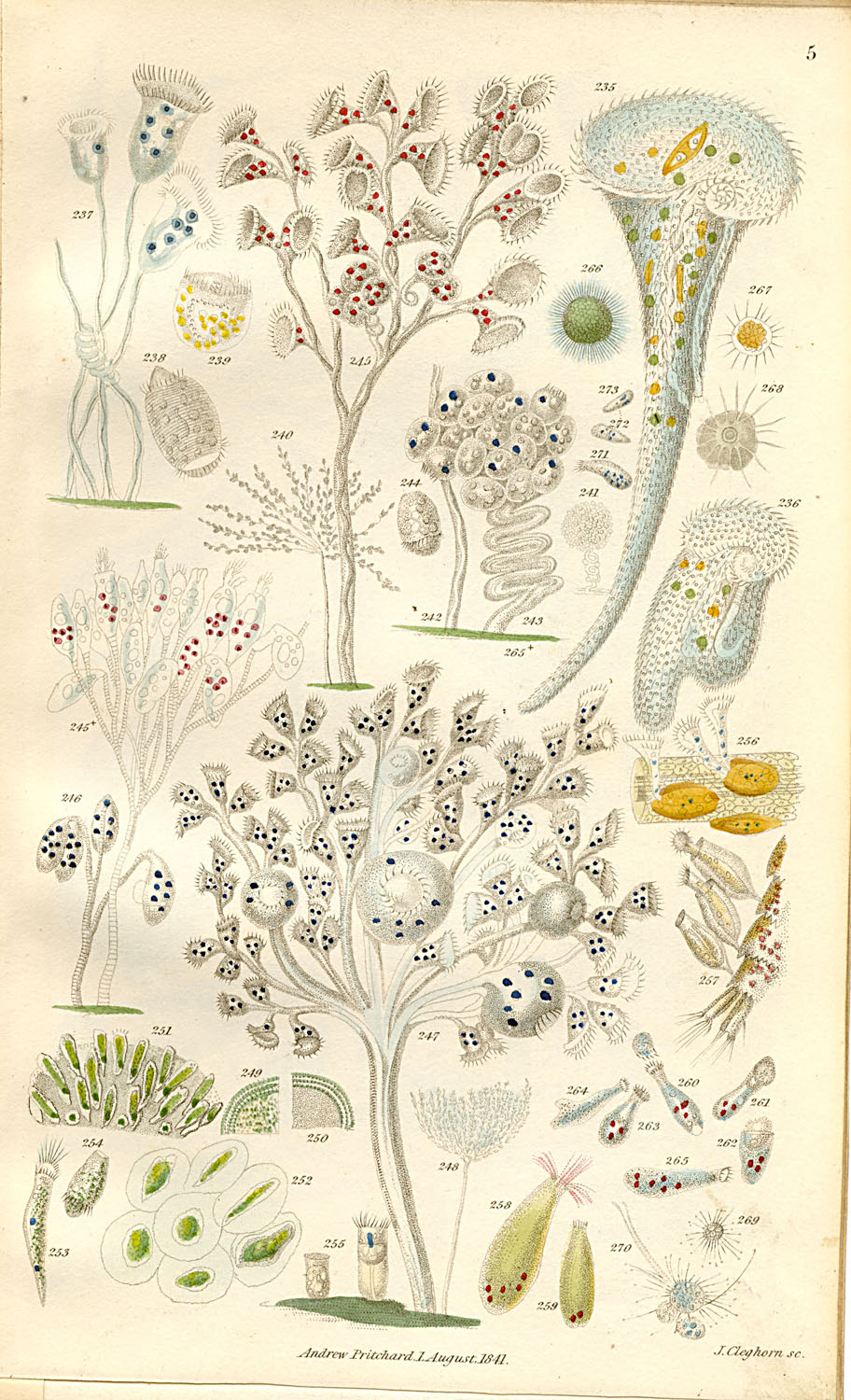
| Plate 5 is obviously made up of ciliates, including Vorticella patellina (237) and Stentor caeruleus (235). The strange beaded macronuclei of Stentor are well shown, as are food vacuoles in these and other species. |
 |
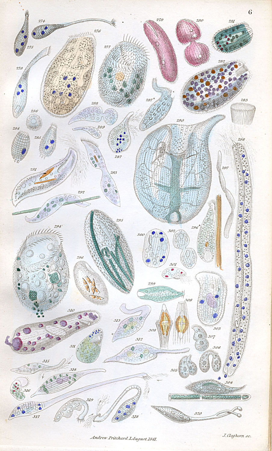
| Plate 6 is also a cilliate plate Figure 291 is Loxodes rostrum and 294 is Bursaria vorticella according to the text, while 297 and 298 represent Spirostomum ambiguum. |
 |
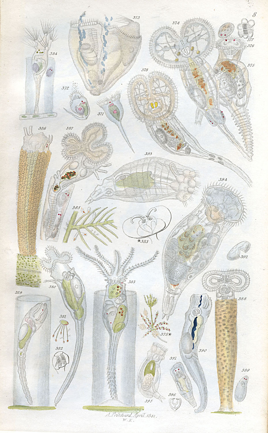
| Finally, Plate 8 is composed of various rotifers. Figure 383 is Stephanoceros eickhornii, according to the text, and 388 shows a tube-making Limnias ceratophilli (probably now in the genus Floscularia). Figure 374 is identified as Megalotrocha albo-flavicans. |
 |