Notes on choosing the correct eyepiece and relay lens for photomicrography with digital cameras

by Ted Clarke, USA

(Note: This is a version for smaller screens and/or printing.
A version for larger screen displays e.g. 1024x768 is here.)


Most older microscopes use compensating optics.  The compensation for chromatic difference in magnification (CDM) and field curvature varies with each microscope manufacturer.  Using a non-compensating eyepiece in a microscope with a large amount of CDM, like the 160 mm tube length Zeiss microscopes is a disaster.  The first image montage below demonstrates this; the second shows the importance of correctly choosing and using a relay lens with the compensating eyepiece.


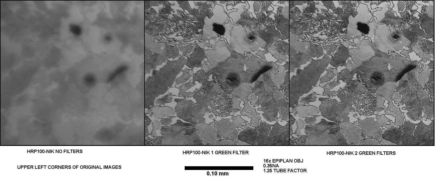
(Resized to fit web page). Click on the image to view the author's original (587 kbytes).
Figure 1. The image sequence above of a metallographic specimen shows what happens when you use a non-compensating relay lens on a Zeiss Universal microscope requiring kpl eyepieces with a monochrome MegaPlus 1.6i/AB digital camera (see footnote 1 and ref. 1).  The filters eliminate the CDM effect by recording only green light but do not correct for field flatness that kpl eyepieces also correct.  The corners of images with a 100x 1.25 NA objective are noticeably blurred when the non-compensating relay lens is used.


(Resized to fit web page). Click on the image to view the author's original (116 kbytes).
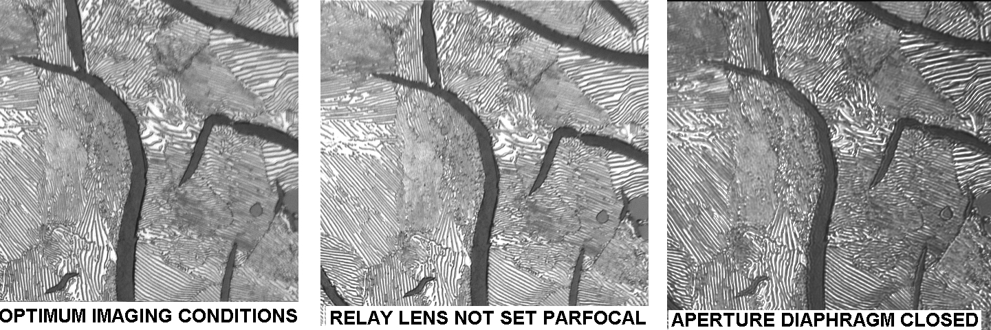
Figure 2 is a montage of digital images of pearlitic gray iron microstructure (see footnote 2). The images were taken with the 40X 0.85 NA Zeiss Epiplan objective to show the effects of incorrect imaging conditions.  Not setting the relay lens to be parfocal with the eyepiece image and focusing with the digital image introduces spherical aberration because the microscope is then not used at the correct 160 mm tube length. Closing the illumination aperture diaphragm down reduces the image resolution because the image resolution depends on both the objective NA and the illumination NA.

Relay lens choice is also important. One suitable relay lens for the Zeiss Universal microscope, is the Thru-Type relay lens as marketed by Edmund Scientific (not tried by the author); it's used with the microscope's eyepiece in the trinocular head.  The Quantimet 720 marketed in the 1970's used such a Thru-Type relay lens.  I have one which was once used with a Zeiss Universal for the Quantimet 720.

All comments to the author Ted Clarke are welcomed.

Related Micscape articles:
Topical tips 8 - a simple way of measuring the eyepoint of eyepieces. Designs with higher eyepoints are recommended for digicam use.

Footnotes:

1) The MegaPlus 1.6i/AB is a monochrome 1.6 megapixel scientific grade camera. Its performance can't be directly compared with a consumer colour digital camera of similar 'pixel count'. Consumer digicams use interpolated pixels from using a Bayer color mosaic filter over the sensor. The consumer camera requires twice the pixel count to achieve the same spatial resolution.

2) The specimen in Figure 2 is a metallographic specimen of pearlitic gray iron.  Clifton Sorby was the first person to see such a microstructure.  The pearlite makes a fine resolution test subject in reflected brightfield illumination because it is composed of alternating thin layers of nearly pure alpha iron and cementite (iron carbide).  This microstructure in a properly polished section is made evident after etching with a 2% solution of nitric acid in alcohol (nital etch) which selectively dissolves the alpha iron leaving the cementite plate edges standing in relief.

Sorby discovered the two-phase structure associated with pearlite in about 1880.  He is probably the one who named it pearlite because his early metallography in the mid 1800's was done at too low a resolution to resolve this two phase structure and it looked "pearly" to him.  His early work was with low power objectives and the vertical illumination introduced by a reflector between the specimen and the objective.  His later work was with the light coming from above a high powered objective with the objective acting as its own condenser.

References
1) 'High-Res Digital Camera Provides Fast, Flexible Defect Imaging'. R&D Magazine, June 1998, 49-50.
An article describing the author's use of the Kodak Megaplus 1.6i/AB digital camera for photomicrography when metallurgist at Case Corp., Racine, Wisconsin.

2) Clarke T, M., 'Digital Imaging in the Materials Engineering Laboratory', The Microscope, 1998, 46(2), 85-100.


Microscopy UK Front Page
Micscape Magazine
Article Library


© Microscopy UK or their contributors.

Published in the February 2002 edition of Micscape Magazine.

Please report any Web problems or offer general comments to the Micscape Editor,
via the contact on current Micscape Index.

Micscape is the on-line monthly magazine of the Microscopy UK web
site at Microscopy-UK

WIDTH=1


© Onview.net Ltd, Microscopy-UK, and all contributors 1995 onwards. All rights reserved. Main site is at www.microscopy-uk.org.uk with full mirror at www.microscopy-uk.net.