|
Gordon Couger USA gcouger@couger.com
|
||
| Click on images for full size images | Nikon CoolPix 995 used unless noted. | |

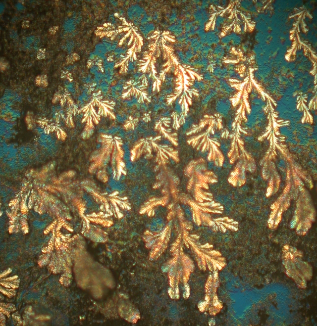
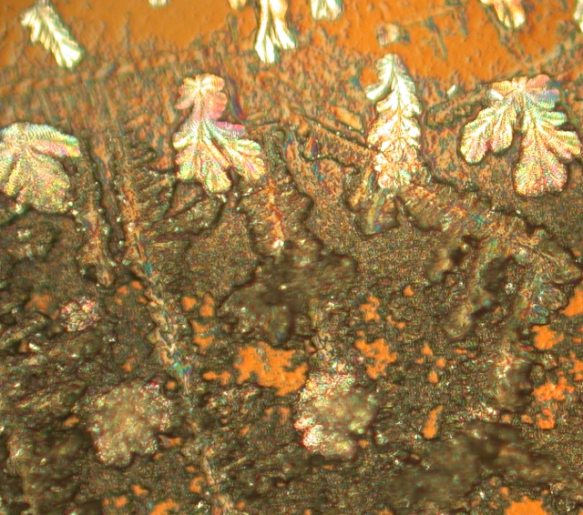
A grain of sand from the Red River magnified about 200 times with a Nachet 300 DIC scope acting as a simple polarized light microscope with crossed polarizing filters to block almost all light not effected by bifringent material in its path. Any microscope will do this with the addition of polarizing filterers. There may be some distortion from strain in the glass between the two polarizing filters but it will still provide striking images if you use high quality filters. The Red River and the Wichita Mountains to the north of are one of the oldest features in the world at the juncture of a failed collision of two plates of the earth's crust. The sand shows it by being very worn down. There is no sand in the area that can be used for concrete or mortar. http://www.waggonerranch.com/Oilprod03.htm
Has a great deal about the geology of the area. The hole in the center
of the ranch is were my mother's family settled and still has a working
ranch.
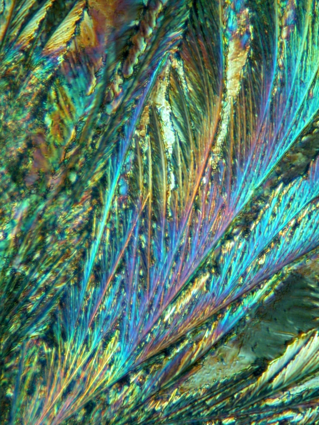
Vitamin C crystals magnification unknown. Taken with the Nachet DIC scope with the DIC acting to give even more color to the already spectacular crystals. Anyone can make these by dissolving vitamin pills in water and letting them evaporate on a clean slide. If you don't have a set of polarizing filters there are inexpensive ones available that can be held in the eyepiece with rubber O-rings and oiled or attached to the condenser with glycerin. This works well even with binocular microscopes with all the reflections in the prisms and beam splitter. Growing crystals is an inexpensive and easy way to get very striking
images from everyday substances Epson salts, photo chemicals, medicines
and many things that will dissolve in water, alcohol, or some other solvent
will leave crystals as when the solvent evaporates.
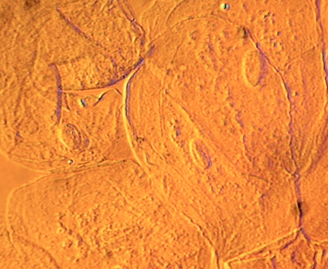
Some simple cells scrapped from the inside of my cheek at a magnification of about 500x. There is no stain of any kind just a wet mount using saliva. DIC is also very useful for pond dipping. The internal workings of transparent creatures are readily apparent. Like all contrast enhancement methods it does creates artifacts that are sometimes difficult to interpret. The 3D effect can suddenly switch to reverse and seem to sink into the slide and if there are bifringent materials in the specimen it is very difficult to interpret them. Many of the images on this page are from the interplay of birefringence of the specimen and the DIC process. It makes nice pictures but they don't have a lot of meaning. |
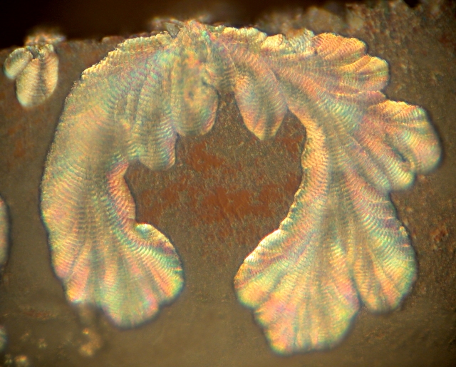
These images were almost lost to the trash when I was cleaning up after some crystal making experiments using silica gel, potasium permanganate and sea salt. The make up of the white flower like growths is unknown. The long ladder like crystals are potasium permanganate. These were purely accidental and the white flowers continued to grow
as the slide dried out. Water glass is gelled with acetic acid and has
very interesting properties when use to grow crystals.
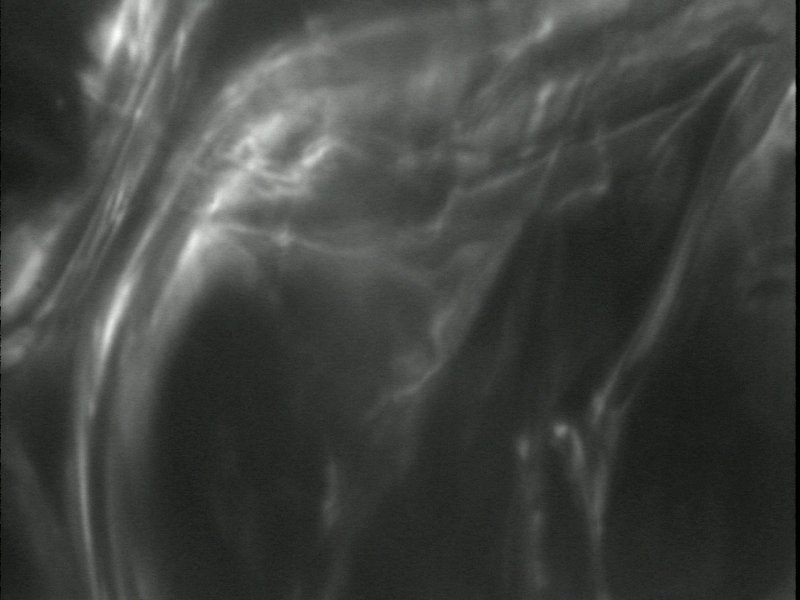
This image was taken on a Leitz Ortholux using a 63x 0.85 Leitz objective
with an adjustable aperture and Leitz darkfield condenser. The onion cell
was dehydrating in a strong sucrose solution. The scope was set so the
image was not completely darkfield. The condenser was too low and the aperture
too wide open on the objective. The image was captured using a monochrome
surveillance video camera with no lens and a 2.5x eyepiece using a Snappy
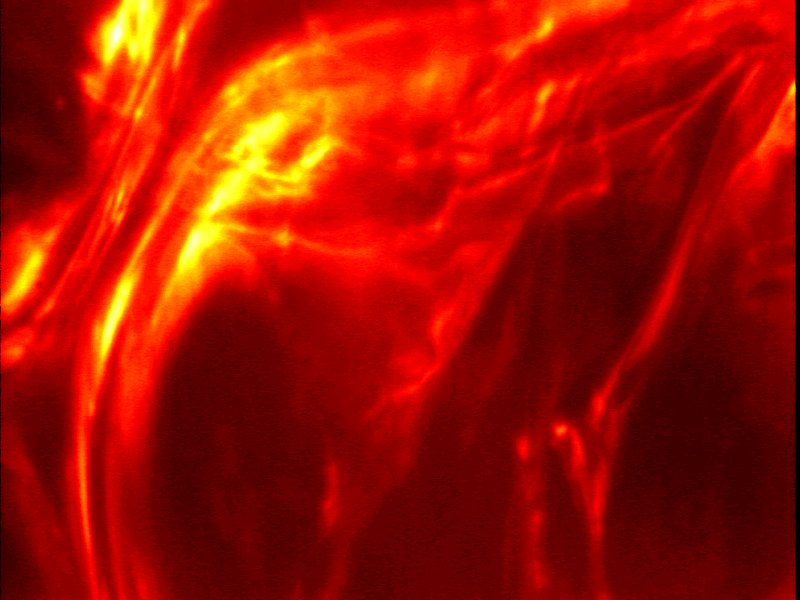
digitizer. The enhanced image was done with what is now ImagJ by shifting
the histogram so the darker areas were stretched and made lighter and a
false color LUT (Look Up Table) was use to replace the gray scale of the
image. A LUT is used to further enhance low contrast areas by changing
the color as well as density of the pixels.
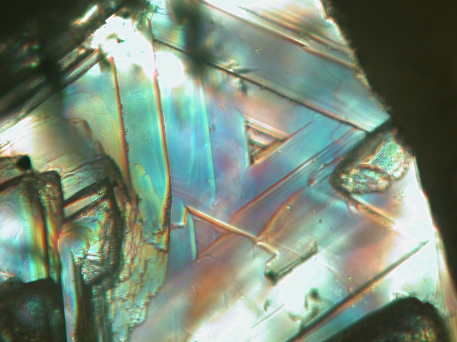
A very small diamond crystal from South Africa magnified 500 times with
transmitted DIC light. The crystal cost less than a dime and will serve
its life as a glass cutter.
Resources ImageJ is a pubic domain program provided by the National Institute of Health. It is a Java program that runs on Unix, Windows, Macintosh and Linux. There is an open interface to write plugins and there are many professionals actively working to expand it. There is a mailing list that supports the programming of plugins. It is the easiest and least expensive program I have found for image processing beyond the simple things provided by Paint Shop Pro and Photoshop. Unfortunately is will not replace these programs. Simple polarizing filters are available from Surplus Shed and Edmund Scientific that are very high quality. For lesser quality you can use filters cut from sun glasses or photo filters. http://www.surplusshed.com/
http://www.edmundoptics.com/
Play, Inc. is bankrupt and Snappy interfaces can be found new on eBay for $25USD and monochrome cameras can be found for less then $50USD, making a very inexpensive imaging solution. There is some problem with the image being a greater magnification than you see in the eyepieces with out an expensive relay lens. I still use it for infra red work and any work involving a video camera. See the Micscape archives for many articles about imaging with video cameras. Techniques - digital & video imaging covers the articles on video imaging. |
|
Microscopy
UK Front Page
Micscape
Magazine
Article
Library
© Microscopy UK or their contributors.
Please report any Web problems or offer general comments to the Micscape Editor.
Micscape is the on-line monthly magazine
of the Microscopy UK web
site at Microscopy-UK
© Onview.net Ltd, Microscopy-UK, and all contributors 1995 onwards. All rights reserved. Main site is at www.microscopy-uk.org.uk with full mirror at www.microscopy-uk.net.