|
|
A Close-up View of the Strange Wildflower "Viper's Bugloss" (Echium vulgare) by Brian Johnston (Canada) |
|
|
A Close-up View of the Strange Wildflower "Viper's Bugloss" (Echium vulgare) by Brian Johnston (Canada) |
One day, while returning from a wildflower hunting expedition, I noticed a tiny blue flower growing in the grass beside the path. The entire plant was not more than 10 centimetres high, and my first impression was not particularly positive. The flower however, was an incredible shade of blue, and I decided that it might be worth investigating. Unfortunately, I was not carrying my National Audubon Society "Field Guide to Wildflowers", and could not identify the plant and research its characteristics. Throwing all caution to the wind, I grasped the stem at the base and attempted to pull the plant, root and all, from the soil. Although this happened many months ago, I can remember the moment vividly. The words pain and shock describe my feelings perfectly. The tiny hairs that I had observed on the stem, were not soft, but sharp, and hard enough to puncture the skin. As well, the tiny plant was anchored so well in the ground that I had to cut the stem to remove it.
Upon returning home, some research revealed the plant to be Viper's Bugloss, or as it is more commonly called, Blueweed. Some refer to it as a "noxious weed" while others call it a "sweet and delicate flower". In North America it is an alien plant, having been introduced from Europe during the late sixteen hundreds. The name Viper is thought to derive from the shape of the seed, which resembles a viper's head. Bugloss is derived from the Greek word for "ox tongue", since the leaves resemble this object.
Although my specimen was very small, Viper's Bugloss can grow up to a metre in height, and prefers limestone soils along roadsides, waste places and pastures. Animals tend to leave the plant alone because of the distasteful bristles. This is probably a good idea since the plant contains pyrrolizidine alkaloids, and can cause liver damage if a sufficient quantity is ingested. A deep, thick taproot assures that it remains firmly in place.
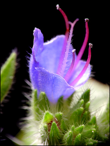
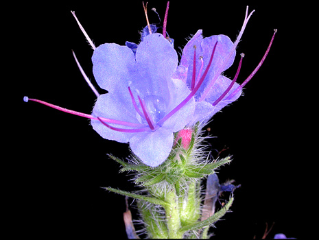
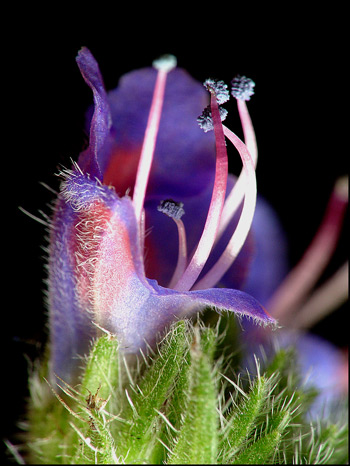
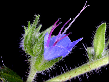
The photograph above shows the main features of the flower. There are five petals which are joined (fused) near the base of the bloom to form a tubular shape. The top petals are larger than the rest, and project out farther. Five stamens, (the male, pollen bearing organ), are usually present along with a hairy stigma, (the female organ where pollen lands). Most flowers are one to two centimetres across. At the base of the flower there is a ring of long, thick, bristly sepals (modified leaves). After each flower has finished blooming, it shrivels up and falls away, leaving the ring of sepals which can be seen in the image below.
In the following image, (which shows the entire plant used in the article), you can see five blooms and at least three flowers that are in the process of shriveling up.
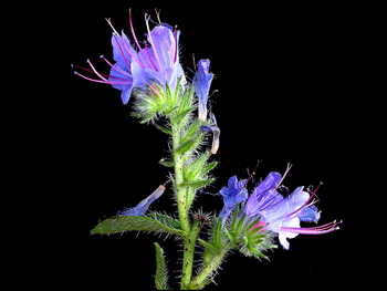
At the centre-left of the image below is a pinkish-red bud about to bloom. Above it is a young flower (pink), and to the right is a mature bloom (blue).
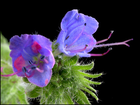
A complete flower can be seen from the front in the following image. The five stamens are obvious but the stigma is partly hidden.
Notice the hairy, two-lobed stigma projecting from the flower on the right.
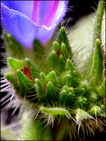
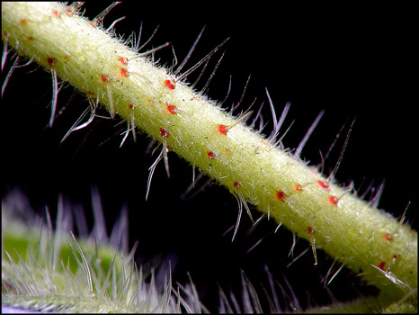
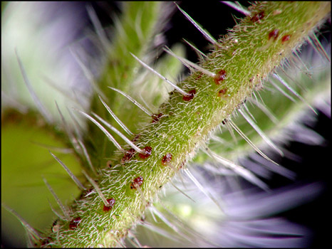
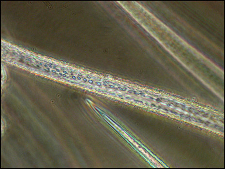
The two images below graphically demonstrate why it is unwise to handle Viper's Bugloss without gloves! In the first of the two, a young stem has very sharp, stiff hairs with reddish, swollen bases. The second image shows an older stem with additional fine, softer hairs.
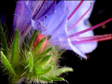
In the following image, notice the hairy underside of the petal and the rough surface of the anthers (the pollen generating organs). The filaments supporting the anthers are smooth and pinkish-red.
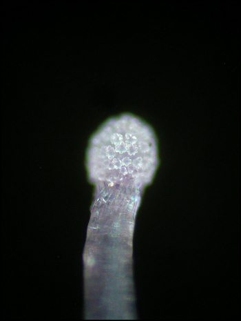
Using a microscope equipped with dark-ground illumination reveals the detail of one of the anthers. Most of the pollen grains appear to be white, and several fine threads can be seen between grains.
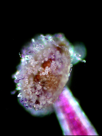
By adjusting the focus of the dark-ground condenser, it is possible to accentuate some of the detail. The filament holding the anther is out of focus in the lower right of the image.
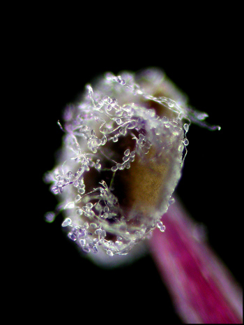
The image below shows another anther that appears to be completely encrusted with translucent pollen grains.
A close-up of the filament reveals that it is not as smooth as it looks to the naked eye. Several pollen grains are visible clinging to the upper surface in the image.
The hairy, two-lobed stigma projects farther out of the flower than the stamens, in most flowers.
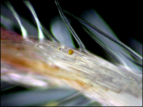
Under the microscope the hairy surface of the stigma is clearly visible. (I believe that the yellow pollen grain in the centre of the image must be from another plant species, because it is much larger, and more coloured than a Viper's pollen grain.)
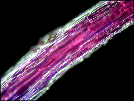
At a much higher magnification, and with the aid of phase-contrast illumination, the spots on one of the hairs of the stigma can be observed.
The object below was also present on the surface of the stigma. I am not certain what it could be. (Perhaps a fungal spore??)
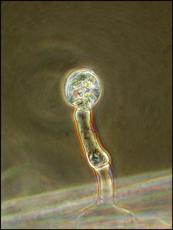
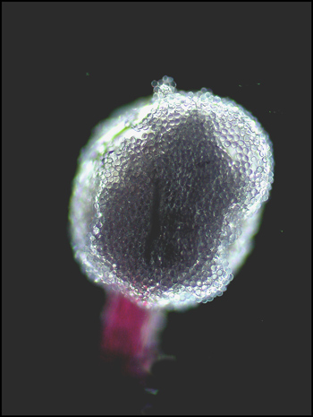
The stigma branches into two lobes. At the end of each lobe there is the bulb-shaped protuberance visible in the dark-ground image below.
First impressions, it is often said, are of paramount importance. The uniqueness of this tiny flower has caused me to re-evaluate my negative view of this bristly weed, and I look forward to finding other (larger) examples to photograph.
Photographic Equipment
The low magnification photographs in the article were taken using a Nikon Coolpix 4500 with natural light and the Nikon Cool light SL-1. Higher magnification images were taken using a Sony CyberShot DSC-F 717 equipped with an accessory achromat which screws into the 58 mm filter threads of the camera lens. (This produces a magnification of from 7X to 9X for a 4x6 inch image.) Still higher magnifications were obtained by using a macro coupler (which has two male threads) to attach a reversed 50 mm focal length f 1.4 Olympus SLR lens to the F 717. (The magnification here is about 13X for a 4x6 inch image.) The photomicrographs were taken with a Leitz SM-Pol microscope (dark-ground and phase-contrast condensers), and the Coolpix 4500.
All comments to the author Brian Johnston are welcomed.
Please report any Web problems or offer general comments to the Micscape Editor.
Micscape is the on-line monthly magazine of the Microscopy
UK web
site at Microscopy-UK