|
|
A
Close-up View of the Wildflower
"Common Mullein"
(Verbascum thapsus)
by Brian Johnston (Canada)
|
PASTORAL
.
.
Oh, grey hill,
Where the grazing herd
Licks the purple blossom,
Crops the spiky weed!
Oh, stony pasture,
Where the tall mullein
Stands up so sturdy
On its little seed!
Edna St. Vincent Millay
This plant, with its very tall
flowering spike, is commonly found along roadsides, and in
fields. Mullein requires full sunlight in order to grow, and is
not found in shady locations. The spike can be very long,
sometimes reaching three metres above ground level. Able to
withstand the worst extremes of Canadian weather, most stalks remain
upright throughout the winter, projecting brown spikes up through the
snow. It is believed that these stalks provide refuge in their
deep crevices for over-wintering insects.
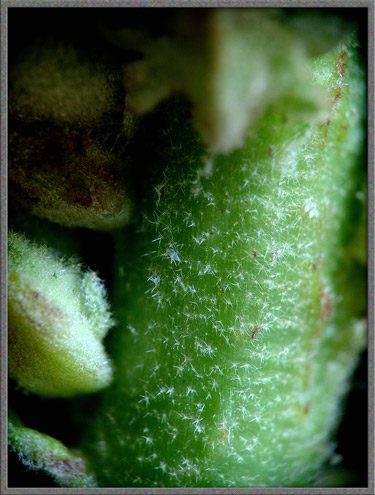
Common Mullein leaves, which are
oblong and stalkless, may be up to 40 centimetres long and 10
centimetres wide. They are densely hairy, and to the touch, feel
like thick felt. In fact, all parts of the plant, including stem
and flower petals share this soft texture. The dried down on
leaves and stem was used historically as tinder, since it ignited with
the smallest spark. Before cotton became the substance of choice
for lamp wicks, dried folded Mullein leaves were used for this
purpose. The old name candlewick plant originated from this
application.
Mullein is the sole member of the Figwort family in the province where
I live (Ontario). It is thought that the Latin genus name of the
plant, Verbascum
originated from a corruption of the word barbascum referring to
barba, meaning beard. (The shaggy foliage was thought to
resemble an unkempt beard.) The species name thapsus is
thought to refer to the ancient Greek city of Thapsos. The plant
has been given many common names over the years, including Flannel
Mullein, Flannel Plant, Feltwort, Velvet Dock and Jupiters Staff.
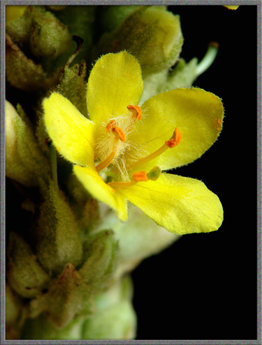
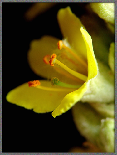
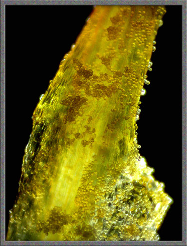
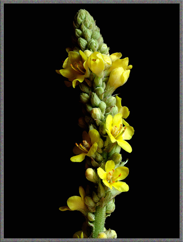
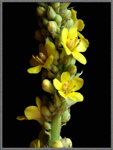
The first image in the article, and
the two below, show a typical flower-head. These structures are
usually from 20 to 40 centimetres long, and consist of a stem holding a
profusion of buds and blooms on extremely short stalks. Unlike
the flower-heads of many plants which bloom from bottom to top, or
vice-versa, Mulleins bloom in a completely random fashion, with only a
few flowers being open at any one time.
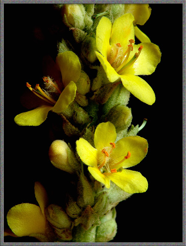
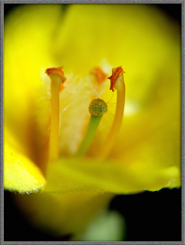
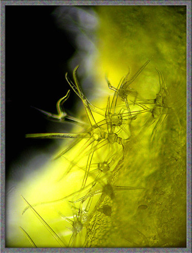
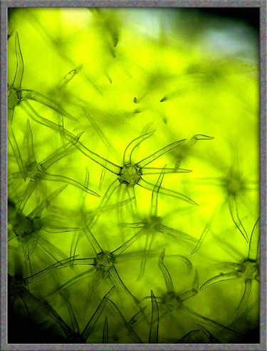
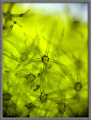
A closer view reveals that some,
but not all of the filaments are completely enshrouded by long white
hairs. (I have not been able to find any information on the
purpose of these hairs, or why they are missing on some of the
filaments.)
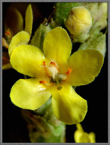
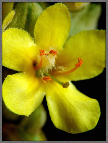
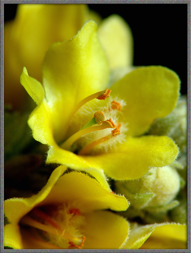
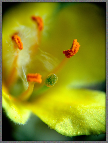
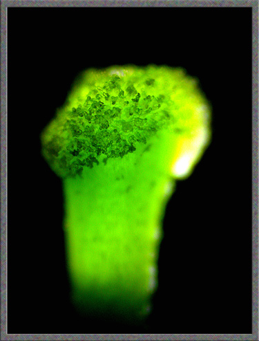
The three images below show
close-ups of the reproductive structures - the five orange anthers and
single green stigma.
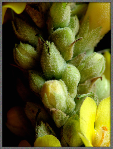
The following photograph shows
several unopened buds, and one which is about to open. The
flowers petals are enclosed by hairy green bracts.
One of these bracts is shown below
at a higher magnification.
The green parts of the Mullein
plant contain small amounts of the chemical compounds coumarin and
rotenone. Coumarin
derivatives, (such as warfarin), are used as anticoagulants to prevent
undesirable clotting of the blood in patients suffering from heart
disease. The molecular structure of coumarin is shown below.
Rotenone
is a more complex molecule that acts as a broad-spectrum
insecticide. The compound works as a contact and stomach poison
by inhibiting cellular respiration. Rotenone is also used to kill
fish as a part of water body management. One such use is to
eradicate exotic fish from non-native habitats. The molecular
structure is shown below. (Both this, and the previous structure,
were produced using HyperChem.)

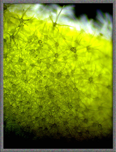
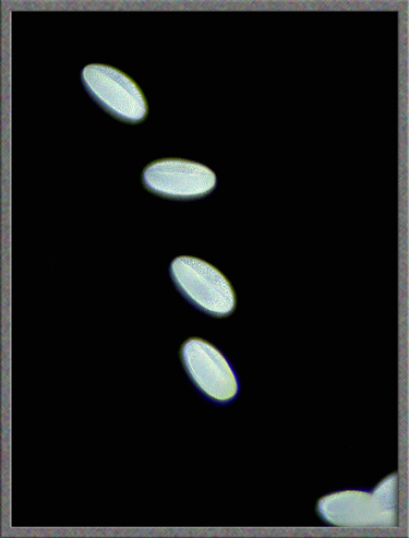
Pollen grains appear to be
ellipsoidal in shape and have two longitudinal grooves.
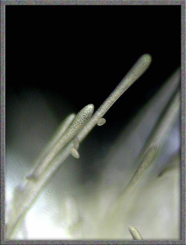
At the base of the style, there are
many hair-like projections with rounded ends. Several pollen
grains can be seen clinging to the surface of one.
The final image shows the irregular
surface of the stigma.
A Common Mullein plant produces a
huge number of seeds during the summer - up to 100, 000! These
seeds, it is said, can survive for almost a hundred years, until
conditions become favourable for their growth. It is no wonder
then, that the striking yellow-green spears are so prevalent in the
environment!
Photographic
Equipment
The photographs in the article were taken with an eight megapixel Sony
CyberShot DSC-F 828 equipped with achromatic close-up lenses (Nikon 5T,
6T, Sony VCL-M3358, and shorter focal length achromat) used singly or
in combination. The lenses screw into the 58 mm filter threads of the
camera lens. (These produce a magnification of from 0.5X to 10X
for a 4x6 inch image.) Still higher magnifications were obtained
by using a macro coupler (which has two male threads) to attach a reversed 50 mm focal length f 1.4
Olympus SLR lens to the F 828. (The magnification here is about
14X for a 4x6 inch image.) The photomicrographs were taken with a Leitz
SM-Pol microscope (using a dark ground condenser), and the Coolpix
4500.
References
The following references have been
found to be valuable in the identification of wildflowers, and they are
also a good source of information about them.
- Dickinson, Timothy, et al.
2004. The ROM Field Guide to Wildflowers of Ontario. Royal
Ontario Museum & McClelland and Stewart Ltd, Toronto, Canada.
- Thieret, John W. et al.
National Audubon Society Field Guide to North American Wildflowers -
Eastern Region. 2002. Alfred A. Knopf, Inc. (Chanticleer Press,
Inc. New York)
- Kershaw, Linda. 2002. Ontario
Wildflowers. Lone Pine Publishing, Edmonton, Alberta,Canada.
- Royer, France and Dickinson,
Richard. 1999. Weeds of Canada. University of Alberta
Press and Lone Pine Publishing, Edmonton, Alberta, Canada.
- Crockett, Lawrence, J.
2003. A Field Guide to Weeds (Based on Wildly Successful
Plants, 1977) Sterling Publishing Company, Inc. New York,
NY.
- Mathews, Schuyler F.
2003. A Field Guide to Wildflowers (Adapted from Field Book
of American Wildflowers, 1902), Sterling Publishing Company, Inc.
New York, NY.
- Barker, Joan.
2004. The Encyclopedia of North American Wildflowers.
Parragon Publishing, Bath, UK.
©
Microscopy UK or their contributors.
Published in the
February 2006 edition of Micscape.
Please report any Web problems or
offer general comments to the Micscape
Editor.
Micscape is the on-line monthly magazine
of the Microscopy UK web
site at Microscopy-UK
©
Onview.net Ltd, Microscopy-UK, and all contributors 1996 onwards. All
rights reserved. Main site is at www.microscopy-uk.org.uk
with full mirror at www.microscopy-uk.net .