|
|
A
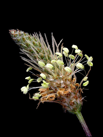
Close-up View of the Wildflower
"English Plantain"
(Plantago lanceolata)
by Brian Johnston (Canada)
|
I
suspect that most people do not consider English plantain to be a
wildflower at all. Its rather drab two to three centimetre
ellipsoidal spike can't compete with the colourful dandelion or
thistle. Nevertheless, this plant is extraordinarily successful
as evidenced by its presence in very large numbers throughout most
urban and country landscapes.
In England, the plant is sometimes
called "Kemp", derived from the Danish "koempe", meaning warrior.
At times in the past, children would use the stalks as swords in a game
of swordplay.
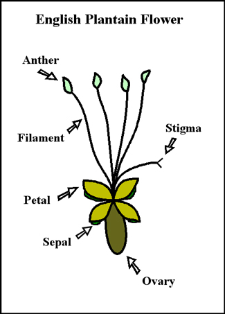
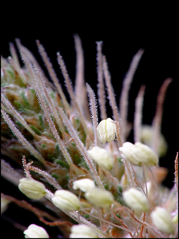
English plantain flowers are packed
so tightly to form the spike, that the actual details of one individual
flower are almost impossible to see. The diagram below shows the
important parts. The most striking feature of the plant is the
four light green anthers, (the male pollen producing organ), suspended
by fragile filaments. Each flower possesses a single hairy
pistil. Four papery petals and green sepals form the base of the
bloom.
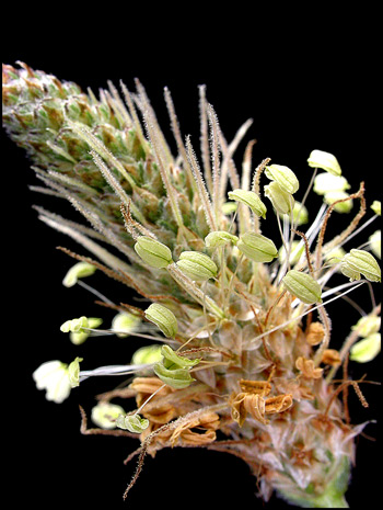
A higher magnification reveals more
details. The flowers at the base of the spike have finished blooming
and have begun to shrivel up and turn brown. Above the blooming
ring, near the tip of the spike, the unopened flowers are protected by
a green assembly of sepals. During the life-cycle of the
flower-head, the ring of green stamens seems to move up the spike from
bottom to top.
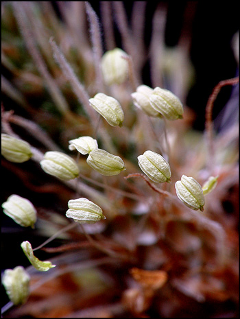
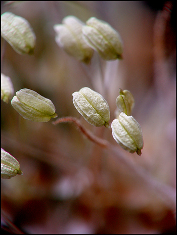
Using a still higher magnification,
it is possible to see the structure of the anthers. Strangely,
pollen grains are seldom seen on the surface of these anthers.
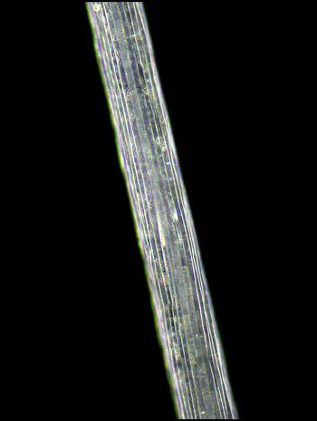
By using a microscope equipped with
dark-ground illumination, still finer details can be resolved.
The left image shows an anther, and the right, a finely grooved
filament.
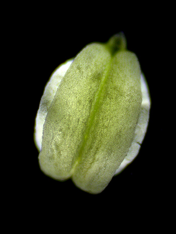
The image below shows the four
beige, parchment-like petals of a typical flower. Under the
petals, the four green sepals are just barely visible. The
reddish pistil protrudes from the junction of the petals and can be
seen to end in a bi-lobed stigma (the female, pollen accepting
organ). Since the flowers at the base of the spike have finished
blooming, the anthers have fallen off, leaving the shrivelled hair-like
filaments that can be seen surrounding the pistil.
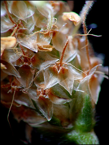
Near the top of the spike, it is
possible to see the still unopened flowers, each protected by a sheath
of four hairy green sepals (sometimes called bracts).
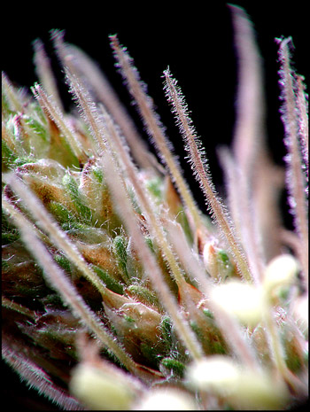
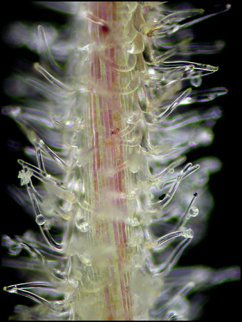
In the image below, notice the
hairy reddish-stalked pistils. At the bottom right, several
pistils have begun to shrivel up, as evidenced by their reduced
diameter and darker red colour.
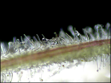
When a mature pistil is examined
under the microscope, a multitude of tiny hairs can be seen. They
are transparent, and many have obviously captured spherical pollen
grains.
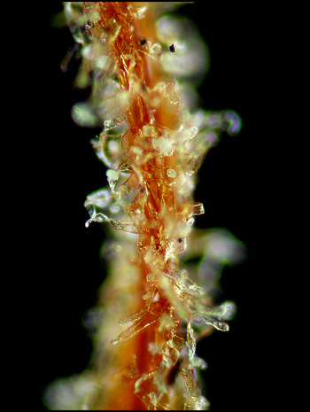
As the pistil begins to shrivel up
as it dries out, the colour becomes darker, and the tubular
protuberances take on an almost ribbon-like appearance.
Although English plantain could not
compete successfully in a beauty pageant with its larger and more
spectacular wildflower neighbours, its form is different and striking
enough to warrant closer attention.
Photographic Equipment
The low magnification photographs
in the article were taken using a Nikon Coolpix 4500 with a combination
of natural light and the Nikon Cool light SL-1. Higher
magnification images were taken with natural light using a Sony
CyberShot DSC-F 717 equipped with a combination of achromatic close-up
lenses (Nikon 5T, 6T and shorter focal length achromat) which screw
into the 58 mm filter threads of the camera lens. (These produce
a magnification of from 0.5X to 9X for a 4x6 inch image.) Still
higher magnifications were obtained by using a macro coupler (which has
two male threads) to attach a reversed 50 mm focal length f 1.4 Olympus
SLR lens to the F 717. (The magnification here is about 13X for a
4x6 inch image.) The photomicrographs were taken with a Leitz SM-Pol
microscope (using a dark ground condenser), and the Coolpix 4500.
©
Microscopy UK or their contributors.
Published in the
February 2008 edition of Micscape.
Please report any Web problems or
offer general comments to the Micscape
Editor.
Micscape is the on-line monthly magazine
of the Microscopy UK web
site at Microscopy-UK
©
Onview.net Ltd, Microscopy-UK, and all contributors 1996 onwards. All
rights reserved. Main site is at www.microscopy-uk.org.uk
with full mirror at www.microscopy-uk.net .