|
Microscopic Seduction by Richard L. Howey, Wyoming, USA |
Let’s begin with a question: In a world overrun by technological gizmos and distractions–TV, iPods, X Boxes, computer games, cell phones–how do you get people interested in microscopy and natural history? After all there are pragmatic, aesthetic, psychological, and ecological reasons for knowing about the wonderfully complex world in which we live–not to mention the fact that microscopy is a hell of a lot of fun. Unfortunately, many people don’t think so as a consequence of having taken a biology course in high school or university. I had the extreme good fortune of having taken only one biology course and that was in high school. It was taught by a wonderfully eccentric old dear with frizzy hair who would swoop around the room with her arms stretched out, flapping, to demonstrate to us how flies spread disease. Or, she would hop across the front of the classroom during our frog dissection trying to get us to examine the extraordinary musculature of frogs’ legs and telling us that if we grasped that, we would understand why they were such a culinary delight. That is not something you tell to middle-class Midwestern teenagers without expecting squeals of disgust at the very thought but, then again, when you’re sitting, poking at a frog that’s probably been dead for 5 years and you are breathing the stink of formaldehyde fumes, such a reaction is not only understandable, but a very sane one. There are, unfortunately, some “teachers” who can spoil even the most wonderful things. In high school, I took a music appreciation class and the aptly named Miss Gore ruined certain pieces of music for me in such a thoroughgoing manner that it was a decade or more before I could listen to them again. I pity the poor student who here at the university had to take the introductory Botany course from the man who was chairman of the department about 30 years ago and who is no longer rustling with us. He wrote an introductory textbook and three times a week he would, students told me, enter the large lecture hall and for 50 minutes read from his book which he also required the students to buy as the text for the course. And we wonder why so many students hate science!
All of this is my long-winded way of saying that if you are an enthusiastic amateur dedicated to intriguing other people and luring them into the endless marvels of the multiplicity of microscopical worlds, then you need to become an intellectual seducer or seductress.
A few of the things I learned in my very first year of teaching are: 1) If you want others to get interested in something, you have to convey your own excitement and passion. 2) You have to get them hooked, intrigued, addicted. 3) You need to develop a repertoire of tricks and show them things that are so dazzling that only the dullest and most plodding can resist their charm. 4) Present things without lecture, with minimal background, so that if they are at all mentally alive, they can’t help but ask questions, giving you the opportunity to begin gently reeling them in. Yes, it is quite like fishing. 5) Keep your answers, short, clear, and to the point. If you natter on, their interest will quickly evaporate.
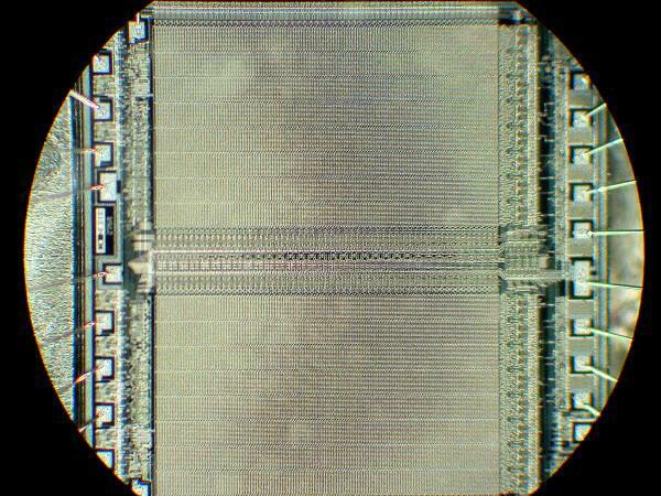
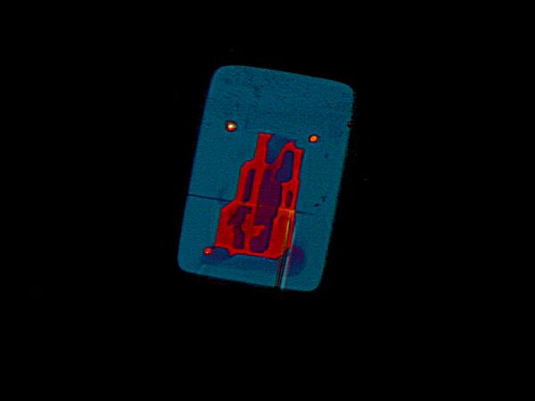
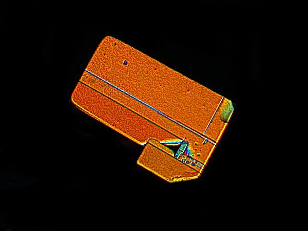
So, the next issue is : What do you show them? If you know something about their interests, then that can give you some clues. I always start with the stereo-dissecting microscope because 1) it’s easy to use, 2) it’s easier to explain how it works (if asked) as against a compound microscope and 3) it allows for a wide variety of specimens with which you can surprise and amaze them by having them first look at the specimen without magnification under the stereo microscope. For example, if you know that they have an interest in computers (who doesn’t these days?), then you can show them a computer chip mounted on a slide and then let them look at it under low magnification.
This is an image of a chip which is at least two decades old and, of course, the new ones are much more elaborate and intricate but, nonetheless, I find this example quite pleasing and amazing.
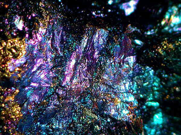
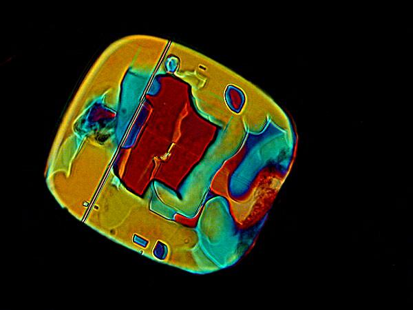
If your intended recruit has an interest in rocks and minerals, then take a small piece of bornite, a copper ore also know as “Peacock rock’, which is splendid to look at just as it is. However, under low magnification, it is even more resplendent.
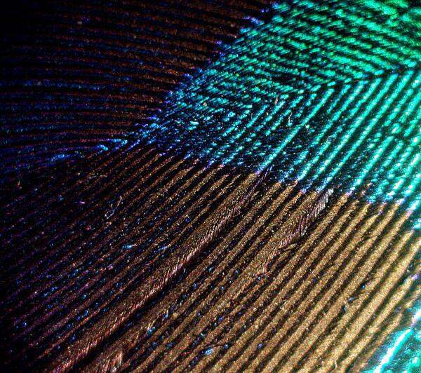
Speaking of peacocks, if your friend happens to be bird-watcher, then a small piece of a peacock feather becomes an astounding experience under a stereo microscope where the individual branches look like they have been wound with metallic threads of different colors.
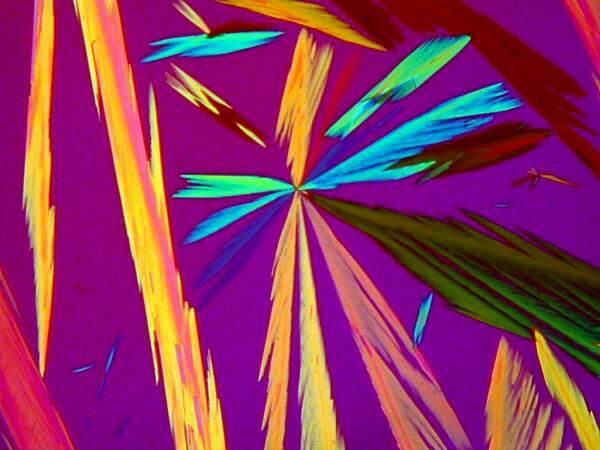
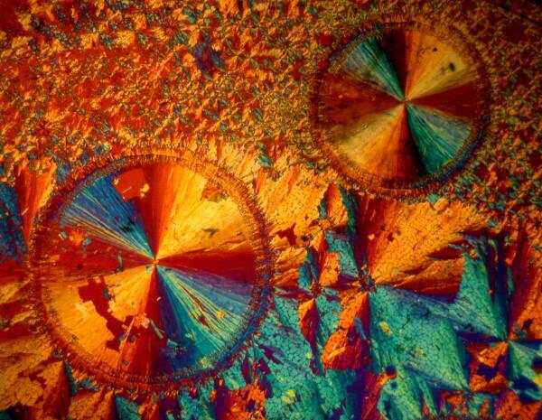
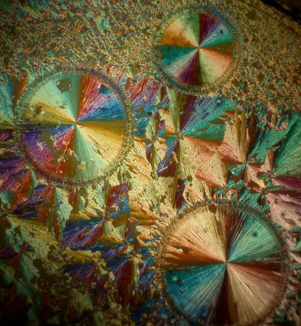
Something almost no one can resist is crystals under polarized light. The biological stain Orange G is particularly suitable because you can rotate the polarizer through its entire range and achieve different color patterns. I dissolve the crystals in 70% isopropyl (rubbing) alcohol, put a drop on a clear slide, get everything aligned and wait until I see crystals beginning to develop at the edge of the drop from the rapid evaporation of the alcohol and then I say to the victim: “Quick! Look at this. You can watch the crystals forming.” This is an especially attractive and almost irresistible display.
If you don’t have a polarizing attachment for your stereo microscope, another stunning crystal display can be achieved with a bit of silver nitrate solution and a tiny sliver of copper which you can trim from a piece of copper wire using a scalpel. Place a shaving or two of the copper on a slide, add a drop of silver nitrate and you will be able to watch the dendritic (tree-like) patterns emerge.
Another almost sure winner is, depending upon the time of year, a recently collected water sample of a rich culture. If it’s still warm enough to collect, you can show them a sample teeming with protozoa, water fleas, ostracods, water mites, and algae depending upon what’s available in a particular location at a given time of the year. People are very much drawn to the motion and busyness of micro-critters. If it’s a hard winter and the ponds and lakes are frozen, then a good Paramecium culture will serve nicely, since they are very active and quite attractive. I always try to keep some Paramecia going year around since they are easy to culture and make good test organisms for various reagents and stains.
By now, your friend should be sufficiently softened up so that you can innocently ask: Would you like to see a Paramecium close-up at higher magnification? Most lay persons are fascinated with the idea of high magnification and so the chances are that you will get an enthusiastic “Yes.”. This will give you an opportunity to introduce him or her to the compound microscope. Set up a slide, check it to make sure that it’s chock-full of active wee beasties and then explain only the minimum–namely, how to use the fine focus, how to adjust for interpupillary distance so that they are getting a single coherent image, and how to use the mechanical stage. Then, sit back quietly, let them explore, and wait for questions–one of which will almost certainly be: How much is the magnification now?
This is an opening for you to explain that as magnification is increased, the field of view is decreased and, of course, with large active organisms this becomes a problem unless you have a means of slowing them down, anaesthetizing them or killing and fixing them with as little distortion as possible. If you have phase contrast or Nomarski Differential Interference Contrast, you can show your friend the different results from different contrast techniques.
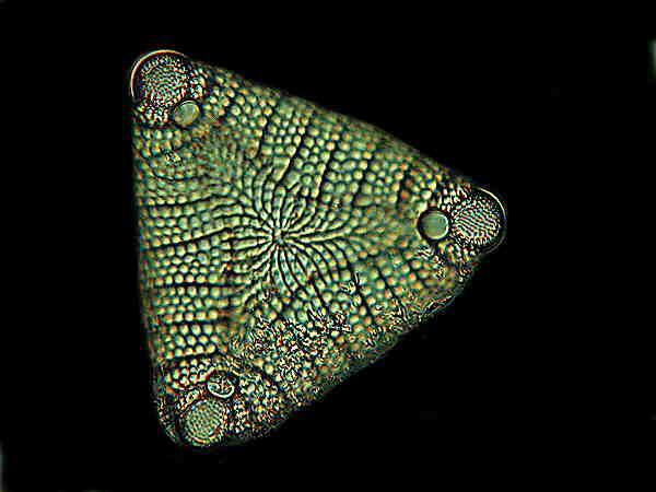
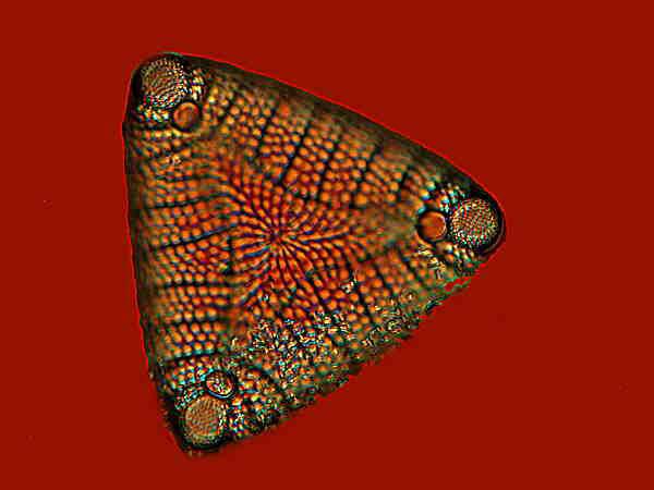
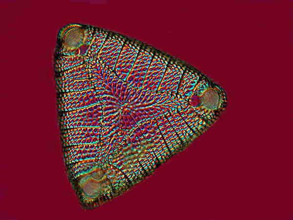
Here are three images of the diatom Etogonia using Nomarski DIC. The variation in colors between the 3 images is achieved by adjusting a prism in the body of the microscope to different positions.
If, at this point, you still have their interest, this is an excellent time to show them a stained, prepared slide such as a cross section of plant stem. This can be a special attention-grabber, especially if it is one which has been prepared using multiple staining and also has birefringent structures that show up impressively under polarized light.
Here I will show you 3 pairs of images of plant material; the first of each pair will be with brightfield illumination and the second with polarized light.
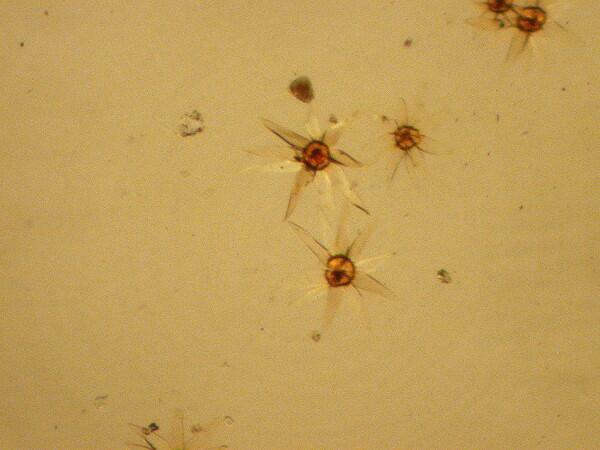
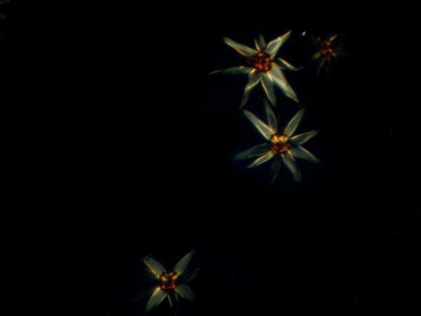
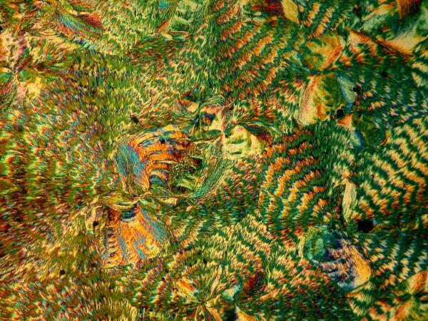
These splendid specimens are fern scales taken from a slide kindly given to me by Brian Darnton who has a special affinity for these structures and has produced some marvelous slides along with his superb work on foraminifera.
This is a transverse section of a root of the Joe Pye weed which has been parasitized by Dodder. Even plants are not immune and some biologists think that there are more parasites than any other type of creature.
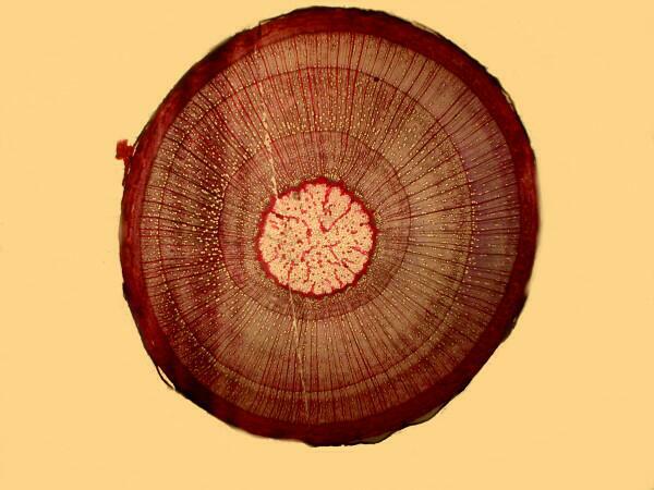
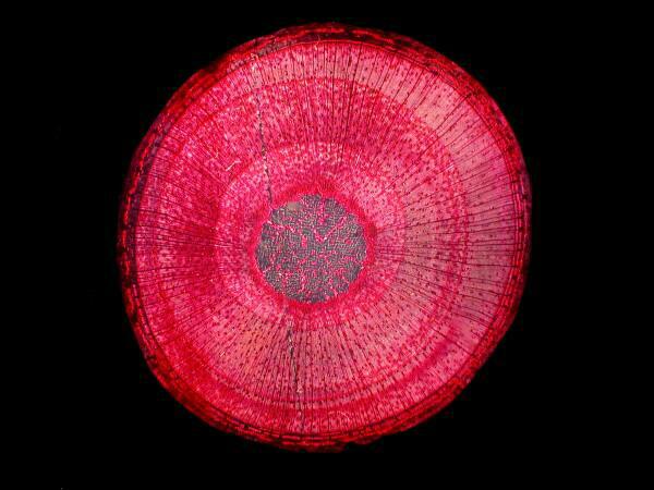
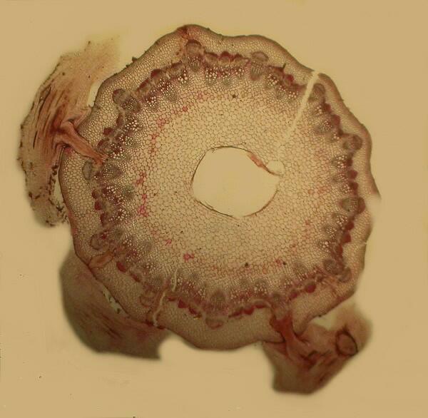
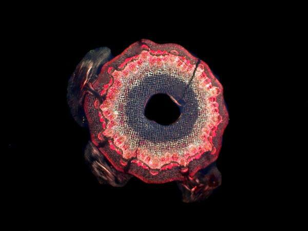
This is a transverse section of the stem of a sycamore maple. As you can see from the first image, it has already been stained using biological dyes.
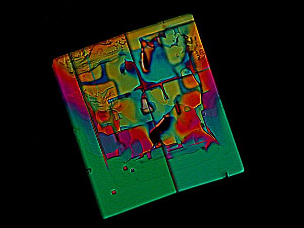
At this point, you should have a good, fairly clear idea as to whether or not this person is a potential convert. If you think, he or she is, then this is a perfect time to introduce birefringent crystals with the compound microscope using polarized light. The diversity of colors, shapes, and sizes present endless opportunities.
A Combination of Magnesium Chloride and the biological stain DAPI
A Combination of Magnesium Chloride and the biological stain Acridine Orange
A Combination of Magnesium Chloride and the biological stain Alizarin Red S
A Combination of Magnesium Chloride and the biological stain Alizarin Red S
If, however, you judge that this person is really not suitably interested, then banish him or her from your house and place a curse upon his or her house.
However, for those who pass the test, issue an invitation to them to join the STMTWSFM (Society To Make The World Safe For Microscopy) and explain to them that: 1)There are no membership fees. 2) There are no meetings. 3) There are no rules or obligations beyond those suggested by the name of the organization and 4) It would be a good idea to buy a microscope and you’ll be willing to help them find one.
To be able to explore with a microscope is an extraordinary gift and privilege, for it opens up new dimensions to us so beautiful and bizarre that even the poets cannot come up with language to adequately describe them. If there are any sins, I would say that to deprive a child of the opportunity to gaze into the mysteries of microscopic worlds, is a cardinal sin.
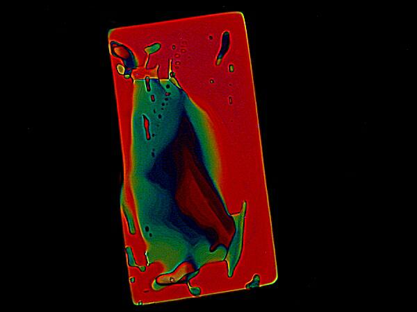
A few final examples of the glories of polarization:
Salacin from a 19th Century slide
Salacin again, this time using a plastic Petri dish as a compensator
Palmitic Acid which is derived from the Brazil Nut Palm. Also a 19th Century slide.
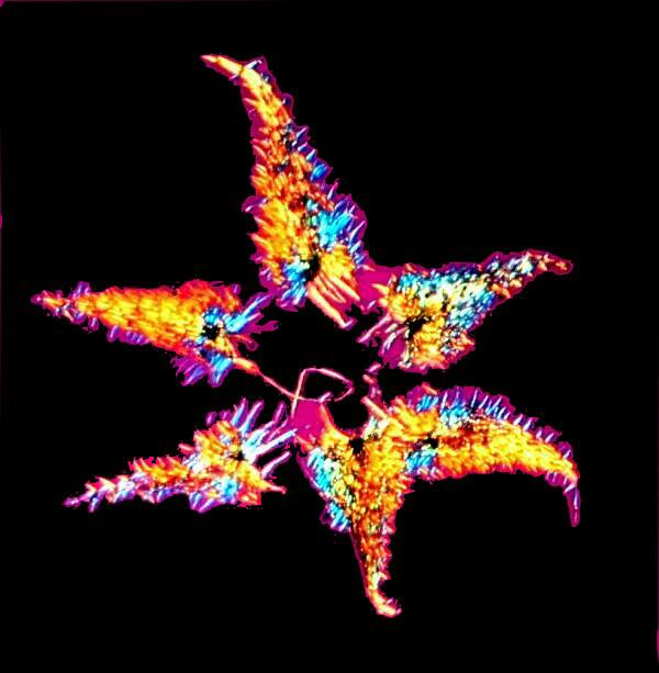
Another of Brian Darnton’s remarkable arrangements of fern scales
A combination of Magnesium Chloride and the biological stains Eosin Y and Light Green
All comments to the author Richard Howey are welcomed.
Editor's note: Visit Richard Howey's new website at http://rhowey.googlepages.com/home where he plans to share aspects of his wide interests.
Microscopy UK Front
Page
Micscape
Magazine
Article
Library
Please report any Web problems or offer general comments to the Micscape Editor .
Micscape is the on-line monthly magazine of the Microscopy UK website at Microscopy-UK .