The Carl Zeiss LUCIGEN Illuminator
by Fritz Schulze, Canada
|
There are
traditionally two ways for microscope illumination1:
critical, where the light source is imaged into the specimen, and the
so-called Köhler illumination, where the light source is imaged
into the near focal point of the condenser (its iris diaphragm).
In the
late 1960s Carl Zeiss Oberkochen introduced a third system: the light
source was directly below the specimen. The system was called LUCIGEN
and was intended mainly for the successor of the popular Standard
Junior, the Standard K, although it could be used equally well with
any Zeiss Standard microscope with a condenser sleeve 39.5mm (Lucigen
S) or a centrable substage (Lucigen Z). The odd model I have is
neither but has an integrated sleeve that screws directly to the
underside of the stage. The sleeve has a helical slot that allows the
illuminator to be vertically adjusted by rotating the main body.
Furthermore, my Lucigen lacks the lamp socket, so, in order to test
it, I had to rig up a suitable light source.
The
instructions2 read: The advantages of this unique
illuminating system are its quick and easy manipulation as well as
its high performance.
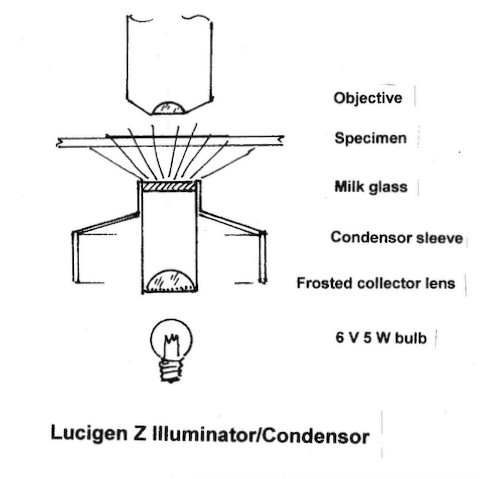
A
special opal-glass (milk-glass) disk of 9 mm diameter , the
heart of the system, is illuminated by a 6V 5W tungsten bulb via a
collector lens and a daylight filter. Control of the illuminating
aperture, and with it the contrast, is achieved by simply adjusting
the height of the Lucigen. Field of view and correct illumination of
the aperture are automatic. Uneven illumination, reduced resolution
due to extreme stopping down, or undesired off-centre illumination
are avoided. The user can concentrate totally on his object, even a
change from a low power 2.5x objective to an oil immersion 100x
requires no additional manipulation: the illuminating aperture ranges
from any dry objective to a maximum of 1.0 (dry) and 1.4 (immersed)
as the opal-glass gives off light in 180 degrees in all
directions. The maximum illuminated field is 9mm = the diameter of
the opal-glass disk.
In fact
this particular illuminator combines condenser and light source in
one, eliminates the necessity of a swing-out front lens or even an
immersed front lens (the limit is, however, the light intensity,
which even at a maximum of 8.2V may be insufficient in extreme cases,
but, then, the system is intended for simple routine work).
The price
in Canada at the time was $30.50 compared to an equivalent condenser
0.6 ($46.00) when a basic monocular Standard K with three objectives
sold for $258.50 plus illuminating/condenser system.
Despite
its attractive price and obvious advantages, the Lucigen never became
popular. That may simply be because of prejudice. Its production was
soon discontinued. Not having a microscope where my Lucigen could be
attached, I rigged up my “vertical optical bench” to
replicate a microscope. By means of double-sided adhesive tape I
attached the Lucigen to the underside of a stage and inserted a small
battery-operated torch from below as light source. As my set-up
lacked a fine focus control and was, admittedly, a bit shaky,
obtaining sharp pictures with the high power was a bit of a problem.
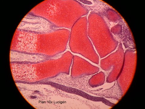
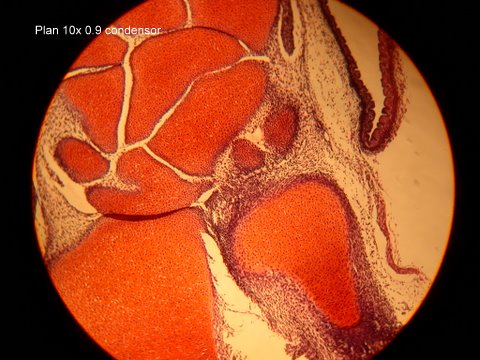
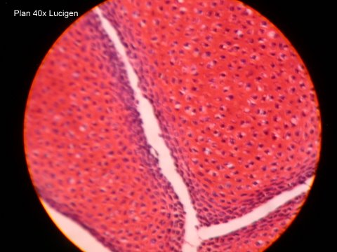
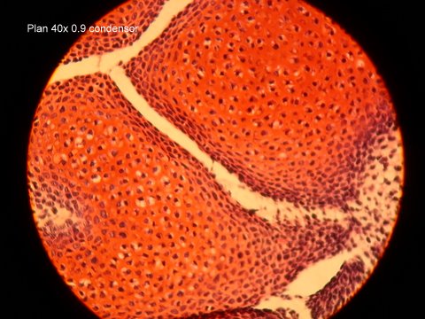
Still, the illustrations show a comparison between a specimen
illuminated by Lucigen and a normal 0.9 condenser (Zeiss Planachromat
10x and Leitz Periplan10x/18 wide angle eyepiece for the Nikon
Coolpix 995). The specimen is the paw (foot) of a 13 day old rat
foetus. The low power picture gives an idea of the object, its
slightly uneven illumination is due to my improvised light source.
Visually there was no discernible difference in resolution or
contrast between the two systems.
I think a
hobby microscopist with an old microscope lacking a condenser could
easily replicate the Lucigen system by using a piece of thin
transparent white plastic directly under the specimen.
All comments to the author
Fritz Schulze
are
welcomed.
|
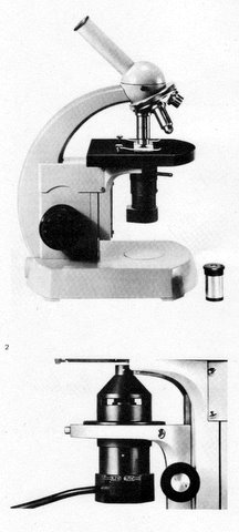
Fig.1 Lucigen S (
top) , Lucigen Z (bottom) on a Zeiss
Standard K microscope
|
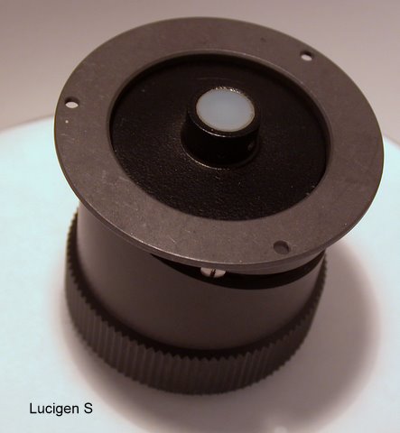
Fig.2 The Lucigen
illuminator
|
|
|
Left. Fig.3 Working principle
|
|
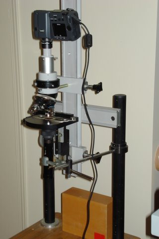
Fig.4 The
experimental set-up
|
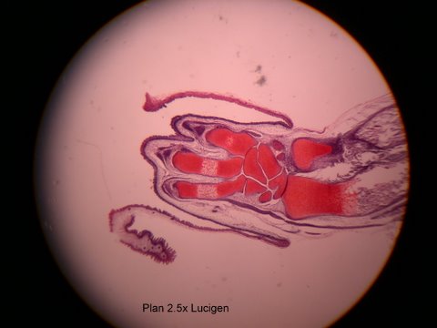
Fig.5 The rat’s paw.
Remaining images are at higher mags, see embedded image captions.
Righthand condenser images taken on a Standard Junior with a 110V 15W illuminator without filter.
|
|
|
|
|
|
|
Microscopy UK Front
Page
Micscape
Magazine
Article
Library
© Microscopy UK or their
contributors.
Published
in the February 2012 edition of Micscape
Magazine.
Please report any Web problems or offer general comments
to the
Micscape
Editor
.
Micscape is the on-line monthly magazine of the Microscopy
UK website at
Microscopy-UK
.
©
Onview.net Ltd, Microscopy-UK, and all contributors 1995
onwards. All rights reserved. Main site is at
www.microscopy-uk.org.uk .