|
Euplectella aspergillum: Part IV. Vandalism on a Glass Sponge by Richard L. Howey, Wyoming, USA |
Visit the Micscape Library for Parts I, II, and III
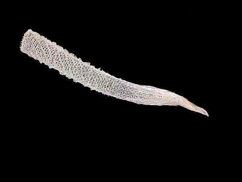
You may recall (two of my friends did and reminded me) that in Part I of this essay on the marvelous Venus Flower Basket sponge, I promised that I would drop a specimen on a concrete floor to see how and if it would shatter and then collect the debris to examine in order to get a clearer idea of the types of spicules and how they were fused together to create this elaborate architectural lattice. In case you didn’t read the other parts, let me show you an image from the first part which gives an impressive idea of the structure of this skeleton.
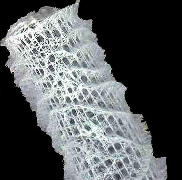
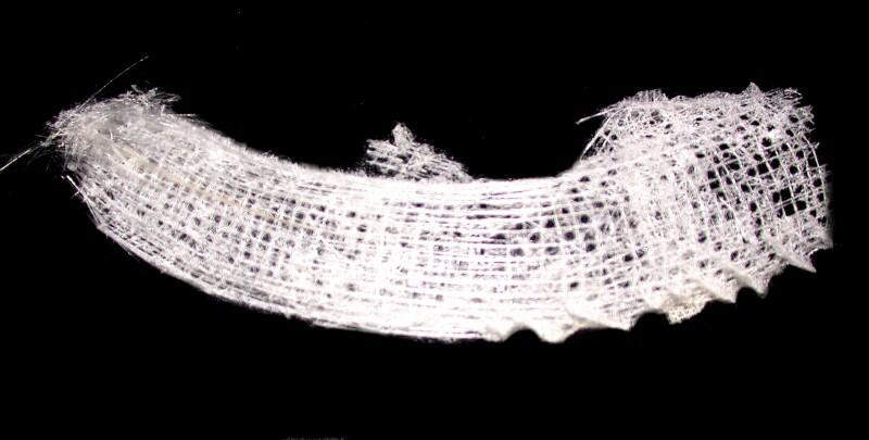
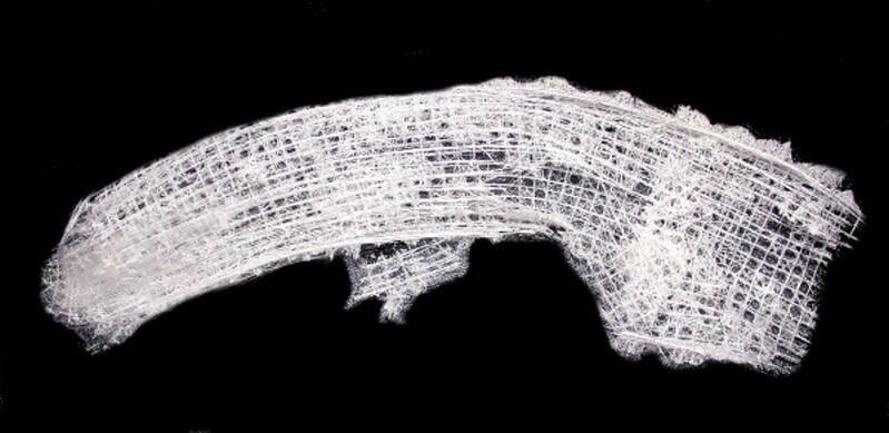
The first image shows the entire skeleton and the second one is a close up of a section of the “body” which demonstrates the remarkable intricacy of this structure.
I anticipated the vast majority of spicules would be hexactal or six-sided since these are the dominant type in glass sponges or hexactinellids. There is a major problem about determining what other sorts of spicules may occur in this organism, since virtually all the specimens which one can obtain from dealers have been subjected to a bleaching and cleaning process to get rid of the grayish-green layers of cells in order to reveal the beauty of the skeleton lying underneath. However, it is precisely this matrix of cells which may provide a “home” for spicules of other types, that is, ones that are not hexactal. I shall be making an attempt to obtain a couple of “natural”, uncleaned specimens in the near future (which will doubtless prompt yet another essay.)
However, back to the vandalism. Quinn Coffeen, one of the friends who reminded me of my unfulfilled promise, was visiting one day last weekend and I said: “O.K., let’s go down the basement to my storage lab (which has a concrete floor) and try it out.” I took with us a long-handled dust pan with a brush to sweep up the debris for later examination upstairs. I opened a drawer and removed a Euplectella specimen and dropped it from a height of 4 feet. Now, one needs to remember that these creatures have a series of long, posterior “threads” that partially serve as an anchor to the substrate and, I deduced, that when these specimens are dredged up the bundle of glass thread is folded up over the base which rests on the sea bottom. This base serves as a kind of collecting point for all sorts of detritus. It tends to become a repository for a wide variety of remains of other organisms and when uprooted in collecting by dredging, it is rather like a ball of mud from which the rest of the skeleton projects.
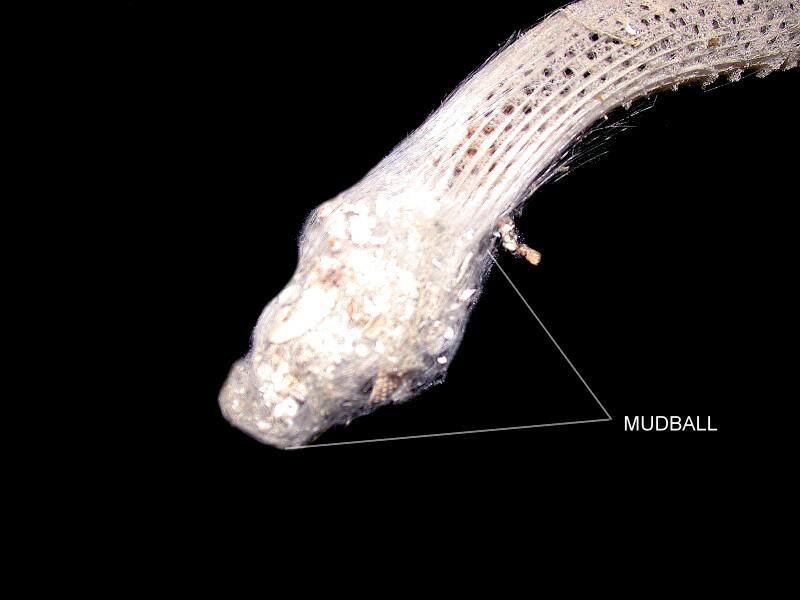
In this ball, I have found a variety of forams, fragments of pteropod (“sea butterfly”) shells, sea urchin spines, spicules from soft corals, bits of mollusk shells, and, in short all kinds of bits and pieces of calcareous and siliceous materials from other species of organisms that have slowly drifted down to form thick layers of detritus. The reason all of this is important is that the “mud ball” forms a weight at the posterior end which sufficiently imbalances the sponge so that when dropped from about 4 feet during the first trial, it landed on the mud ball and then fell gently over to come to rest on the cement floor with no damage. Just look at the image above again and you can easily imagine how this happened. So, for trial #2, I tossed it up in the air to a height of about 8 feet expecting to have to get the dust pan to sweep up the shards. However–you guessed it–the same result as before; no damage! Clearly this called for bolder measures, so for trial #3, I hurled the sponge from a height of 4 feet, with a force appropriate to a superhero, oriented so that it would hit the cement at the anterior end where the osculum (sieve plate) is located. Remember that this skeleton is composed of tiny bits of silica (in other words, minute shards of glass) and so, after throwing it, I turned away and covered my face, not wanting to inhale powdered glass for I expected a small cloud of fragments. Result–no damage! At this point, annoyance led to trials #4, 5, 6, and 7 with the adrenaline increasing my superhero strength with each hurling blow.–NO DAMAGE (maybe, a strand or two from the posterior end, but nothing obviously noticeable).
At this point, annoyance gave way to extreme irritation and I picked up the sponge, put it in a one gallon plastic bag, took a hammer and proceeded to pound the hell out of it. And here is the result which we all expected much earlier, a pile of powdered spicules.
CAUTION: At this point, I am going to make several very strong recommendations. When working with Euplectella or any of its parts (or any other form of glass sponge):
1) Work in an area which you can easily clean if you happen to spill any of the material and make sure it is an area which any pets you have do not have access to.
2) Wear goggles.
3) Wear a small face mask over your mouth and nose.
4) Wear sturdy, but flexible rubber gloves or if you have only thin latex ones, wear 2 pairs. As much as possible, handle any of the material with forceps and not your gloved fingers and when you are finished with a session, carefully wipe any tools you have used and place the wipes in a plastic bag to be discarded.
Now, this may strike some as a lot of fuss about nothing but, remember what this material essentially consists of is minute fragments of needle-sharp glass and powdered glass (after you have smashed it). If any of this material gets into your eyes, it could do serious damage permanently affecting your vision. If it gets into your nostril, mouth, or lungs, it can create severe irritations which the human body is not very well designed to cope with and can lead to serious infections. As for the skin, embedding glass needles in your hands is not an accepted from of acupuncture. One afternoon, I was working with a stereo-microscope in an area where I didn’t think there were any fragments. Well, I was wrong and I got a tiny glass sliver embedded in one of my fingers. At first, it was just a mild itching sensation but, the more I rubbed at it to try to remove it, the sharper the pain became. Finally, with the help of a magnifier, I was able to locate this tiny, glass sliver and with the help of a pair of very sharp micro-forceps, remove it. So the message is–take all proper precautions!
Here are some images of the results.
This is just what we would expect; a pile of smashed silica. However, I cheated a bit. Now, these fragments were indeed taken from that plastic bag but, they don’t quite tell the entire story. This “pile” is an extreme close-up of material taken from the bottom of the bag taken at about 60 magnifications with a stereo-microscope. Incredibly, the rest of the skeleton was simply two flattened, connected strips which were highly flexible and I could bend them back and forth without their breaking or showing any signs of fracture. For a minute, I wondered if I had been duped and that someone had sold me a plastic replica–unfortunately the world is full of counterfeits of an incredible range of objects. This specimen, however, turned out to be astonishingly real.
At this point, I wished that I knew one of those relatively new brands of researchers who is a materials scientist. After some poking around on the Internet, I found some who were examining, of all things, the structure of Euplectella from the level of nanostructure to macrostructure. (So, that will mean yet another article). However, in the meantime, I will use what modest mental resources I have to suggest a few possibilities regarding this extraordinary structure and then later on see what the experts have come up with.
However, first a couple of brief digressions; any of you who have read any of my previous essays have learned to expect digressions and you might even find these two worthwhile and I get to have fun since I love rambling off on tangents.
1) Euplectella are often given as wedding presents and while they are extraordinarily beautiful, they also have great symbolic significance. There is a pair of small shrimp which enter the sponge in a larval stage when they are still small enough to pass into the interior. They are Spongicola japonica and they provide us with a nice example of symbiosis in that the shrimp help keep the interior of the sponge clean which also benefits the sponge and the shrimp have a constant supply of food in the flow of plankton plus the added benefit of a well-protected house with elegant architecture. There is an additional marvel here; there are enormous numbers of bioluminescent bacteria in and on the sponge and since they live at depths where there is little if any light, they act as lanterns attracting positively phototrophic species of planktonic organisms thus acquiring additional food for themselves and the Spongicola. Now, just imagine how glass fibers that are light conducting function in transmitting the light throughout the entire skeleton. This information made me want to rush right out and buy a deep sea submersible so that I could observe this phenomenon first-hand, but my wife wouldn’t let me; she’s claustrophobic.
There are also reports that the shrimp themselves are bioluminescent; however, this could be simply a result of their ingesting quantities of the bacteria while performing their functions as cleaners.
My guess, based on no evidence whatsoever, is that they might enter through the osculum, the sieve plate, at the top which we will be talking about a bit more when we get to the second digression. I suggest this because the openings in the osculum are larger than those in the columnar “body” of the skeleton and also are less likely to have cellular material and detritus partially blocking the openings. In any case, it sorts out in such a way that a mating pair of shrimp larvae enter the sponge and then having, in most cases, an abundant supply of food being filtered through to them, grow sufficiently large that they are no longer able to leave the sponge. I doubt whether anyone knows whether there are larval shrimp battles to establish a dominant alpha male or whether the shrimp simply sign a prenuptial agreement. In other words, I don’t think anyone knows much about what takes place–I certainly don’t–because such behavior would be extremely hard to study in a controlled environment. As far as the symbolic significance is concerned, if you are an optimistic Romantic, you can regard the shrimps’ permanent companionship as an eternal commitment which allows them to create new larvae small enough to escape the confines of the sponge and go off to find their own special Venus Flower Basket. If, however, you are a pessimistic Cynic, you can regard this as an eternal bondage from which there is no escape, no divorce. I am sure that the traditional giving of these sponges as wedding gifts is meant to be directed to the former.
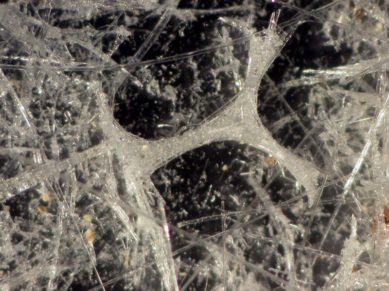
2) During my pummeling, most of the osculum or sieve plate came loose and it was an easy matter to separate it virtually intact. What struck me immediately was that it has a quite different character from the main body of the skeleton even down to the texture and color.
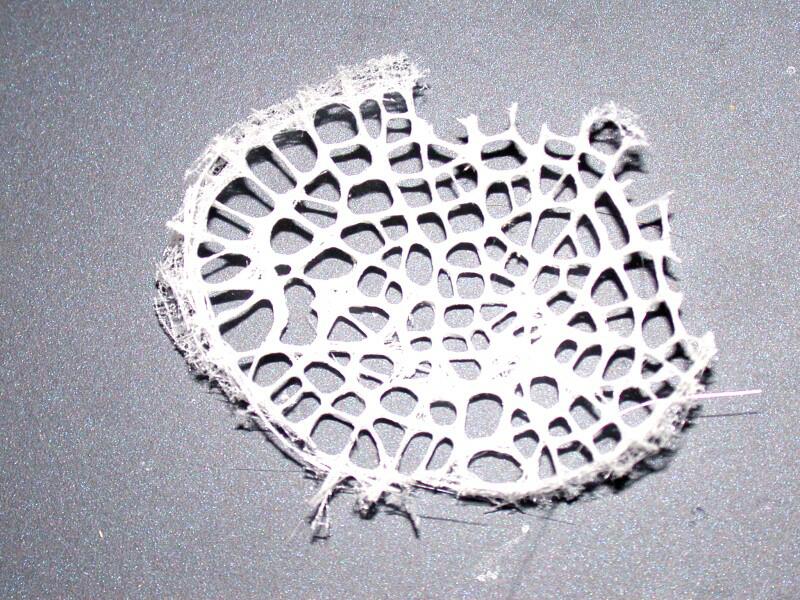
The appearance is less crystalline almost giving the impression that it had been painted with a coat of flat white enamel. The cross pieces are thicker and have a less delicate appearance than those of the main skeleton. This makes me wonder if the cells which produce this structure are programmed differently than those which produce the main body of the skeleton.
In the main body, we can observe a series of circular, spaced rings which get smaller as they descend away from the osculum. In addition, there are a series of “V” shaped, spiraling ridges on the outer surface which are especially prominent on the upper half of the main body. The first image shows the lower part of the skeleton with the basal strands (or hairs) extending upward. Here at the lower aspect of the skeleton, the circles are most clearly visible as an ascending series.
The image below shows the “V’ shaped ridges which spiral down the body and are most prominent on the upper two-thirds of the skeleton.
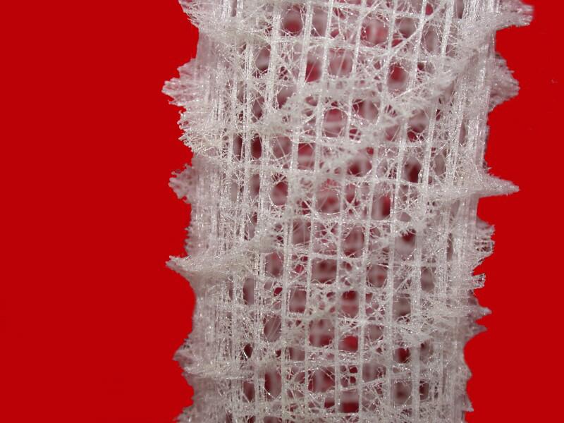
I am convinced that it is this remarkable arrangement of rings and ridges that provides Euplectella with its extraordinary structural flexibility and resilience.
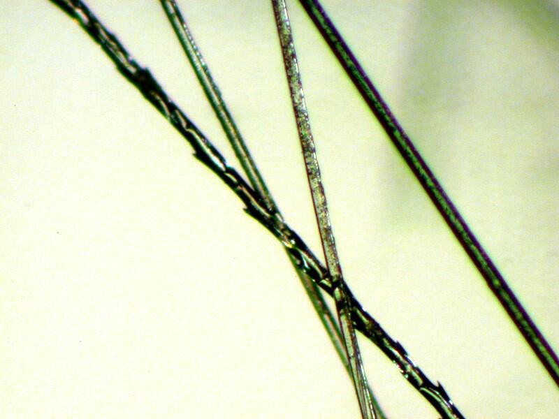
As we descend to the base, we find new puzzles. When I speak of the base, I am referring to the “mud ball” that seems to be the anchor for the organism. However, what then do we make of the series of long, clear individual silica strands emanating from the base?

My inclination is to regard these strands as anchoring devices rather like the roots of a tree but, that is by no means certain because, when one buys specimens, they have been commercially dredged, cleaned, and I suspect that these strands are then wound around the “mud ball” base for shipping to keep them from breaking off. To my naive mind, these strands, which, incredibly, are better than the fiber optics which we can produce at the moment, can reach a length of over 8 inches and are amazingly flexible, suggesting the possibility of yet another set of cells with yet another kind of programming directing them to produce these slender, glass fibers.
In addition, there is the mystery of how cells extract silica from sea water. Clearly, the ratio of silica to organic material is quite high, that is, the weight and volume of the skeleton significantly surpasses the cellular mass. Furthermore, even a cursory examination tells us that the center of the skeleton remains largely free and open which allows the cells to filter relatively large quantities of water and micro-plankton which is, of course, an advantage for the shrimp since this is their only source of food. (When I find a pair of blissfully imprisoned shrimp in a Euplectella, I will perhaps once again commit an act of vandalism to isolate them to try to rehydrate them with a mixture of water, glycerine, and alcohol and then examine them).
As far as spicules are concerned, I have thus far encountered almost exclusively hexactons and a scattering of monaxons and triaxons. However, given the way in which spicules are fused together in the structure of this skeleton, it is very difficult to figure out exactly what configurations are involved. Perhaps some soft X-ray radiography would be revealing but that is beyond my ken, my skills, and my budget.
It is, naturally, desirable to examine a good number of specimens in order to look for anomalies. If you have access to only a single specimen, it is possible even though somewhat unlikely, that you got a weird mutant form. This is not as impossible as one might think. Out of a dozen or so specimens I have examined, I found 2 in which there were distinct irregularities in the main body of the skeleton. It was as though Dali or Escher had programmed a few of the cells to create surreal architectural alterations in the usual beautifully symmetrical terraces and balconies that struck me immediately as out of place.
As science has progressed, we have discovered that genetic misinformation occurs much more often than previously thought. Human aberrations were often exploited in circus sideshow as were animals–two-headed calves or five-legged dogs. To human beings seeking order and rationality, these variants are disturbing, in part because they suggest a kind of unpredictability.
I am also curious about whether or not sponges which have not been cleaned have, as I think I already mentioned, some additional types of spicules which are embedded in the fabric of the cells. From the few accounts I have been able to find regarding the cleaning process, it is rather drastic. Initially the specimens are placed in a strong solution of sodium hypochlorite (bleach) which eliminates most of the organic material. Then, after rinsing–an important step to avoid poisoning by chlorine gas–the sponges are placed in a strong solution of nitric acid–in other words, the whole procedure is a rather nasty business. However, the current research techniques involved in order to study the ultrastructure of these spicules using Scanning Electron Microcopy (SEM) are even worse. The team headed by Johanna Aizenberger at the Wyss Institute at Harvard uses Hydrofluoric acid to etch the spicules thus eroding portions to give better access to parts of the segments to reveal their detailed structure. These results are impressive. You can find a number of images of close-up structure of Euplectella by following this website link.
However, an amateur should NEVER, NEVER, NEVER use Hydrofluoric acid!!! This acid is so reactive that it “eats” glass and the vapors violently attack mucous membranes so that even a small inhalation or spill on the skin can prove life-threatening. Even professional researchers must have careful training and the proper safety equipment to even consider using this extremely dangerous chemical.
Using these techniques and a variety of other approaches, what recent research has revealed is that the spicules of glass sponges have a variety of forms and, in some cases, are laminated or fused and, in other cases, are interconnected by networks of organic material rather than a direct fusion of the siliceous spicules.
Some accounts suggest another interesting possibility which, if true, adds yet another dimension of strangeness. A few investigators suggest that, depending on the size and age of the sponge, there may be a fundamental change in its structural material composition. The claim is that younger and smaller specimens tend to be more flexible and less brittle, whereas older and larger ones are likely to be more rigidly crystalline and as a consequence, more fragile and susceptible to damage. I might, I suppose, try to understand this in terms of an analog to the degradation of the calcium phosphate in the arthritic bones of my knees, hips, and back but even though I have a vast ignorance of the chemistry involved in biomineralization and degradation, nonetheless, my intuitions, my limited vandalism and my misguided egoism all conspire to make me somewhat skeptical about this particular claim. I’m sure that someone cleverer than I (with a lot of more sophisticated equipment) will one day solve this problem. An ordinary adult specimen is estimated to be about a foot in length including the slight curve of the body. The specimens I have range from 8 to 12 inches and the one I attacked so vigorously was about 10 inches long. With a structure of this design and composition, it is not difficult to accept a significant degree of variation of flexibility versus rigidity in different areas especially considering the proportion in specific segments of organic overlays and underpinnings in those areas. In the specimen I demolished, such variations were modest and, as I mentioned previously, the osculum remained intact. The central two-thirds of the body, though radically flattened, remained largely connected and flexible and the portion that seemed to be most susceptible was the lower part immediately above the “mud ball”.
What I want to try next on a new specimen, very carefully using micro-scissors and micro-forceps, is to attempt to work my way down from the top, first removing the osculum and then trying to isolate some of the rings which define the main cylinder of the skeleton and, at the same time, see if I can separate portions of the “V”-shaped spirals that wind their way down the outer part of the central tube. When I get down o the “mud ball”, I will have an investigative orgy and taxonomic nightmare and, even then, that’s not the end of it, for there still remain those mysterious thin glass fibers which so intrigue the material science researchers. The one thing I do know about these fibers it that some appear quite smooth and some possess tiny barbs.
As you have already guessed, I am at this point, sufficiently obsessed with these issues so that you will have several more essays from me on these subjects which you can quietly ignore.
All comments to the author Richard Howey are welcomed.
Editor's note: Visit Richard Howey's new website at http://rhowey.googlepages.com/home where he plans to share aspects of his wide interests.
Microscopy UK Front
Page
Micscape
Magazine
Article
Library
Please report any Web problems or offer general comments to the Micscape Editor .
Micscape is the on-line monthly magazine of the Microscopy UK website at Microscopy-UK .