HIGH RESOLUTION PHOTOGRAPHS
WITH CIRCULAR OBLIQUE LIGHTING.
By Yannis Tsamouris, ATHENS GREECE
My passion with microscopy started when I was at the first grade of high school and I looked at a microscope preparation of living protozoa during a biology class. From that time on I became obsessed with microscopy and I bought my first toy microscope soon after. It was a Japanese microscope with glass lenses and from the first moment I experimented to achieve a better picture with higher magnification trying to achieve what I was seeing in the pictures of my biology books. Eventually my experiments ended with a broken microscope thrown into the garbage.
I started again many
years after this wonderful experience when I had the spare
time and the money to become involved with more professional
equipment. eBay was a great help and for the last 15 years
I've been gathering a lot of hardware and experience of
manipulating this intricate machinery. I have to admit that
this magazine was a great inspiration to me and gave το me access to many aspects of amateur
microscopy and how to find practical solutions to problems
that any amateur microscopist faces every day in handling
these wonderful instruments. I experimented a lot with all the
current techniques like dark field, Rheinberg light staining,
Phase contrast and Differential Interference contrast (DIC).
One of the first accessories I acquired with a Leitz
Ortholux microscope was the Heine condenser. When I started
taking photographs with a CCD camera I observed that I could
take very clear pictures with high resolution. After many
trials I ended up with the following results that show the
hidden capabilities of circular oblique lighting that
according to my opinion is capable of providing high
resolution pictures without even immersion objectives. The
microscope I used is a modified Zeiss Photomicroscope III with
a Heine condenser adapted to it, a special illumination setup
made by me which can provide quite parallel rays of light and
a Zeiss Axiocam MRC5 CCD camera which gives superb results.
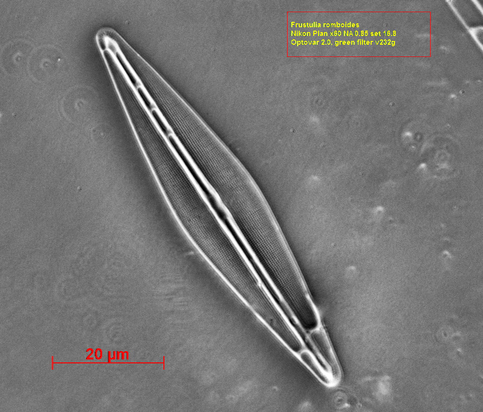
The photographs that follow show that with this arrangement
you can resolve even with a dry lens (Nikon Plan x 60 NA 0.85) the striae of Frustulia
rhomboides which according to D.B.Murphy (Fundamentals
of Light Microscopy pp 94) has a period of 0.29 μm/stria. According to theory this
resolution can be achieved only through oil immersion lenses.
The preparation I used is the 8 FORM TEST SLIDE and the 100 FORM SLIDE by K.D.KEMP
Frustulia rhomboides Nikon Plan X 60 NA 0.85 dry green filter
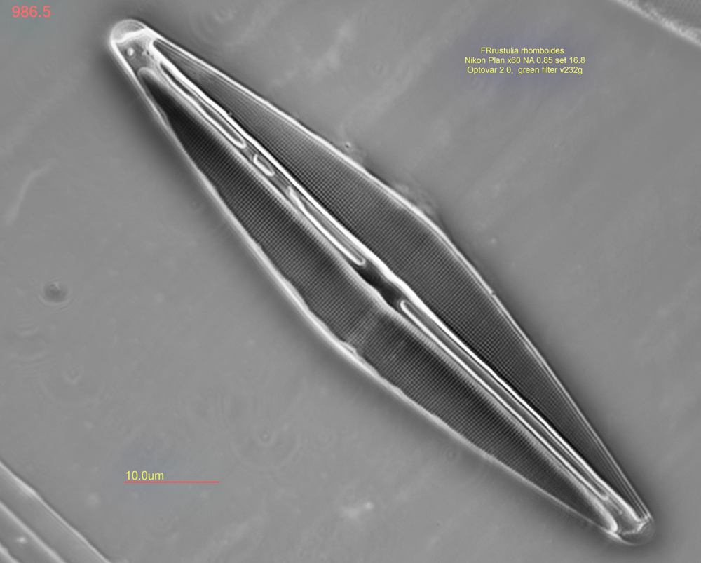
As you can see from this photograph,
although the resolution is inferior compared to the microscope live image, not only the striae
are resolved but also the pores. This is made more clear in
the following picture taken with a 15MB photographic camera (OMAX) and processed with the
Adobe Photoshop.
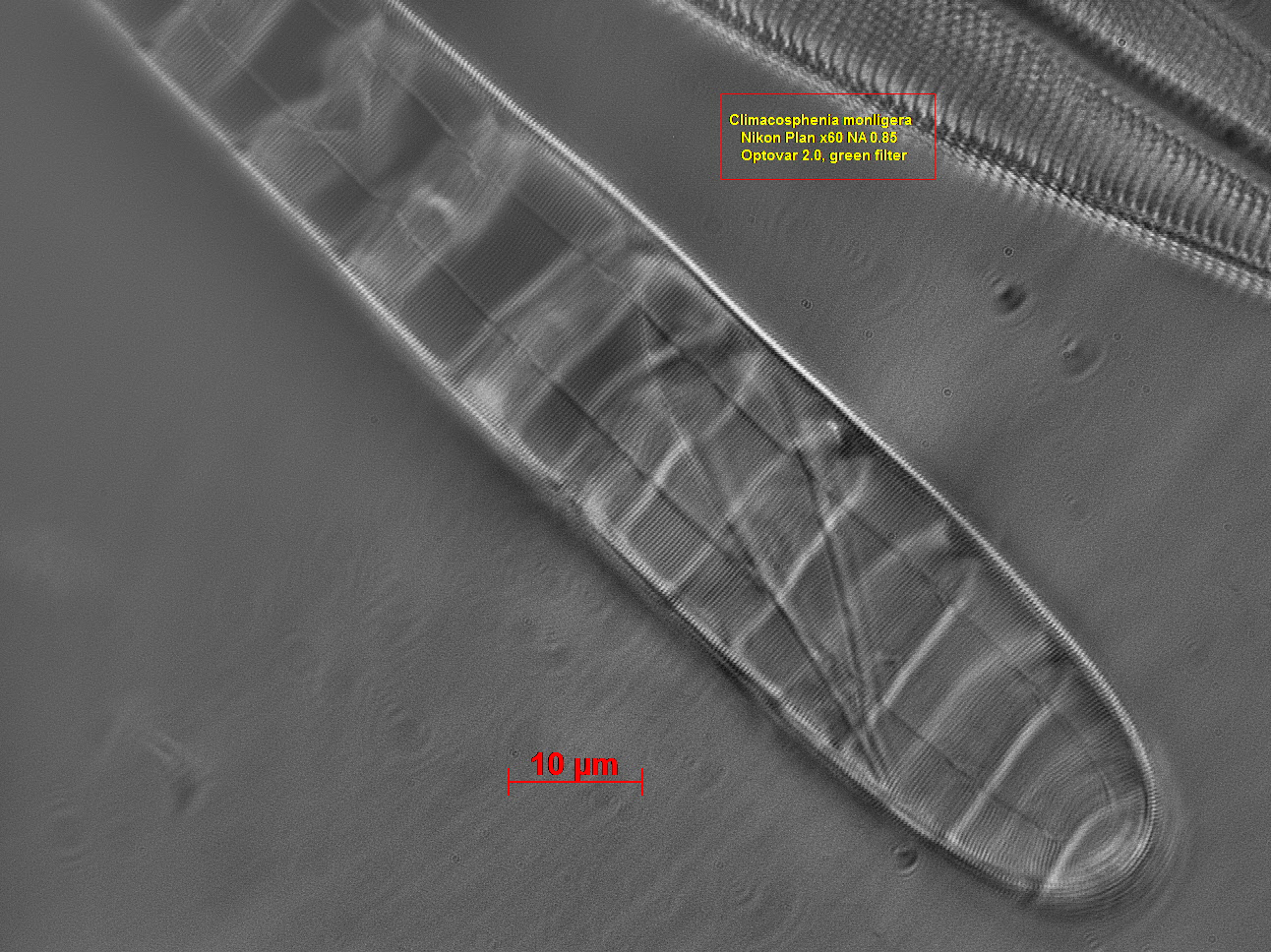
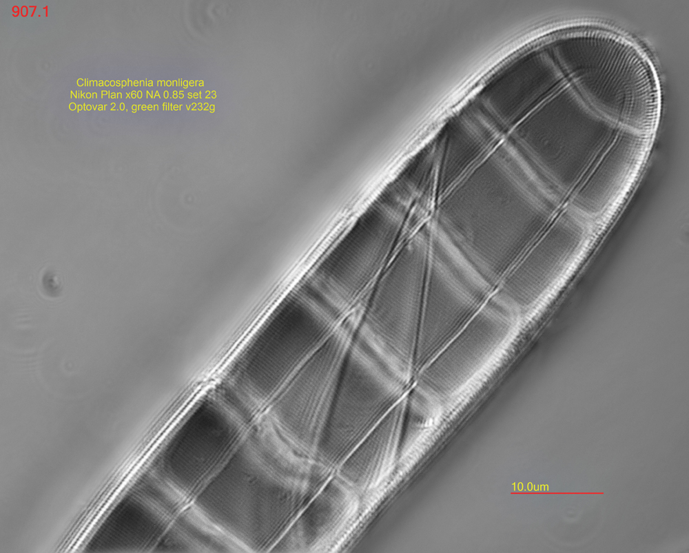
The most striking
photograph I achieved was the resolution of the individual
pores in the striae of the following diatom (Climacosphenia monligera) for which I
calculated three (3) striae per μm
and about four (4)
pores per μm. The microscope
image is very clear but the resolution of the
MRC5 camera (pixel 3.4 μm)
is not enough to capture such small details. The
sample is from Klaus Kemp 100 Type diatom slide.
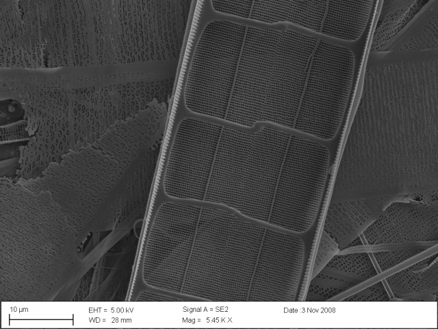
The SEM image which is hosted at Protist Central (photo credit Chris Lobban) shows a striae spacing of 0.40 microns and a pore spacing
of 0.33 microns.
With the same
technique and moderate optics the resolution and the depth of
field is great.
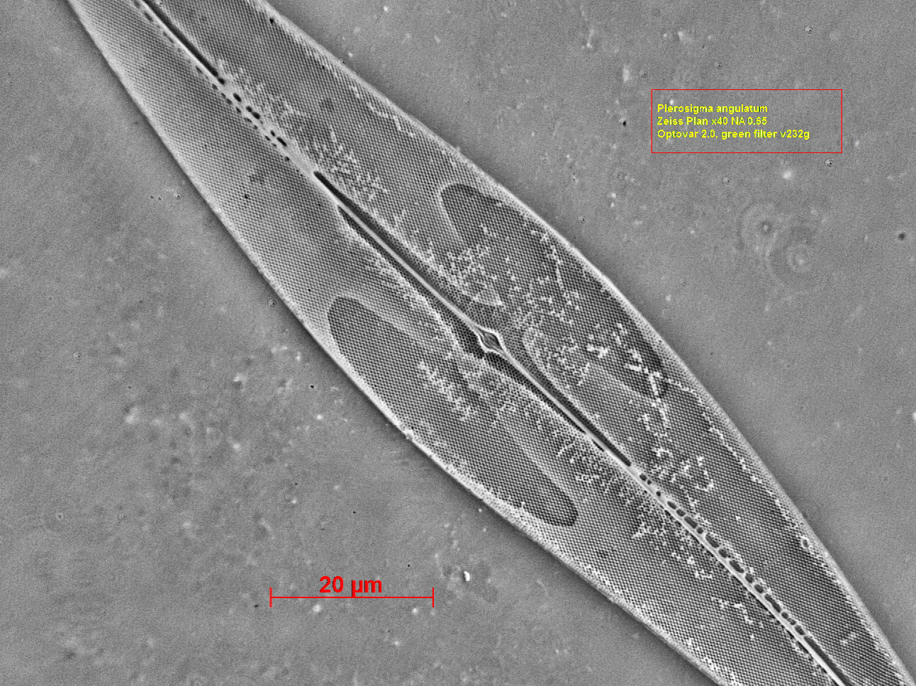
Zeiss Plan x 40 NA 0.65 dry, green filter. Pleurosigma Angulatum 0.53 μm / stria
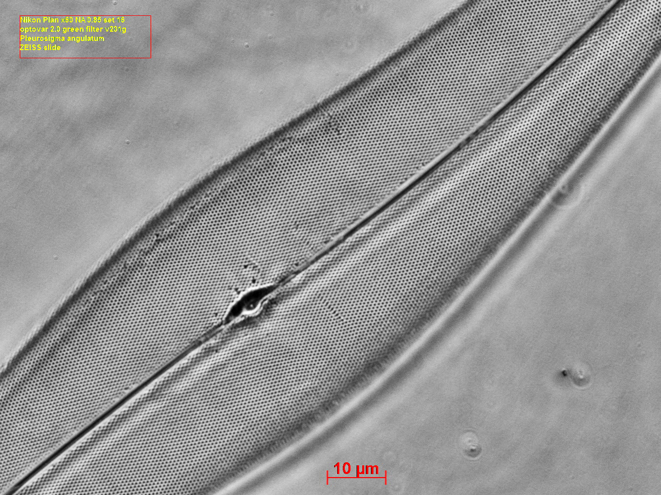
Pleurosigma angulatum Nikon Plan x
60 NA 0.85
With an oil immersion
objective the results are more striking.
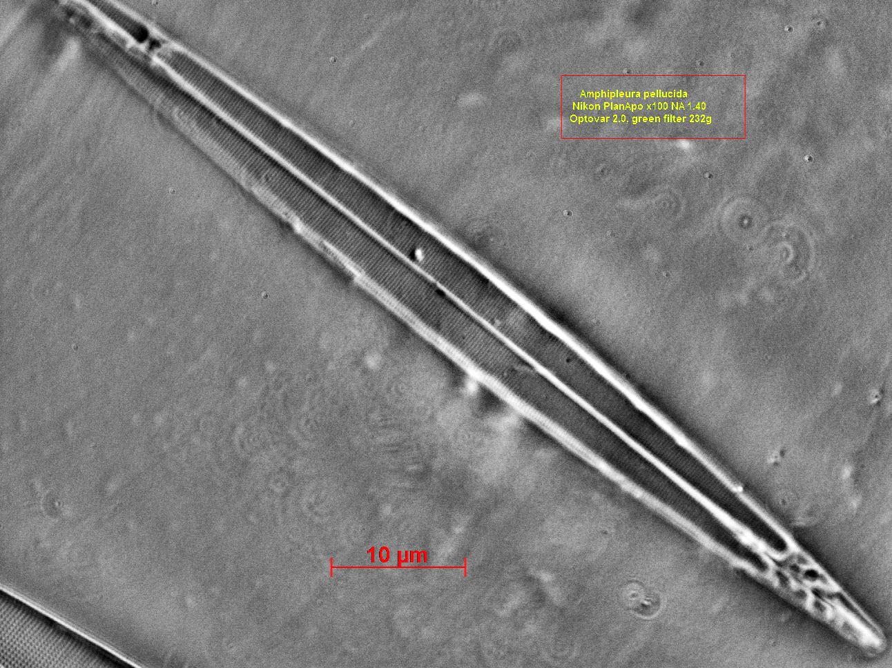
Nikon PlanApo X 100 NA 1.40 oil green filter. Amphipleura pellucida Period 0.25 μ/stria
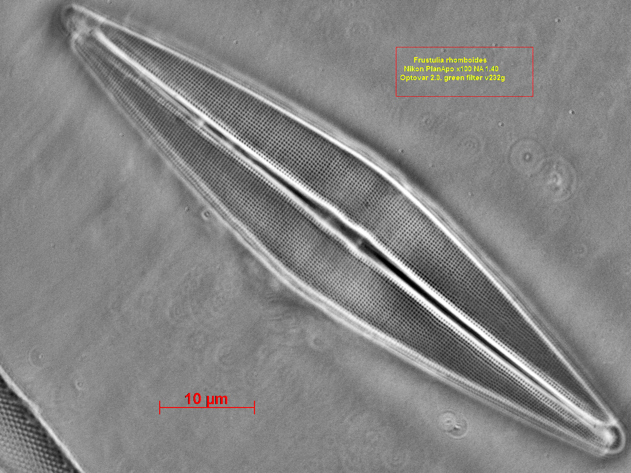
Nikon PlanApo x100 NA 1.40 oil, green
filter. Frustulia
rhomboides
Period 0.29 μm/stria
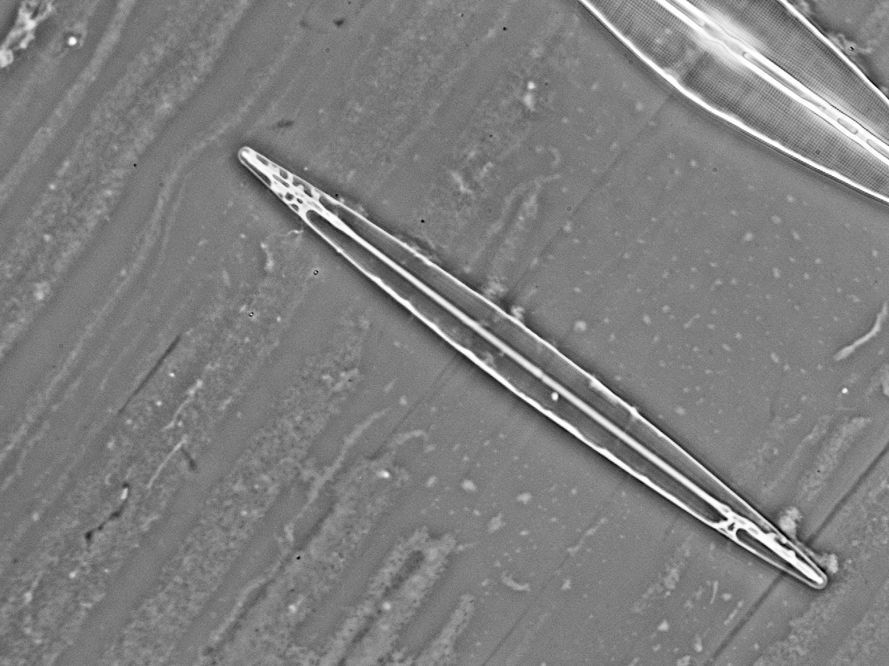
Nikon Plan Fluar X 40
NA 1.30 oil, green filter. Navicula hennedyi
Nikon PlanApo X 100
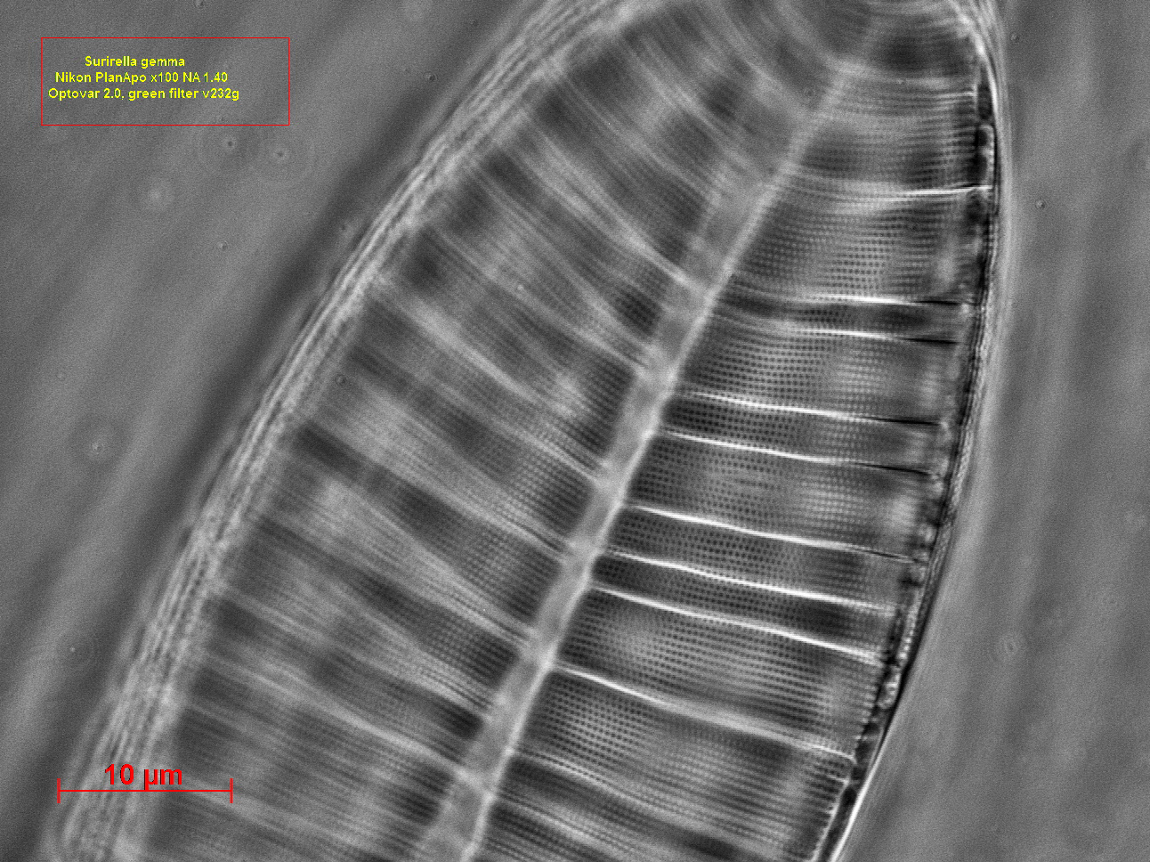
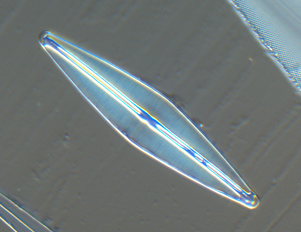
NA 1.40 oil green filter. Surirella gemma Period 0.50 μm / stria
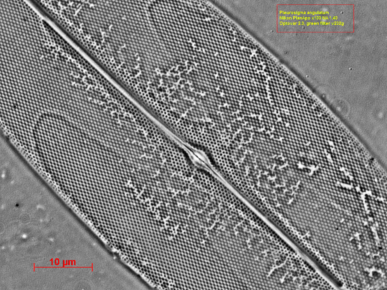
Nikon PlanApo x 100 NA 1.40
oil, green filter. Pleurosigma angulatum Period 0.50 μm/stria
If we compare the
above photographs with the results that we get with the
customary techniques of Phase contrast and DIC the results
speak for themselves.
Zeiss Phase PlanApo x
60 NA 1.40 oil green filter, Zeiss Phase condenser NA 1.40
oiled. Amphipleura
pellucida.
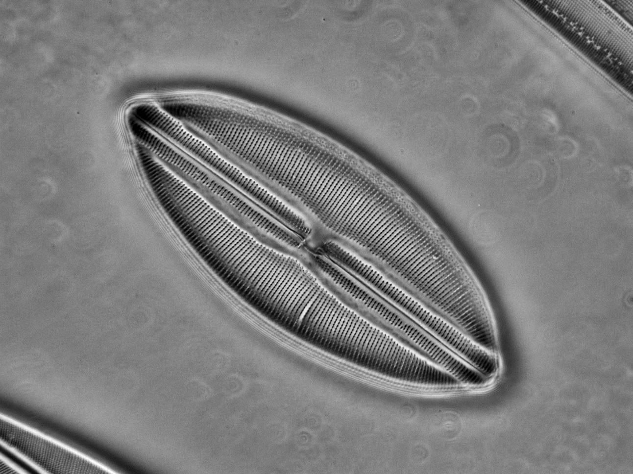
Zeiss PlanApo x 60 NA
1.40 with DIC slide, blue filter. Frustulia rhomboides.
Some more photographs
give us a glimpse to the beauty of the crystal palaces where
the diatoms live. (Olympus x 100 NA 1.30 oil).
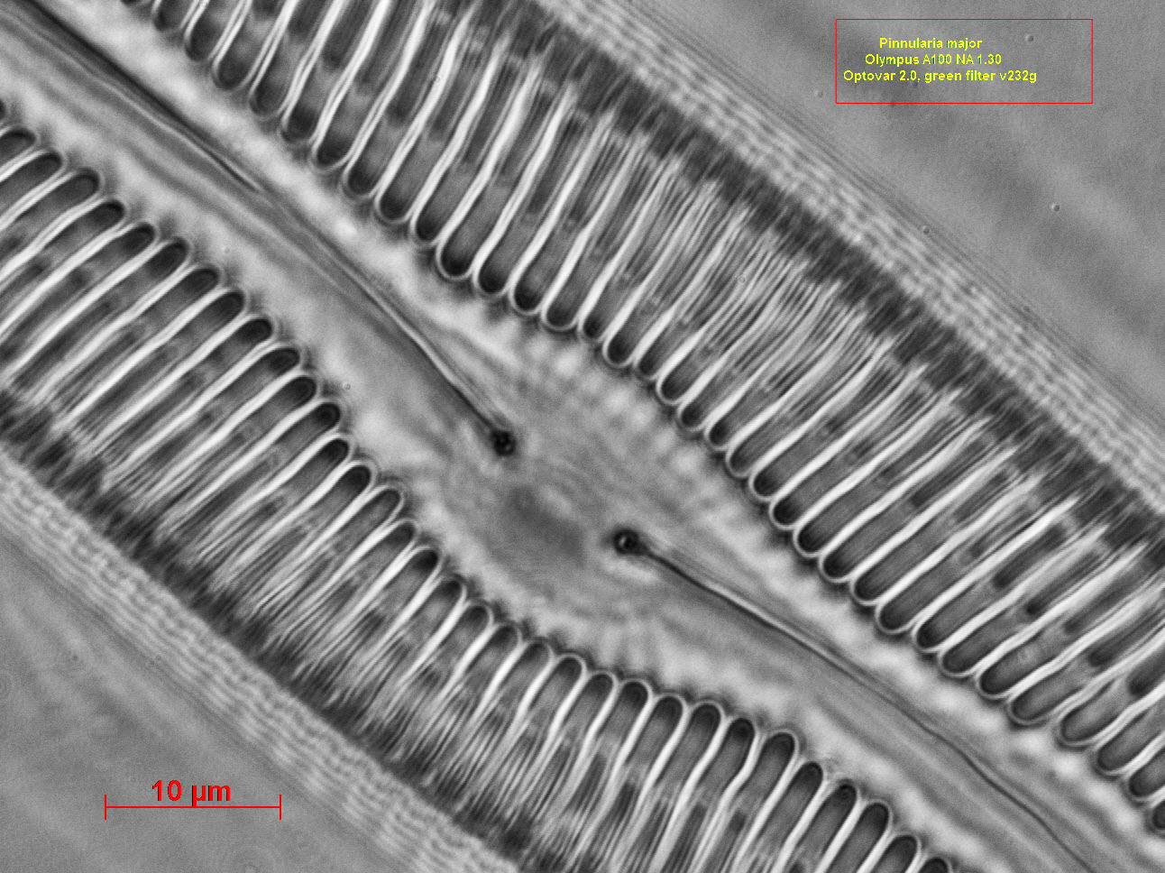
Pinnularia major
Olympus A100 NA 1.30 oil green filter
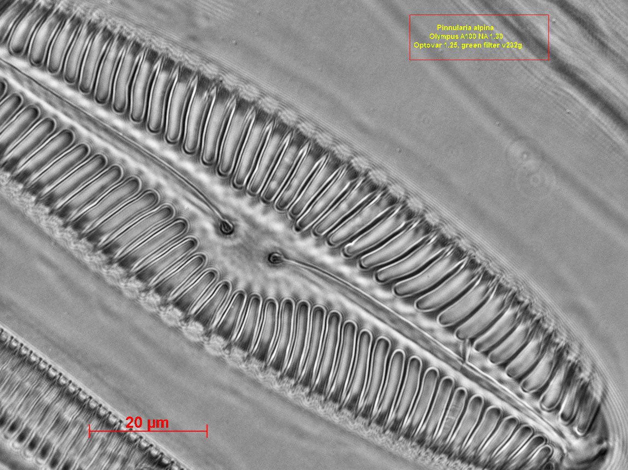
Pinnularia
alpina Olympus A100 NA 1.30 oil green filter
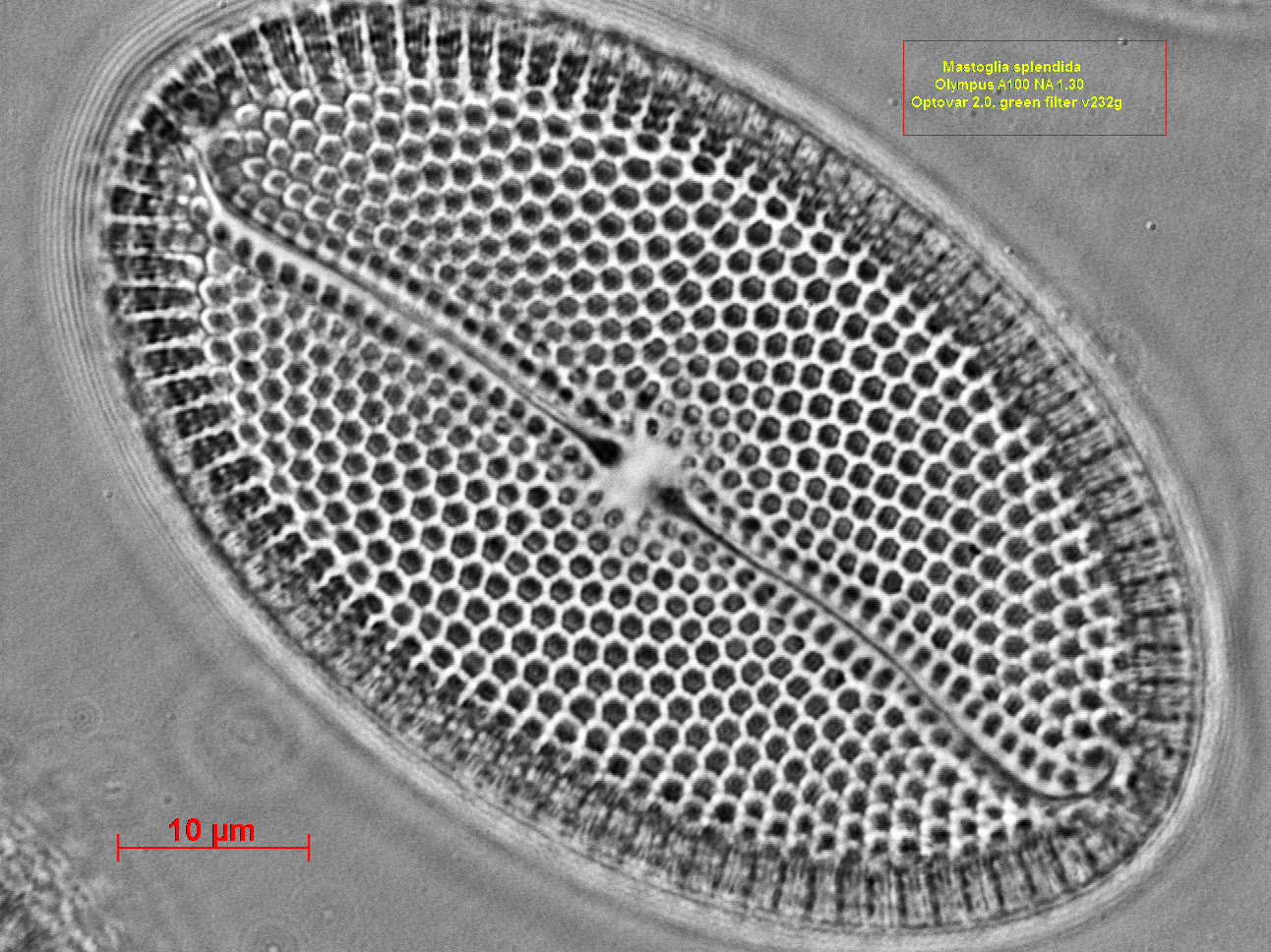
Mastoglia
splendida Olympus A100 NA 1.30 green filter
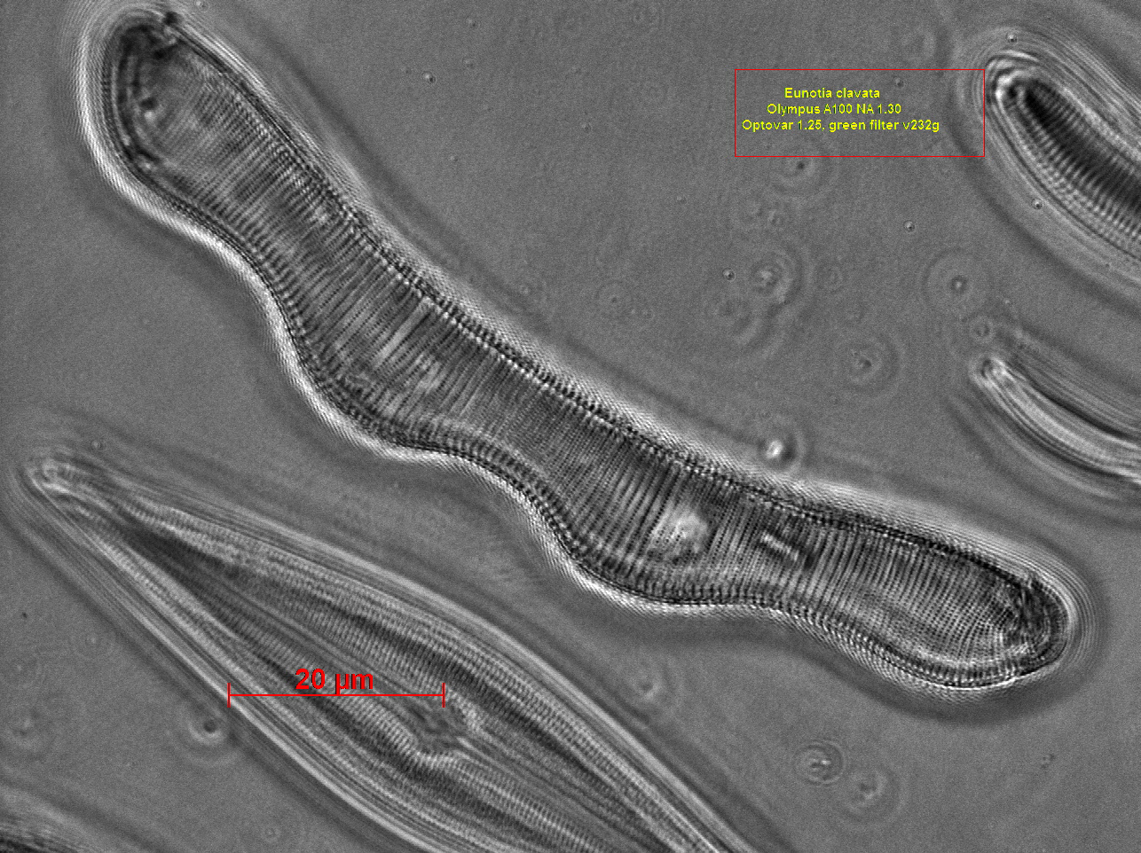
Eunotia clavata, Olympus A100
NA 1.30 oil, green filter v232g
If you study the photographs by zooming in you can appreciate the details they contain.
I dare to make the
assumption that in the light that comes out of the objective
lens after its interaction with the specimen there is a lot
more information than we are used to suppose. It’s up to us to
find the way to gather it. The COL provides an easy and cheap method to get the highest
possible resolution with great depth of
focus.
REFERENCE
Douglas B. Murphy “Fundamentals of light microscopy and electronic imaging”
A JOHN WILEY & SONS INC., PUBLICATION
All comments to the author are welcomed.
Microscopy
UK Front Page
Micscape
Magazine
Article
Library
Published in the February 2013 edition of Micscape Magazine.
Please report any Web problems or offer general comments to the Micscape Editor .
Micscape is the on-line monthly magazine of the Microscopy UK website at Microscopy-UK .
© Onview.net Ltd, Microscopy-UK, and all contributors 1995 onwards. All rightsreserved.
Main site is at www.microscopy-uk.org.uk .