|
Desiccated Stuff Part 2: More on Echinoderms by Richard L. Howey, Wyoming, USA |
Last time, we left off with echinoids (sea urchins) and there is much left to explore about them. In fact, we have barely scratched the surface. Recently I saw a scanning electron photomicrograph in a Zeiss catalog and it was an image of a tiny section of sea urchin test and the degree of fenestration was staggering. Swiss Cheese–eat your heart out!
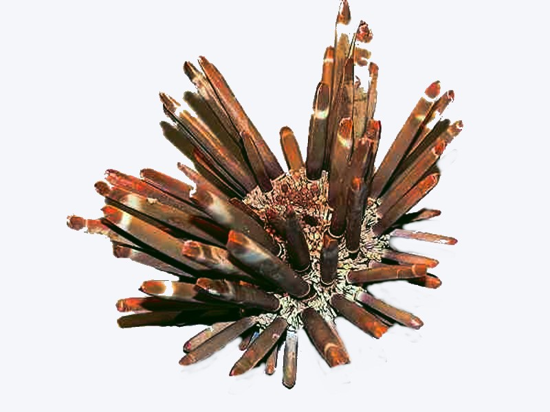
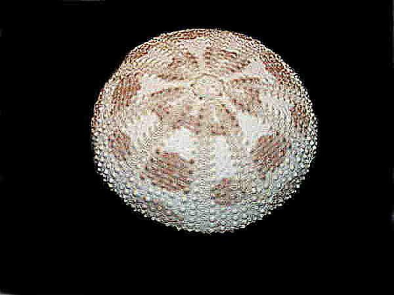
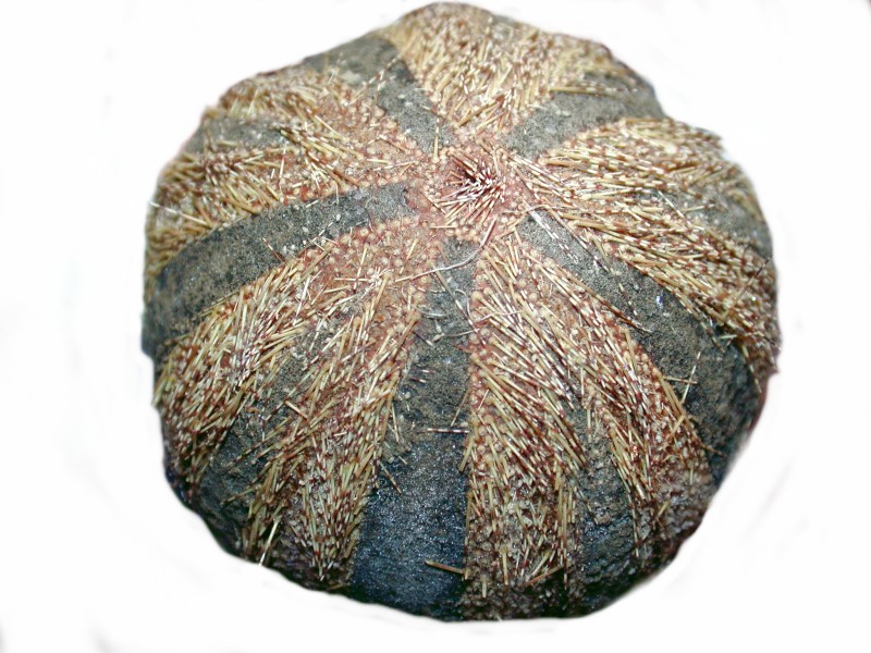
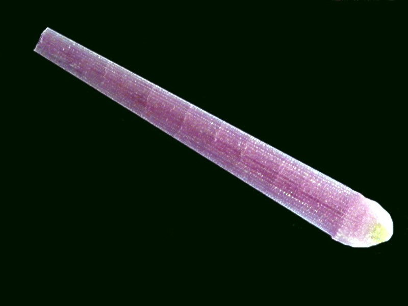
However, before we get to tests and the detailed structure of other structures, I want to show you a few more examples of complete dried urchins including one that might be called the “king of the echinoids”–Heterocentrotus mammilatus–a “slate pencil” urchin with thick spines (and remember that these are essentially chalk), which was used in that very way–as slate pencils in coastal school rooms. It’s real, it works, I’ve tried it.
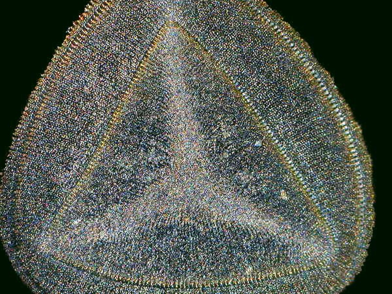
These very impressive spines, by the way, have an almost triangular cross section.
My personal advice regarding making such sections: DO NOT TRY THIS AT HOME! It is a documented fact that 97.3% of microscopists who tried to produce thin sections of sea urchin spines went insane. You might have to take that figure cum grano salis since all philosophers are liars and I am a philosopher, therefore I am telling the truth. Or consider Mark Twain’s remark (which was a popularization of Benjamin Disraeli’s phrase): “There are three kinds of lies: lies, damned lies, and statistics.” I’ve always loved to indulge in making up statistics–people are so deliciously gullible. However, in this case, there is no question that trying to produce such sections is enormously frustrating and requires saintly patience unless you either have an indentured lab assistant who enjoys drudge work or some very high-tech equipment.
It is astonishing how many extraordinarily fine slides of cross sections of sea urchin spines were produced in the last half of the 19th and the early 20th Century which might explain the great increases in the population of madhouses in England and Germany during that period.
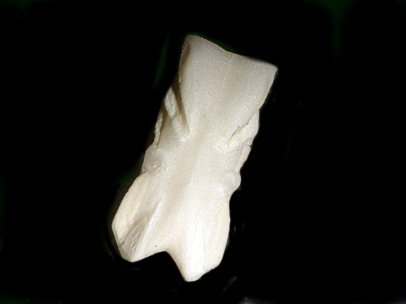
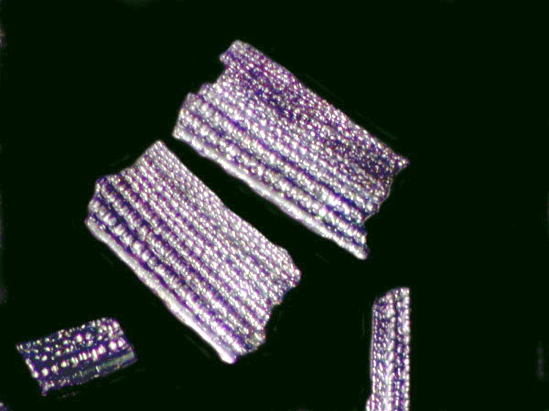
My few attempts ended in broken bits usually just when I thought I was reaching a point where I had a usable thin section. So, now I have come to a compromise when I want to look at the internal structure of spines; I break them into very small pieces, select out the most promising, polish them a little with an emery board, give them a quick bath in diluted acid, rinse and examine. Below is an example of such a preparation of a spine of Strongylocentrotus purpuratus.
For truly fine thin sections, I now rely on eBay and occasionally I even come across a few I can afford and manage to snag them and I’d like to share them with you–by way of images, that is,–you’re NOT getting the slides.

While we’re looking at neat pictures, let’s look at some sea urchin tests since we’ve been talking about them off and on.
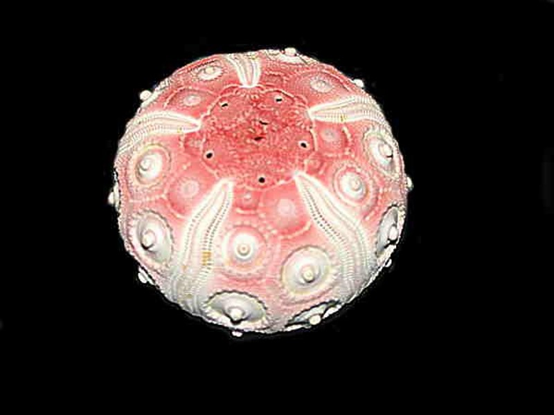
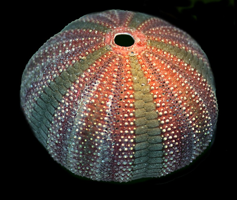
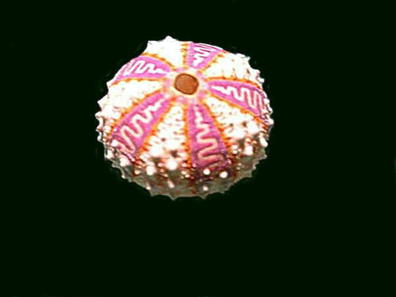
The tests pose many puzzles, posers, and conundrums like the spines. How are they constructed? Are there specialized cells involved in the deposition of the calcareous crystals? Clearly, there must be different sets of genetic coding between species to produce such radically different tests.
Let’s start by taking a closeup look at a small section of the test of a Colobocentrotus. The first image shows 5 small, flat, perforated plates attached to a larger plate.
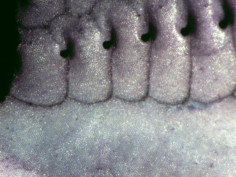
I don’t know why but, for some reason, they remind me of the foot pedals of a pipe organ.
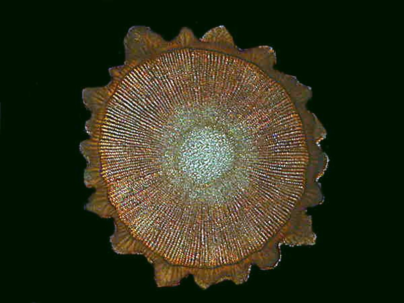
I’ll show you a closeup of the smaller plates. Here we get a clear indication of their crystalline structure from the way the light glistens off the surface.
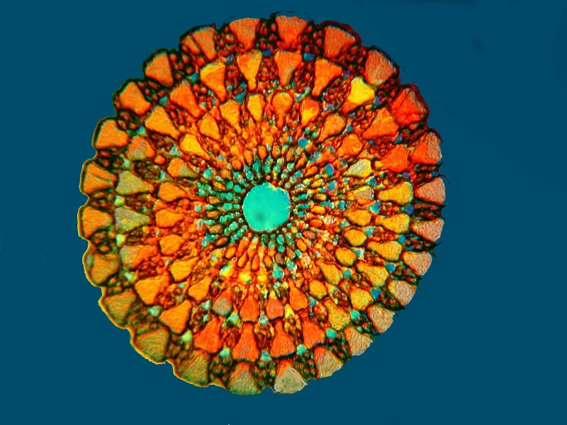
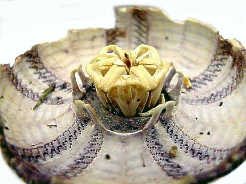
To give you an idea of what an incredible engineering project a sea urchin is I’ll show you a picture of the bottom of a test which I cut open–another act of Rampant Vandalism just to satisfy your insatiable curiosity (and mine own, of course). It is justified by the fact that it opens up a whole new set of perspectives.
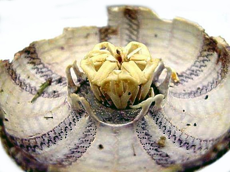
In the center, we can see the altar to Poseidon where all the cells come to worship. Just kidding. Actually, it’s much more interesting than that. That structure is known as Aristotle’s Lantern and the ancient Greek philosopher was the first to describe it over 2,000 years ago. Like you, Aristotle was possessed by boundless curiosity and wrote extensively on Natural History.
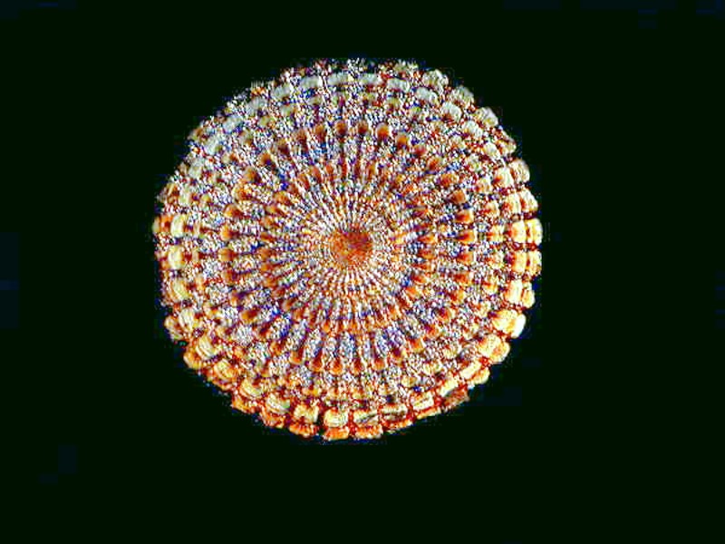
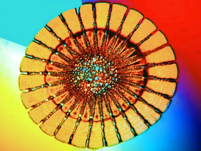
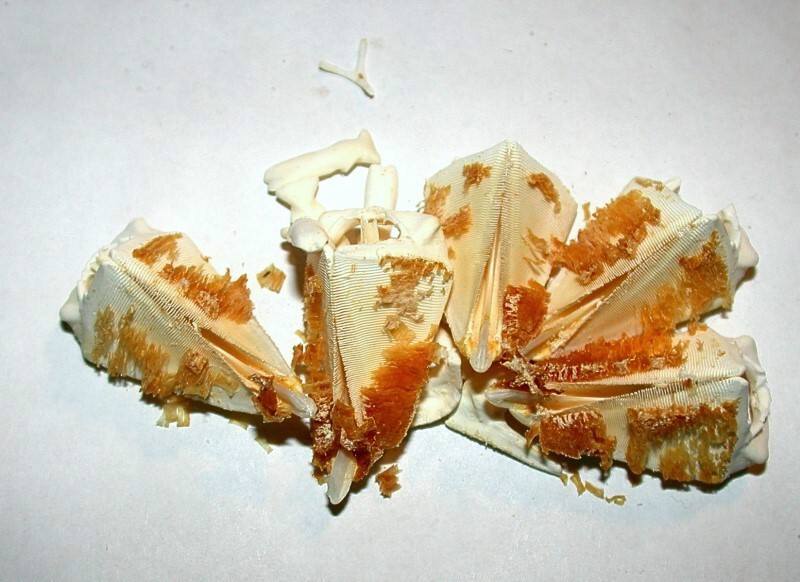
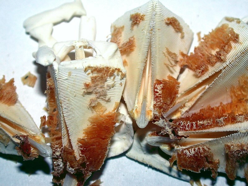
And here are 2 images of a lantern which has been isolated. It is from an urchin of the genus Toxopneustes.
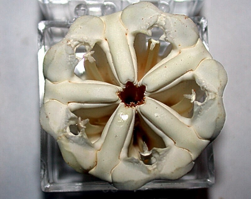
This apparatus consists of five teeth and a series of plates, supports, and cross bars which comprise the lantern–usually about 40 pieces. Below are 2 images of the pieces of a disarticulated lantern from Toxopneustes.
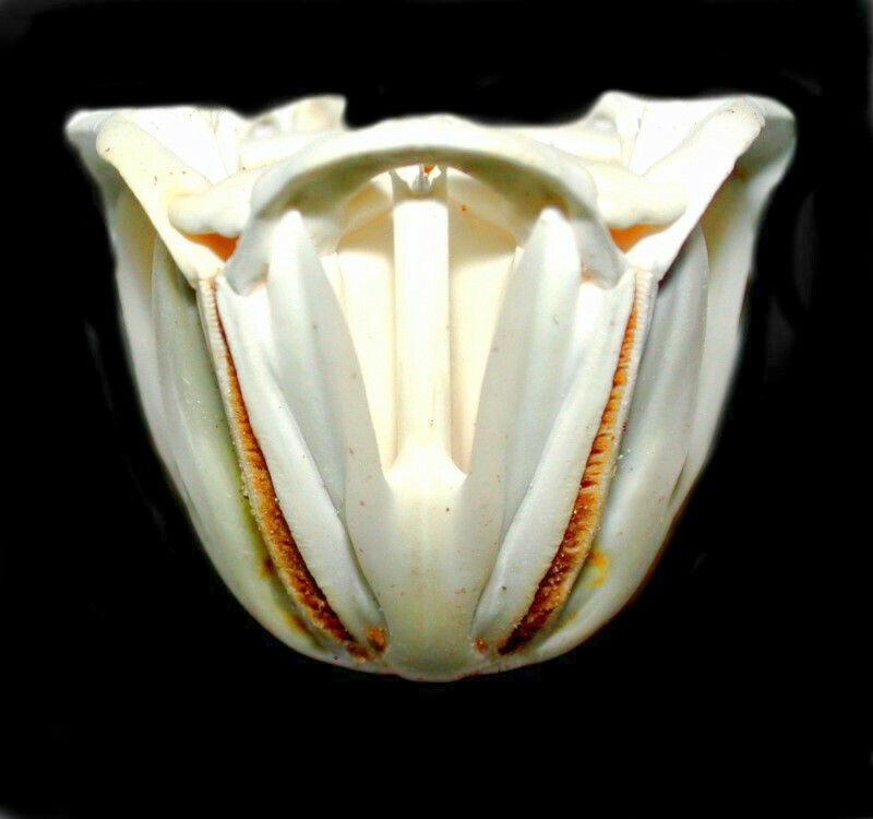
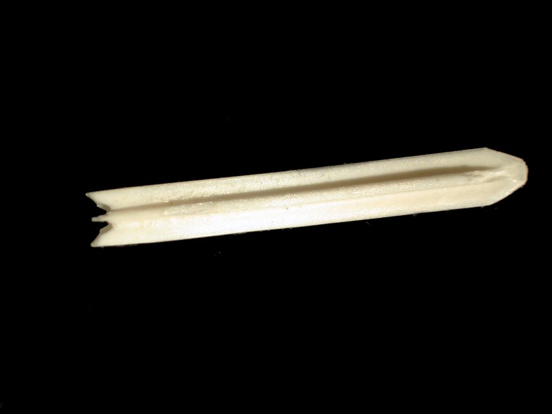
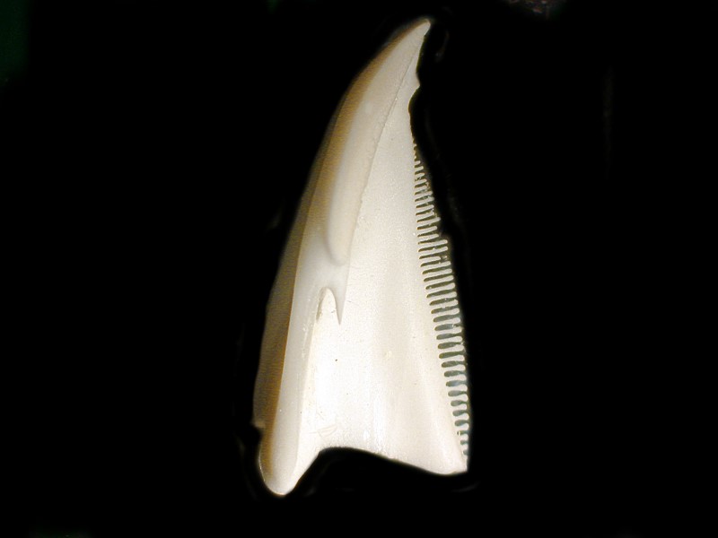
Next, I’ll show you a tooth and 3 major types of components of a lantern from Strongylocentrotus purpuratus. First, one of the teeth.
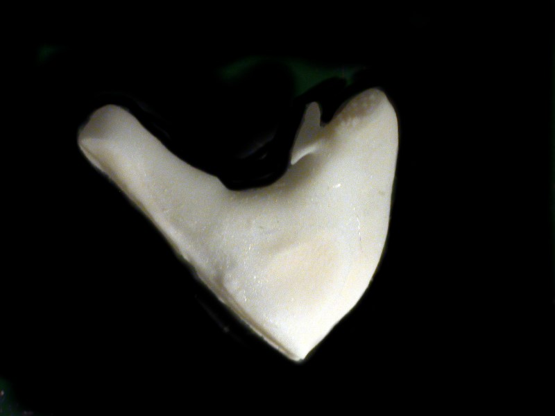
There are 4 other usual types of structures: pyramids, epiphyses, auricles, and rotules. Here are examples of 3 of them.
These can be arranged on a piece of stiff black card putting the matching structures in rows. This makes an informative and attractive display.
In the first image of the lantern, you can get a sense for how it extends through the test and the ends of the teeth are visible from the other sides. Going back to the original image of the lantern on the half shell, we can also see part of the rows of perforations through which the tube feet extend.
A few spines almost always break off under the rigors of being shipped and I always save them and set them aside. Later, I sort through them and select a suitable variety and mount them in a Riker box.
Spines vary widely in size and construction.
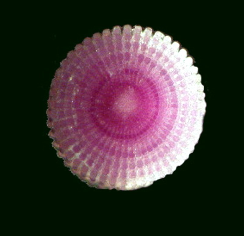
Here are some of the tiny spines on the test of a Mespilia, that little “Christmas” urchin that we looked at in Part 1. Note that they are banded with alternating pale green and red. The reason for such coloration is unknown.
Coelopleurus tends to have long, bowed spines that are 4 or 5 times the length of the diameter of the test and even more remarkable has tube feet that can extend out even beyond the tips of the spines.
Strongylocentrotus franciscanus, along with a number of other species, has a fascinating arrangement of crystals along the outer surface of the spines. The crystals are arranged in such a way that their tips all point to the top end of the spine. As a consequence, if you slide your thumb and finger from the base to the tip, your fingers move smoothly. However, if you reverse direction, you will find yourself unable to move down the spine due to the tips of the crystals impeding such motion. The outer part of the spines is like a long stack of tiny crowns gradually diminishing in size as they near the tip.
Some members of the genus Diadema have spines slightly over a foot long which have poison glands near the tip, and which can, when triggered, produce a toxin which is excruciatingly painful to humans who are unfortunate enough to have a close encounter of the spine kind.
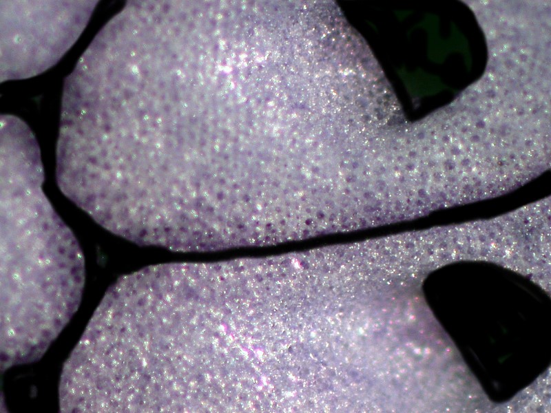
If we look closely at a portion of a spine from Strongylocentrotus purpuratus, we can learn more about the structure of the spine. There are very thin lines of crystals running from the base up toward the terminus, but even more interesting is the fact that the spine consists of a series of thick disks piled on one another rather like the construction of a Greek column.
If we take a tiny piece of this spine and crush it, we can get a glimpse into the way the crystals are aligned.
Habitat can play an important role in terms of the characteristics of spines in some genera. Colobocentrotus, for example, tends to reside in coastal areas where there is heavy surf action. As a consequence, the spines are greatly flattened which prevents the urchin from being easily dislodged by the pounding surf. This modification is a significant one and shows up dramatically.
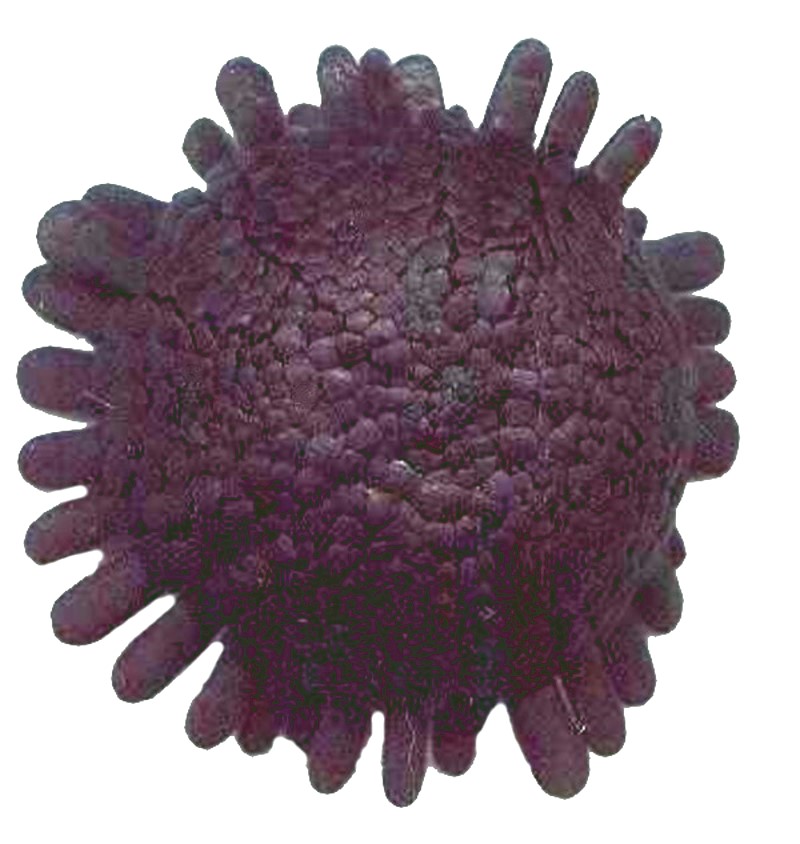
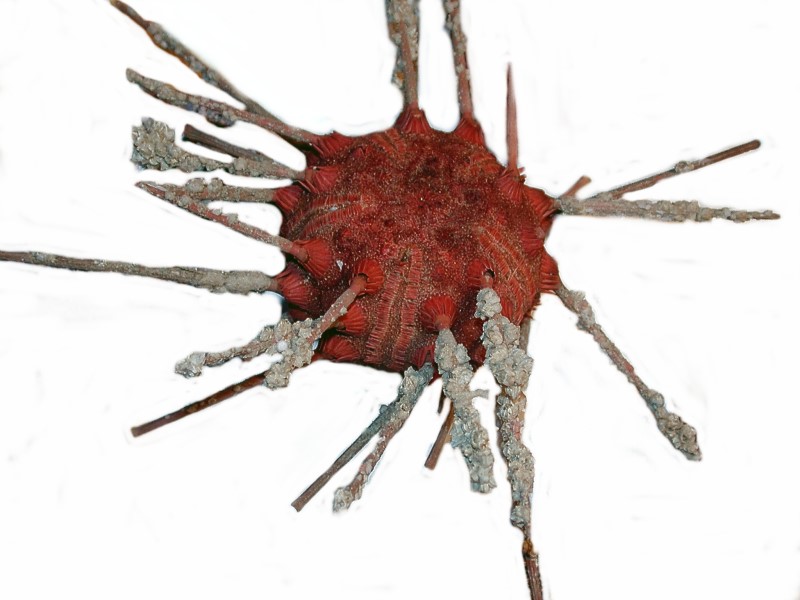
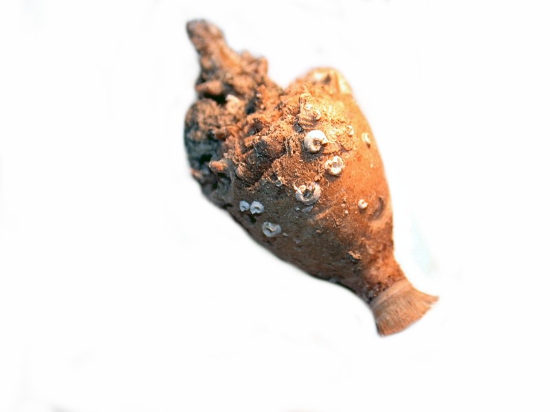
Another attractive and highly interesting echinoid is Stereocidaris granularis var. rubra. Notice the incredible number of “hitchhikers” which have attached to the spines.
I have been haunted by those bizarre spines of Psychocidaris and so I have decided to soon return to them and break 1 or 2 apart to see what I can discover. I’ll try to remember to let you know what I find. In the meantime, here is a close up of a single spine which looks rather like an ancient urn pulled out of an archaeological dig.
This essay is getting a bit long, so some other wonders such as pedicellaria and sphaeridia, along with the whole range of irregular urchins, will have to wait until Part 3.
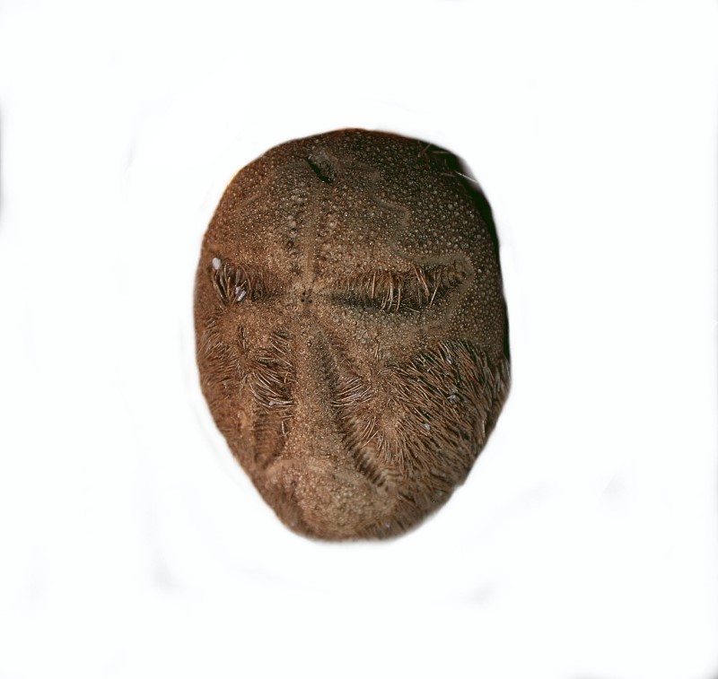
However, just to show you how much fun irregular urchins can be, let me show you an image which with its 2 “eyes” and “extended tongue” could be an African mask of comedy.
All comments to the author Richard Howey are welcomed.
Editor's note: Visit Richard Howey's new website at http://rhowey.googlepages.com/home where he plans to share aspects of his wide interests.
Microscopy UK Front
Page
Micscape
Magazine
Article
Library
Published in the February 2017 edition of Micscape Magazine.
Please report any Web problems or offer general comments to the Micscape Editor .
Micscape is the on-line monthly magazine of the Microscopy UK website at Microscopy-UK .
©
Onview.net Ltd, Microscopy-UK, and all contributors 1995
onwards. All rights reserved.
Main site is at
www.microscopy-uk.org.uk .