|
Further microscopy on stamps - electron microscopes.
compiled by David Walker, UK
|
Updated Mar. 11th 2019 with new references.
The number of stamps and philately related items featuring some aspect of microscopy, whether microscopes, microscopic subjects, microscopists are now into the many hundreds, so collectors of this thematic like myself often select small themes within the topic. See the Resources section below for topics that have been previously shared.
Seeking a new thematic to collect, electron microscopes piqued my interest as the Nobel Prize for Chemistry in 2017 was awarded to three scientists for their work on cryo-electron microscopy where new issues to celebrate the winners are guaranteed by some countries. Electron microscopes are a well established sub-theme in microscopy thematics. William Wergin in his seminal five part series of colour illustrated articles in the Proceedings of the RMS in 1990 had an attractive plate showing a selection of seven with discussion (1). The late Bill Heathwood catalogued eight in his list of all stamps depicting some form of microscope in 1997 (2). More recently, Thomas P Johnston notes on his webpage that he writes a regular column for Health Physics News entitled A Philatelic Look at Health Physics History. His article on electron microscopes shows eight stamps with an EM in the design.
Update Mar. 11th 2019. Thank you to John Delly who kindly let me know of and sent copies of two illustrated articles that he had published showing microscopes on stamps in 1976 and 1978. See refs. 3 and 4.
Those shown below are based on both Wergin's and Heathwood's listings plus more recent issues found by searching delcampe.net, a major dealer portal for thematics. I would be interested to hear from readers who are aware of others and also if readers recognise a model shown as some are very detailed. The gallery does not include stamps where only photographs taken using an EM form part of the design. This would expand the thematic considerably and could form a sub-theme in their own right. In-line links are to relevant Wikipedia articles unless stated otherwise.
I've never been fortunate to be instructed on and operate an electron microscope, either scanning or transmitted but was indirectly familiar with their capabilities. As a chemist working in the petrochemicals industry involved with new heterogeneous catalyst and adsorbent development, electron microscopy (TEM / SEM) was an invaluable service provided by the on-site analytical division to help characterise their structure, morphology and elemental analysis of selected areas using EDAX. I enjoyed sitting in with the microscopist as they explained features of note on samples submitted.
|
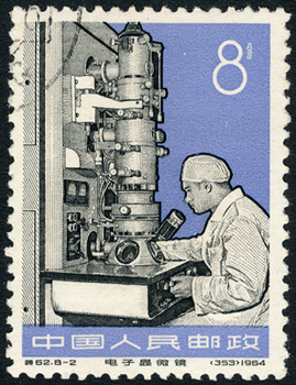
China 1966. Wergin notes that this was the first stamp to depict an EM. It is also one of my favourites because of the fine engraving detail. It is one of four celebrating what the dealer notes as 'new industrial machinery'.
|
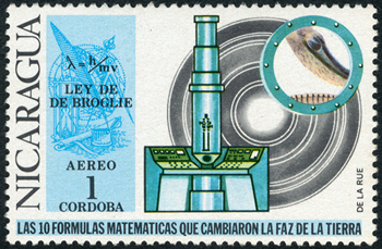
Nicaragua 1971. This forms part of a very popular set of ten where each features a famous maths formula with a related design. De Broglie's wave equation is shown (with TEM and electron diffraction pattern) and is key to the principle of an EM. The electron can act as a wave as well as a particle but with much smaller wavelengths than light they can resolve far beyond a light microscope.
|
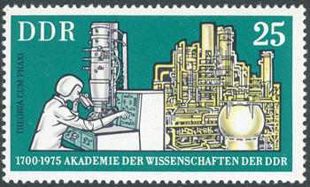
Germany 1975. An electron microscopist (with TEM) and a chemical plant. It was one of four showing different designs to celebrate the 275th anniversary of the German Academy of Sciences.
|
|
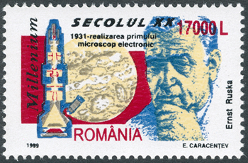
Romania 1999. Design credit E. Caracentev. This stamp celebrates the work of Ernst Ruska who is credited with the design of the first EM. He was awarded half of the Nobel Prize for Physics in 1986 for his work on electron optics.
|
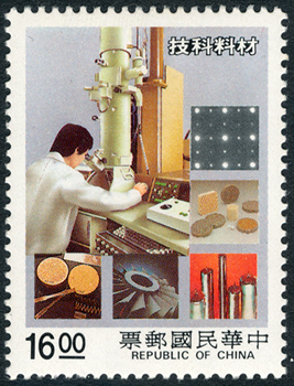
China 1988. The dealer's description notes that this celebrates material science and shows a model in use with typical subjects likely to be studied inset.
|
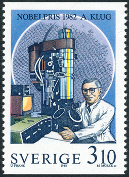
Sweden 1988. A stamp celebrating the work of Aaron Klug a British biophysicist (Lithuanian born) who won the 1982 Nobel Prize in chemistry "for his development of crystallographic electron microscopy and his
structural elucidation of biologically important nucleic acid-protein
complexes."
|
|
|
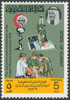
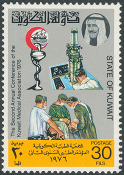
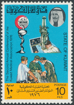
Kuwait 1976. A set of three celebrating the 'second annual conference of the Kuwait Medical Association'. The EM reflects the importance of the microscope in medicine for both research and diagnostics.
|
|
|
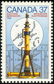
Canada 1988. Electron microscope 1938. One of a set of four for Canada Day featuring inventions with dates, the others were Marquis wheat, kerosene and cobalt therapy. A Google search for this microscope reveals that it was the first made in Canada by two physics graduates at the University of Toronto. Ari Gross has a fascinating illustrated page describing the EM's design and history. He notes that it is currently at the Canada Science and Technology Museum and that at the time it had the finest performance with a mag of 20 000X and resolution of ca. 140 Å.
|
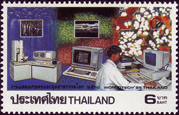
Thailand 1995. Celebrating 'World Tech 95'. One of a set of four featuring different designs.
|
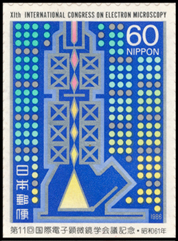
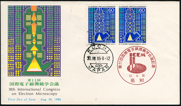
Japan 1986. Celebrating the XIth International Congress on Electron Microscopy (ICEM). An attractive stylised design of the electron beam path and its focussing.
Some attractive covers were also issued with further EM designs on the covers, see below.
|
|
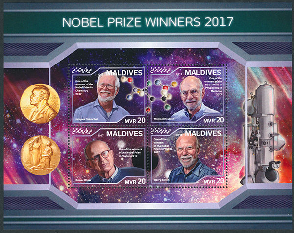
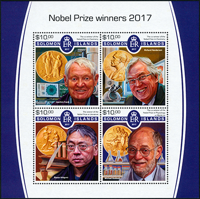
Nobel Prize for Chemistry 2017. The twelve stamps shown above are to date all that have been previously catalogued plus those issued since lists published, pending further finds. The Nobel Prize for Chemistry in 2017 (link to Nobel Prize site) has increased that by at least six and are shown below.
The Prize was awarded jointly to Jacques Dubochet (University of Lausanne, Switzerland), Joachim Frank (Columbia University, NY,USA) and Richard Henderson (MRC Laboratory of Molecular Biology, UK) "for developing cryo-electron microscopy for the high-resolution structure
determination of biomolecules in solution."
The Nobel site has some splendid documents that describe the background and techniques both for a popular readership and more in depth.
|
Some small countries and states are noted for their prolific output of thematics on many subjects including the Nobel Prizes. Such stamp issues can be a rather thorny topic in philately circles as the subjects have no relation to the countries. Each collector is advised in publications to make their own mind up of what to collect. My own approach is that if it is a very narrow thematic with few examples, as here, they can be a valued part of a collection. They are often my favourites as the mini-sheet format when chosen allows more detailed and informative designs. Some of the other narrow themes I collect would not exist without these attractive issues from smaller countries.
Prizes are announced in early October of each year so the associated stamp issues are usually early in the next year although some countries publish promptly in the award year.
|
|
Maldives. One of two. A modern EM features in the sheet's background design.
|
|
|

|
Maldives. Two of two. An attractive mini-sheet with an EM in the background and a modest stereo microscope at the other extreme of mag (and cost!) in the foreground.
Mini-sheets allow more extensive design features to be added beyond what can be fitted into a typical stamp size.
|
|
|
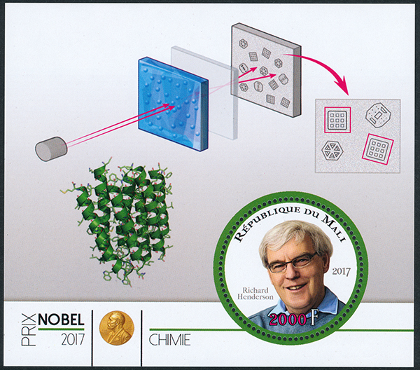
Mali. One of three. In recent years Mali has adopted the sheet style shown for a variety of thematics. Two of the three do not show an EM but do show aspects of the prize winners' work so included for completeness. The designs are often based on figures from the Nobel's detailed publications. Although some aspects of this design do not feature.
I haven't yet sourced the upper design feature. The lower is based on Figure 3 from the 'Popular Science' document with the caption: "In 1990, Henderson presented a bacteriorhodopsin structure at atomic resolution."
|
|
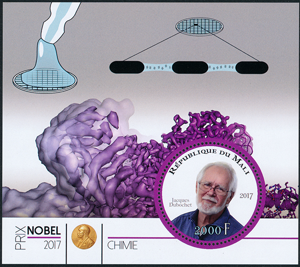
Mali. Two of three. The upper sheet design shows part of Figure 5 from the 'Popular Science' document showing "Dubochet's Vitrification Method".
The lower design is from Figure 9 in the 'Scientific Background' document and shows "The resolution progression of cryo-EM" using glutamate dehydrogenase as an example. The resolution at right is noted to be 1.8 Å.
A key aspect of the power of cryo-EM is the ability to instantly freeze aqueous based samples so that the water adopts a vitrified state. If the water turned to ice its structure shown by the electron beam would confuse that of the subject image.
|
|
|
|
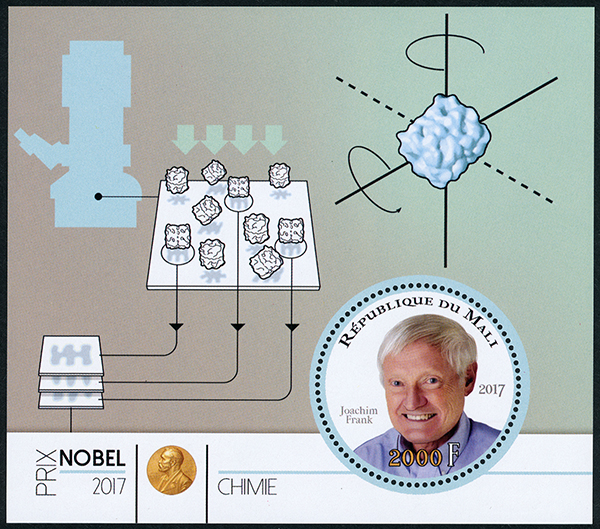
Mali 2017. Three of three. The sheet design reproduces part of Figure in the Nobel 'Popular Science' document. It shows "Frank's Image Analysis for 3D Structures".
|
|
|
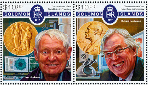
Solomon Islands. Left and detail above. EMs are shown in two of the stamps.
|
|
|
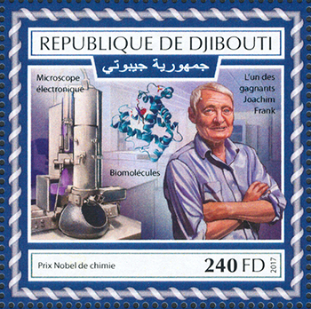
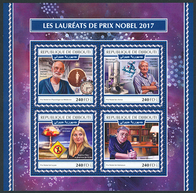
Republic of Djibouti. Left and detail above.
|
|
Covers - the remaining images are examples of covers or postcards where an EM features in the design on the cover (known as the cachet). This collection are those found to date and could be many more.
|
|
|
|
Japan 1986. An attractive cover to accompany the stamp described above.
|
|
|
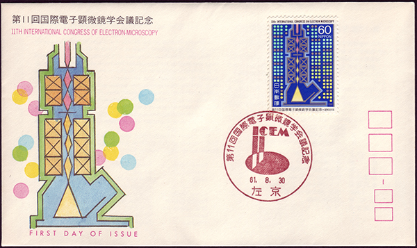
Japan 1986. Another attractive cover to accompany the stamp described above.
|
|
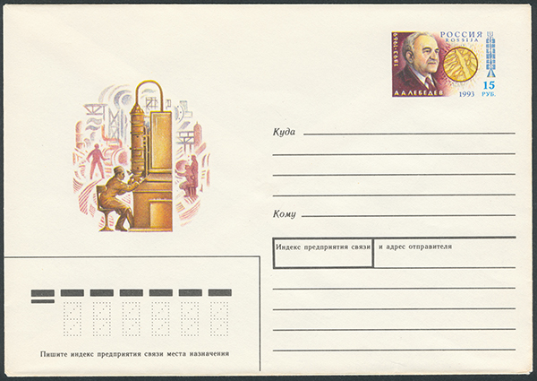
Russia 1993. A preprinted envelope, the stamp showing the Russian physicist A A Lebedev (1893 -1969) and electron beam diagram.
|
|
|
|
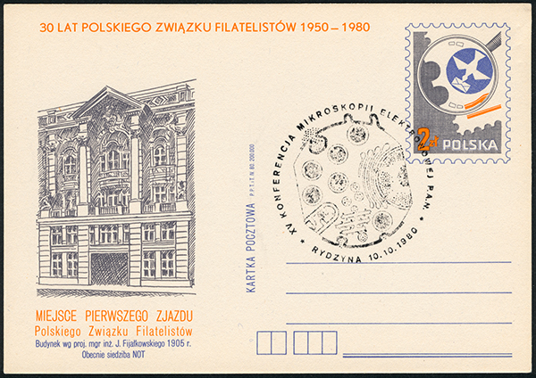
Poland 1980. Although I remarked that electron micrographs as the sole feature on designs were not included, I make an exception as was intrigued that in this example it formed the postmark design. It shows the classic cell internal structure by TEM and notes that it celebrates the XVth conference on electron microscopy in Rydzyna.
|
Comments to the compiler are welcomed.
Micscape Resources
Micscape articles on microscopes / microscopy on stamps.
Selected topics in microscopy, photomicrography, diatoms, radiolaria, Van Leeuwenhoek / Robert Hooke, marine plankton / oceanography and further photomicrography by
David Walker.
Microscopes
and Stamps by Fritz Schulze.
Acknowledgements
The basis of the collection above relied on the published illustrated gallery of EMs by William Wergin and lists by Bill Heathwood (references below) and extended with recent issues.
External resources
1) 'Microscopes and Postage Stamps' by William P. Wergin, Proceedings of
the Royal Microscopical Society (now published as 'infocus Magazine'), 1990,
volume 25, pages 115-121, 212-214, 249-253, 324-327, 416-422. A series of
definitive articles and coloured plates. The last set of pages is a list of
stamps depicting microscopes up to August 1990. The list includes the country,
date of issue and the Scott catalogue number. Also provides references to
articles published prior to 1990 on similar topics. The Electron Microscopes plate and description feature in part 1, p.116.
2) 'Microscopes on Stamps: A Checklist to 1996' by W Heathwood. Quekett
Journal of Microscopy, 1997, 38(1), pp. 37-47. This invaluable list provides the
SG catalogue number, the face value, year of issue and a brief description of
the stamp. Useful guidelines on starting a collection are also provided in the
accompanying article. The now sadly late Bill Heathwood kindly sent me an updated checklist to June 1997 and exchanged some enjoyable letters on a shared interest.
3) 'Microscopes on Stamps' by John Delly, The Microscope, 1976, vol. 24, 279-290.
4) 'Microscopes on Stamps - Part 2' by John Delly, The Microscope, 1978, vol. 26, 97.
Image acknowledgements. The copyright of the stamps and envelope designs remain with the postal issuing authorities and/or the designers and have been credited where known. They are presented here at a limited size solely as an educational resource on this
not-for-profit website. If a copyright holder wishes to contact the author please use the email above. Thank you.
Sourcing stamps. For readers interested in seeking out one or more of the stamps on the above or other topics, one of the best portals for thematics is delcampe.net. Catalogue numbers aren't needed and often not stated. Searching for country with year and keyword of theme can usually show all the dealers offering them. Many dealers are now aware that EMs are a thematic and often included in the auction title. Where not
offered on delcampe.net, eBay is the second choice.
©
Microscopy UK or their contributors.
Published
in the February 2019 edition of Micscape.
Please
report any Web problems or offer general comments to
the
Micscape
Editor
.
Micscape
is the on-line monthly magazine of the Microscopy UK web
site at
Microscopy-UK
©
Onview.net Ltd, Microscopy-UK, and all contributors 1995
onwards. All rights reserved.
Main site is at
www.microscopy-uk.org.uk.