|
WHAT SEX ARE YOU? |
||
|
We seem to be living in an age where sexual identity has become confused in the young generation. I have no doubt that there are genuine cases both born out of either physchological issues, or growing up in a social system where everything is being questioned down to a miniscule level. And then there are the more scientific or biological issues where a baby is born with both sets of sexual organs complete: male & female. In these
cases,
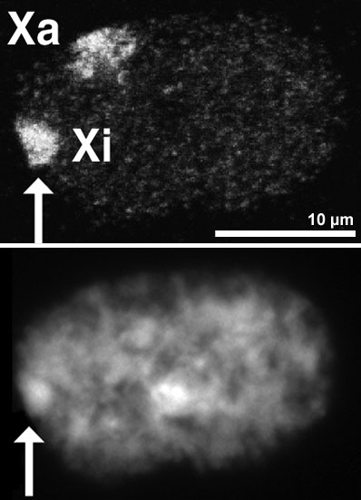
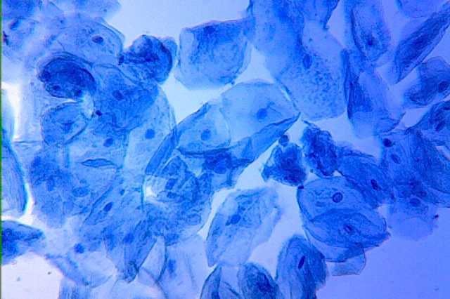
what does a doctor, or surgeon do to determine the sexual identity of the new born person? This is where microscopy comes in! It might be interesting to discover what the deciding factor is as it comes down to genetics: the machinery of creating life. Somatic cells are any cell in the body that are not gametes (sperm or egg), germ cells (cells that go on to become gametes), or stem cells. Essentially, all cells that make up an organism’s body and are not used to directly form a new organism during reproduction are somatic cells. The word somatic comes from the Greek word σὠμα (soma), which means body. In the human body, there are about 220 types of somatic cells. There are many different kinds of somatic cells in the human body because nearly every cell found inside and on the surface of the human body, with the exception of cells that become sperm and eggs, is a somatic cell. In addition, mammals have many organ systems that specialize in specific functions, so there are many different specialized cells. Cheek Cells are epithelial cells serve a similar function to all epithelium, which is as a lining and barrier separating the underlying tissues from the body's cavities and the external world. These cells also secrete mucus that adds protection, lubrication and a barrier against drying out. Some epithelial cells have secretory, gas and nutrient transport, absorptive functions or mechanical movement facilitated by cilia. But the simple squamous epithelium is primarily a barrier to infection. Look at you own cheek cells? |
 |
|
|
Human Cheek Cells - stained |
||
|
|
||
|
|
||
 |
||
|
Cheek cells stained with iodine |
||
|
Microscopy UK Front
Page © Microscopy UK or their contributors.Published in the February 2020 edition of Micscape Magazine. Please report any Web problems or offer general comments to the Micscape Editor . Micscape is the on-line monthly magazine of the Microscopy UK website at Microscopy-UK . ©
Onview.net Ltd, Microscopy-UK, and all contributors 1995
onwards. All rights reserved.
|
||