N.A. 0.10 ![]()
![]()
![]() 3370mm X 2250mm.
3370mm X 2250mm.
N.A. 0.25 ![]()
![]()
![]() 1350mm X
1350mm X ![]() 900mm.
900mm.
N.A. 0.65 ![]()
![]()
![]()
![]()
![]() 337mm X 225mm.
337mm X 225mm.
N.A. 1.25 ![]()
![]()
![]()
![]()
![]() 135mm X 90mm.
(oil-immersion).
135mm X 90mm.
(oil-immersion).
THE
EXAMINATION OF MICROFOSSILS, NANNOFOSSILS AND OTHER MICROSCOPICAL
OBJECTS USING CELLULOSE LACQUER ROCK PEELS.
BY KEITH W. ABINERI, UK.
42
West Borough, Wimborne, Dorset BH21 1NQ, UK
Tel. 01202 885547
Introduction.
During the mid l980's an amateur technique was developed by the
writer to prepare cellulose lacquer peels from samples of
Mesozoic sedimentary rock collected along the Dorset coast. This
simple technique was based on the use of a proprietary rapid
drying cellulose lacquer ('Humbrol' nitrate cellulose dope for
model aircraft). The lacquer was applied to a small prepared area
of rock surface, using a clean small nylon artists' paint brush,
with a self-adhesive paper label, cut in the form of a small
frame. The frame was used to remove the film from the rock after
drying for about 1 hour at 30° - 40°C. The "flow"
properties of the lacquer were found to be crucial for the
best results.
The lacquer formed a tough smooth film which could be mounted on
a microscope slide under a glass cover-slip. These mounted peels
were only about 1 cm2 in area and were found to be
crowded with minute detail, in some examples down to <1 micron
i.e.<1/1000 mm. in diameter. By sealing the mounted microscope
slides carefully with shellac it was possible to use the high
power oil-immersion objective for close examination of the peels.
With my Nikon microscope, using the binocular eyepiece, a visual
magnification of circa. X 1500 was achieved under these
conditions.
The simple peeling of the tough dried lacquer film from the rock
formed, either accurate replicas of the surface minute features,
or, in many cases, removed thin layers of softer sediment from
the rock surface. Both these processes could occur, even on the
same peel, and, of course, depended on the nature of the rock
sample, as well as its ingredients. Excellent examples of these
varying results were obtained from the many Dorset Kimmeridge
Clay beds, where the peels showed numerous minute marine
nannofossils (coccoliths and coccolithophores), a variety of
microfossils (dinoflagellate cysts, calcispheres etc.), plant
microspores (pollen grains), microscopic grains of pyrites,
layers of kerogen with buried fossil wood fragments (Mesozoic
conifer fusain) etc. All this tends to confirm the variable
influx of land swamp debris to the sea at the time of deposition
of the Kimmeridgian marine microplankton.
More recently, in the late 1980's, stained cellulose lacquer
peels were prepared by combining the basic method, described
above, with a simple well-known staining procedure for carbonate
rocks. (See Adams, MacKenzie & Guilford, 1984.). This method
can distinguish between carbonate rocks with regard to
composition and texture, and produce individual differential
staining of microfossils and nannofossils, on a minute scale,
helping in their detection. This technique will be discussed
later in the series of articles.
The six accompanied illustrations show a selection of unstained
peels with differing magnifications, as examples of what can be
seen with an optical microscope, using the basic technique only.
The Visual Microscopy and Photomicrography of Cellulose
Lacquer Rock Peels
I have been especially fortunate in possessing a very fine well
equipped Nikon "Labophot" microscope, which has been
the main instrument used for the examination of my slides with
the mounted peels. Nevertheless I have used an old Leitz
microscope and more recently a low power Meiji stereoscopic
instrument for detecting the larger objects on the peels. Any
students' microscope giving a combined magnification of X 500 to
X 600 would be suitable for much of the observation, discussed
here. However the advantages of my Nikon are considerable for
both visual and photographic work and include the following:
I). A trinocular eyepiece tube type F. This allows 100% of the light to be diverted from the binocular eyepiece to the camera, with a quick simple movement.
II). A range of objectives, N.A. 0.1, 0.25, 0.65 and 1.25 (oil-immersion), giving visual magnifications of X 60, X 150, X 600 and X 1500 respectively with the binocular eyepiece
( X 15).
III). Nikon FE camera body with connecting tube and mounting, fitted with a projection lens.
IV). Phase contrast turret condenser with additional positions for brightfield and darkfield microscopy. Facilities for the use of polarizing plates.
V). Illumination brightness control, field diaphragm control, condenser aperture diaphragm control and a calibrated mechanical stage.
VI). A range of filters for visual and photographic observations.VII). Calibrated eyepiece micrometers.
VIII). Coarse and fine focusing controls.
The main film used has been Fujichrome DX ASA 100, to produce 35 mm colour slides. The approximate fields covered by these slides with the various objectives were as follows:-
N.A. 0.10

N.A. 0.25
N.A. 0.65
N.A. 1.25
A day-light correcting filter was used generally
with photography with the illumination brightness control at
maximum. The Nikon camera was used with automatic exposure.
Obviously all these extra facilities have added to my own
enjoyment and gradual increasing understanding of the wonderful
microscopic world of micropalaeontology, with all that this tells
us of the Earth's history. I must, however, point out that a good
quality students' microscope, used with care and ingenuity will
show much of the detail discussed here, when applied to these
cellulose lacquer rock peels.
Examples of Unstained Lacquer Peels. i.e. Figures (1) to (6).
| Figure (1).
Pyrites microcrystals from the Lower Jurassic Belemnite
Marls. Lower Lias. Map reference : SY.380.927 East of
Charmouth. Dorset coast. Objective N.A. = 0.1. Total field area of the image = ca. 2140 micron X 1440 micron. |
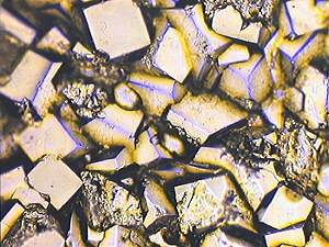 |
Figure (1) is mainly a replica type image, because of the hard nature of the pyrites microcrystals. Figure (1) exhibits brightfield illumination.
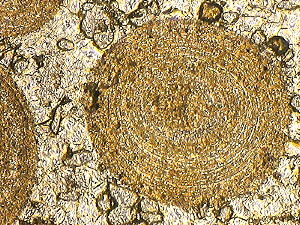
| Figure (2).
Oolith sections in the Upper Jurassic, Upper Corallian,
Osmington Oolite Limestone. Map Reference : SY.742.814.
Bran Point near Osmington, Dorset
coast. Objective N.A = 0.1. Total field area of the image = ca. 2140 micron X 1460 micron. |
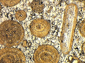 |
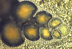
Figure (2) is an example of both replica and removed thin layer type of images. The diameters of the oolith sections range from circa. 290 microns to 930 microns on this peel. Mainly they show the characteristic fine layered structure, due to the gradual growth of these spherical mineral calcite grains by the slow action of regular gentle movement in water, which contains some soluble calcium bicarbonate, which produces layers of insoluble calcium carbonate with the liberation of carbon dioxide. This situation is associated with lagoon conditions. The growth of these ooliths may start around a nucleus of a fine grain of sand, or quartz, or fragment of shell, or other organic debris. Figure (2) shows brightfield illumination.
Figure (3). This shows an enlarged picture from the same peel used with Figure (2) but with phase contrast illumination. Here the objective N.A. = 0.25 and the total field area of the image = ca. 880 micron X 640 micron. The phase contrast image accentuates the fine layered structure. The real diameter of the main oolith section is ca. 605 microns on Figure (3).
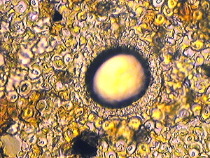
Figure (4). A calcisphere embedded in a layer of coccoliths from the Upper Jurassic. Upper Kimmeridge Clay, Rope Lake Head Stone Band, Map Reference : SY.926.775. Rope Lake Head, East of Kimmeridge, Dorset coast. Oil-immersion objective N.A. = 1.25. Total field area of the image ca. 120 microns X 80 microns.
Figure (4) is an example of both replica and removed thin layer type of images. Clearly it is a high resolution cellulose lacquer peel. (Limiting resolution <1 micron.) The real diameter of the calcisphere, including its complex outer structure is circa. 37 microns. The real diameters of the background coccoliths range from circa. 3 microns to 7 microns. The numerous coccoliths were derived from the surfaces of unicellular marine plants (phytoplankton) known as coccolithophores, or coccospheres. The coccoliths are minute calcite plates of variable crystalline structure and show characteristic optical figures when observed with crossed polarized plates. They are classified as calcareous nannofossils and are widely distributed, with marked fluctuations and variations in form, during the last 200 million years of Earth history.
The Rope Lake Head coccolithic limestone band appears to mark one of a number of intense periods of coccolithophore marine algae "blooms", which occurred in Kimmeridgian times. Clearly good SEM pictures are really needed here. Figure (4) shows brightfield illumination with plane polarised light (PPL).
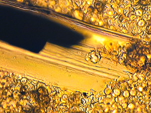
Figure (5). Part of a minute fragment of Mesozoic conifer wood embedded in a layer of coccolithic limestone, from the Upper Jurassic, Upper Kimmeridge Clay, Freshwater Steps Stone Band. Map Reference : SY.943.772. Freshwater Steps, West of Hounstout Cliff, Dorset coast. Oil-immersion objective N.A. = 1.25. Total field area of the image = circa. 110 microns X 80 microns.
Figure (5) is an example of both replica and removed thin layer type of images. The Mesozoic conifer wood fragment is mainly a replica, but some area of fusain (carbon) is shown to have been removed as a thin layer. Although Figure (5) was derived from a high resolution cellulose lacquer peel, some of the background coccoliths are out of focus with the oil-immersion objective due to very low depth of the focus. The width of the wood fragment is estimated as only about 51 microns. This is an example of the influx of ancient forest swamp debris into the Kimmeridgian sea at a time when the North American Continent was very close to Europe, and extensive low lying Mesozoic conifer forests grew in both land areas. Many other wood fragments have been identified at various horizons in the Kimmeridge Clay, on numerous peels, together with pyrites grains and kerogen, mixed with marine microfossils and nannofossils. Many of the wood fragments show microscopic "bordered pit" structures, characteristic of conifer wood. Figure (5) shows brightfield illumination with plane polarised light.
Figure (6). Heterohelix, a biserial foraminifera from Cretaceous, Upper Chalk, Actinocamax Quadratus Zone. Map Reference : SY.851.802. West of Arish Mell on the cliff, Dorset coast. Oil-immersion objective N. A. 1.25. Total field area of the image = ca. 100 micron X 60 microns.
The small features on Figure (6) are out of focus damaged chalk coccoliths due to the small depth of focus available. The diameter of the single apex chamber is circa. 18 microns on this image. Foraminifera are unicellular organisms belonging to the rhizopod Protozoa. Their protoplasm, during life, is emitted in the form of retractile "pseudopodia", through very fine pores in the chamber walls, which can be seen in this highly magnified image. They are used in catching prey, in locomotion and in the building of successive chambers in the skeleton, or test. A large variety of Foraminiferans occur not only in the Upper Chalk, but throughout geological times, from the Devonian (some 400 million years BP) to present times. "Forams" are very important microfossils, since they provide valuable information on the dating of strata and the reconstruction of sedimentary environments. It is, of course, quite remarkable how these minute simple unicellular animals have evolved over hundreds of million of years, to produce an enormously varied series of skeletons, or tests.
Figure (6) is an example of both replica and removed thin layer type of images. It was obtained with brightfield illumination with plane polarised light.
Notes and References
Micropalaeontology is concerned with microfossils and
nannofossils. These objects must be studied with the light
microscope or the electron microscope. Nannofossils (or
nanofossils) are microfossils with a diameter less than 50
microns (mm). (1 micron = 1/1000 mm.)
Mesozoic: Life during the Triassic, Jurassic and Cretaceous
periods. Estimated dates of these periods are as follows.
Triassic : circa 248 - 213 million years ago.
Jurassic : circa 213 - 144 million years ago.
Cretaceous : circa 144 - 65 million years ago.
The following papers and books may be of interest : -
Abineri K.W. 1986. The preparation of cellulose
lacquer rock peels. Microscopy (The Journal of the Quekett
Microscopical Club). 35, 451-59.
Abineri K.W. 1988. Notes on methods of staining lacquer rock
peels. Microscopy Bulletin. (The Quekett Microscopical Club). 11.
July 1988, 3-5.
Abineri K.W. 1989. Photomicrographs of cellulose peels from the
Mesozoic rocks of Dorset. Proc. Geol. Ass. 100(2). 161-74.
Adams A.E., MacKenzie W.S. and Guilford C.
1984. Atlas of Sedimentary Rocks under the Microscope. Longman.
Bignot G. 1985. Elements of Micropalaeontology. Graham &
Trotman Ltd.
Brasier M.D. 1980. Microfossils. George Allen & Unwin.
House. M.R. 1993. Geology of the Dorset Coast. The Geologists'
Association. guide No. 22.
Editor's note: The Editor thanks Keith Abineri for sharing his knowledge and interests on this fascinating subject on Micscape. Note that some of the quality of the author's original 35mm slides is lost in the scanned and compressed web images. Comments to the author are welcomed, who can be contacted at the above address or comments can be passed on via the Micscape Editor, see contact details on magazine index.
Article prepared for the Web by Dave Walker.
Published in the February 1999 edition of Micscape on-line and archived at http://www.microscopy-uk.net/mag/artmar99/kamast2.html
Please report any Web problems
or offer general comments to the Micscape Editor,
via the contact on current Micscape Index.
Micscape is the on-line monthly
magazine of the Microscopy UK web
site at Microscopy-UK