Micscape Article for Amateurs:
Deep-field Amateur Microscopy
by Maurice Smith. April 1996
Updated March 2001 with software
links that do the job!
Image Enhancement
A lot of software abounds these days to help the amateur and professional
scoper get the best out of computer images 'sourced' from their microscopes.
There is a particular aspect though that - to my knowledge - has not been
considered by software writers, which could prove a massive advantage to
the amateur microscopist and educationalists. This aspect is concerned
with deep-field microscopy.
Deep-field?
Yup! When I first heard this term, I found it confusing too. Deep-field...
sounds more like a rural or farming expression rather than anything to
do with 'scoping'. What does it mean? Well, here's the important bit: it
relates to the property of lenses. If you have done a lot with your camera
in ordinary photography, you will probably understand that all lenses have
a 'built-in' distance at which things seen through them will be in focus.
In the optical microscope, these distances are very fine indeed. Often
you will only to be able to focus on a specific detail of a specimen on
a slide, it's top surface for example, and everything below this depth
will be out of focus and will appear blurred. To examine most specimens,
you need to view the object or subject at varying depths (different
focus levels) to interpret true structure and form.
This limitation of any lens/focusing system is known as its 'depth of
field'. It is possible to extend the working focus minimum and maximum
distance of a lens, but you eventually reach a point where you need extremely
bright illumination to maintain an image. Again, photographers will know
that you can achieve a crisper image over a better 'field of distance'
by stopping the aperture of the camera down to a smaller and smaller setting,
where only a tiny hole is left to let the light - carrying the image information
- through and onto the film. You need nice bright lighting or very sunny
days for this.
Photomicrography
With a stereo microscope, it is possible to obtain slightly better visual
interpretation of a specimen. One set of lenses can be focused on a specific
plane in the subject while the other lens can be set to view a slightly
deeper level. Two such images taken this way are below: F1 is focused on
the nearer surface of the diatom frustule and F2 on the furthest surface.
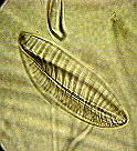 Diatom F1
 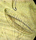 Diatom F2
The problem comes when you wish to record the specimen to camera, either
by normal photographic methods ('still' shots) or by using video recording.
The second method can be used to record different focus levels of the subject:
you simply keep altering the focus of the microscope when 'filming'. This
at least enables the viewer to get a clearer understanding of the structure
and processes of the specimen.
Diatom F2
The problem comes when you wish to record the specimen to camera, either
by normal photographic methods ('still' shots) or by using video recording.
The second method can be used to record different focus levels of the subject:
you simply keep altering the focus of the microscope when 'filming'. This
at least enables the viewer to get a clearer understanding of the structure
and processes of the specimen.
No such solution exists for 'still' photography. The only answer is
to take several photos at varying levels of focus (slices) and then present
the complete set of pictures to anyone who may wish to see them. It would
be great if the amateur had a way of recording a single image where all
crisp detail from both slices (images) were combined to form a focused
composite image!
Intrigue
A year or so ago I became quite intrigued by this limitation of an optical
scopes. As an amateur microscopist I was aware that these problems have
been beaten for the professionals since they can use Scanning Electron
Microscopes or Confocal Microscopes to obtain images where all the structures
in the specimen are clearly focused. But what about us amateurs? We can
not afford the extraordinary costs of this technology. I thought a solution
might be found by using software and the old home PC, and being a programmer
- decided to 'play' with the problem and see if I could build something
which might help.
Fuzzy Logic
Where to begin? I'm not that good at maths (surprise, surprise) so I had
to find a method which allowed me to understand things a different way
than by mathematical concepts. The simplest way would be to sit at the
microscope with a suitable subject on the stage, and then keep refocusing
the scope and try to observe what happens to the image itself as the focus
control is altered. I could also 'sample' some slices by rigging my video
camera up to the PC and 'grabbing' some images. After several long hours
I decided that a set of fuzzy rules could be considered as workable. These
could then be used as a basis for a set of software algorithms which could
work at a pixel level on the sampled images.
The Quest
The trick is get the computer to work out which pixel in any slice is 'most'
focused relative to its neighbours (pixels) in any given slice. These pixels
(or at least, their colour values) could then be used to rebuild a final
composite image where only the sharpest elements from the source images
would be present. Here are some of the thoughts and observations which
became rules for selecting the correct pixels:-
-
A pixel of high contrast to its neighbours has 'sharpness' priority.
-
A pixel remaining the same value or within a variable tolerance threshold
as its relative pixel in other slices will have sharpness priority.
-
Pixels within a 3x3 block which have a high rate of change in their comparative
colours and contrast will be given a higher 'sharpness' priority than a
relative 3x3 block in any other slice. ('Busy' areas take precedence over
common tone ones).
-
A smoothing algorithm will be applied to the final image, the composite
one, to minimise spurious errors. This should take out any isolated one-pixel
dots: a pixel that is in stark contrast to adjacent pixels which themselves,
have the same or similar colour colour value. The isolated dot will be
replaced with a randomly selected neighbour.
I built the software to work with 256 colour images to a maximum size of
640x480 pixels. I included several switches to allow selectable tolerance
levels to be applied during the image analysis: it became apparent in
a couple of quick experiments that this would be necessary to help the
software work on different subjects. For example, transparent or semi-opaque
subjects present a different set of problems from solid subjects.
Did it work?
It worked far better than I thought it would. To simplify the task, I set
a limitation that it will only work with 2 slices (images) for now. I'll
show you an example in a moment but don't get too excited because the software
is not for sale. It was just an experimental piece of work which, without
more development, is not very user-friendly; the main difficulty being
what switch parameters (numbers) should be included in the command line
to get the best result on a particular image. With all my other commitments
right now, I doubt if I will get much time this year to develop the program
but you never know.
Another solution
By sheer luck, another possibility for solving the depth-of-field problems
has come to light. I purchased a very low priced bundle of software called
Double Vision (around 18.00 pounds). The software turns computer
images into 3-D images, you know the sort you look at with those cardboard
(red/blue) glasses. Normally you would use the software to combine a left
image and a right image from a stereo pair and turn it into a composite
3-D anaglyph. In microscopy use, you can take a photo of a specimen (or
video still), move the specimen slide slightly to the left or right a bit,
and then take another shot. Once the two images are in the computer, the
Double Vision software will soon change it into a true 3D image which can
be viewed on the monitor or after printing out in colour, using the red/blue
glasses supplied with the product.
I was fooling around with the software - taking images through the scope
and then turning them into 3D, when I had a mini-brainwave: instead of
moving the slide to take the second shot, why not just refocus the scope
and capture a deeper slice of the specimen. The software can then use the
two separately 'focused' images to construct a deep-field image. I tried
it out and hey presto it works! More than this, you can move
the slide, refocus, and then take the second shot to get a deep-field 'true'
3D image!
I'm not here to promote the software but if, like me, you are an amateur
scoper and a computer user, you should really rush out and buy this cheap
bundle. I reckon its great! See our 3-D
section to find out more.
And remember, if you go out and get the software, when you get a result,
send one or two of your 3D Microscopy or Nature Images into here and we
will publish them on our site for you. This way other amateurs and students
can enjoy them too.
Examples
I promised you an example, so here it is. The row of images below are of
two Diatom frustules (cases) which I shot under the old scope at about
400x mag. (ish). Working from left to right: the first image is taken at
one level of focus and the next image along at a another level - deeper
into the slide. Note how details in one image are missing from the
other! The third image is the result of my software attempting
to combine them into a true all-detail-focused image. The forth and final
image to the right (slightly shorter) is what Double Vision software
made of them when combining them into a type of 3-D image. You will
need red/blue 3-D glasses to see this properly! I have reduced
the images by about 50% from their original size and converted them into
256 colour from 24 bit (64000 colours), resulting in a loss of detail.
Watch out in our 3D section and our Image library for them, because I might
load the larger versions of these images into these two sections.
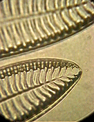
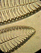
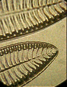
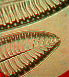
   Focus level 1      Focus level 2      Result with      Result with

                                         MY software     Double Vision

                                                        (3D glasses reqd.)
... and I think Double Vision produces a better result although you do
need to wear those funny glasses!
Footnote: If any budding programmer out there wants to consider working
on this problem with a view to building some user-friendly software for
the amateur scoper, I will be happy to part with any experience or know-how
gained 'playing' with the ideas to-date. Please contact me - The Editor
update!
March 2001: And a very good
programmer did do a wonderful job!
Having
interests at both extremes of the visual spectrum, micro and telescopic,
my interest became intense when I realized that there is a program which
will do the things required as outlined by your article, and more; plus
the best part - it is absolutely, positively - free! The name of the program
is AstroStack. It can be found at several FTP - Astronomy Sites. I have
forgotten exactly which site I downloaded it from; however I suspect a
web-search using the title "AstroStack" would turn up several in short
order.
This marvelous piece of
software is used to compile an image from several images to increase depth
of field, correct contrasts and stabilize the poorer images. I've played
with it a little using photos from telescopes. The results are indeed remarkable!
I'll try it with some micro-photos soon. Thanks to you - my memory clicked
so as to cause me to remember the program. The program is fairly small
and not to difficult to use. If I can find out which site I downloaded
it from, I'll send you a link.
That way you can see for
yourself how cool the program is not too mention the other manipulations
possible. Just think - one could take a subject - shoot 5 or 10 shots depth-wise,
move the slide ever-so-slightly to the left or right- shoot 5 or 10 more
depth-wise pics then combine them all for a very detailed true 3D composite
image; much like you intimated in your article. A image on "steroid", so
to speak.
Thanks again for all
the wonderful articles.
Regards,
Rick Comer
And
Rick kindly took the time to locate the link to the software...
I
believe in the motto I've seen posted all over the internet. "Share what
you know... Learn what you don't," so by all means feel free to share the
information. I did do a search and found the site for AstroStack it is:
http://utopia.ision.nl/users/rjstek/
Copyright www.microscopy-uk.net and their contributors. Originally published
in ®Micscape Magazine,
the official web magazine of Microscopy
UK - 'Home' of Amateur Microscopy on the Web!
WIDTH=1
© Onview.net Ltd, Microscopy-UK, and all contributors 1995 onwards. All rights
reserved. Main site is at www.microscopy-uk.org.uk with full mirror at www.microscopy-uk.net.