|
|
|
|
William Ells
Coniferae, Walnut Tree Lane, Loose, Maidstone, Kent. ME15 9RG.
SUMMARY
Brief descriptions & some new figures of rare &
interesting desmids from North West Sutherland, Scotland,
including a new record for the British Isles.
INTRODUCTION
The author has for several years been interested in the ecology
& distribution of desmids, listing species from various
habitats in Britain. Sutherland with its wonderful 'flow country'
is known to be one of the best areas for desmids, not only in
Britain but in the world. The paper which follows provides some
comments on just four of the many rare and interesting desmids to
be found there.
OBSERVATIONS
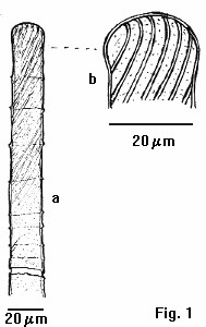
Penium spirostriolatum var. amplificatum Schmidt
(Fig. 1a & b). In a recent paper on new or interesting
British desmids, Williamson (1991) has reported the occurrence of
this desmid from the island of Yell, Shetland. Some authorities
such as Prescott et al. (1975a) and Ruzicka (1977a)
express doubts about the validity of the var. amplificatum,
the latter stating that for it to be accepted, its distinctive
capitate apices must be a constant feature in the entire
population. Williamson found such a population in Shetland where
none of the nominal variety was present. It is therefore worth
reporting that a similar but smaller population has been recorded
from North-West Sutherland. The specimens examined in 1989 were
of additional interest in that the comparatively broad diagonally
disposed striae go into and apparently curve right over the
apices, (Fig. 1b), a condition which does not seem to have been
previously reported for the British species. The girdle bands
were seen only as faint lines on the surface of the cells, but
clearly seen at the margins as protrusions, rather than as
shallow indentations as illustrated by previous authors. The
length of specimen illustrated (semi-cell Fig. 1a) was 296.0 um
breadth at the median suture 20.0 um. (Note: 'um' = micrometre).
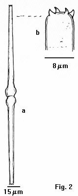
Pleurotaenium tridentulum (Wolle) West var. tridentulum (Fig. 2a & b). This taxon reported by W. & G.S. West (1904, p. 209) to be "one of rarest British species of the genus," has been found in one of the Sutherland samples. The specific epithet tridentulum, is something of a misnomer since there are always four apical teeth on each of the cell apices rather than three. It should be pointed out, that normally only three are seen in face view. As the Wests state, the four apical teeth are not all in focus at the same time, and it must be for this reason that Wolle (1883) gave the species its misleading name. It is interesting to note that Prescott et al. (1975 b) list this species as Pleurotaenium sceptrum and according to them 'Roy described this as Docidium sceptrum in 1883, Wolle described it as D. tridentulum in 1884, not 1882 as West & Krieger state. Therefore sceptrum is the earliest name for this species with four teeth'. This has not been universally accepted.
The specimen illustrated had a length of 273.0 um breadth at the basal inflation 14.5 um and at the apices 8.0 um. Brook (pers. comm.) and Williamson (pers. comm.) have also found this species in samples from the West of Scotland; the latter also from the West of Ireland. Although not abundant in any sample it would seem to be less rare than the Wests indicate.Haplotaenium rectum var. rectissimum (Gronbl.). Genus novum = Pleurotaenium rectum var. rectissimum (W & G S West) Gronb. was abundant in the sample.
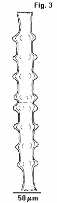
Pleurotaenium nodosum var. borgei (Gronbl.) Krieg. P. nodosum is the most striking species of the genus and although said by W.&.G.S. West (1904 p.215) to be rare, it would probably be more correct to say that it has a restricted distribution, for though never abundant, it has been found on numerous occasions from various localities in Sutherland and Invernesshire (Brook pers.comm.). However none of the type variety have been found by the author, var. borgei differs from the type by the occurrence of distinctive straight sections of the cell wall between each ring of nodules around the cell (Fig. 3). The variety is newly recorded for the British Isles, though Ruzicka (1977b) expresses doubts about the validity of the var. borgei, as noted above none of the type variety were found in the samples, more specimens need to be examined to explore the range of forms which exist within the species. Specimen illustrated: length 375.0 um; breadth at nodules 48.6 um.
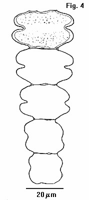
Desmidium pseudostreptonema West & West (Fig. 4) Croasdale et al. (1983) state this species is of very rare occurrence. First described from Ceylon (W.&.G.S. West 1906), it has previously been reported once in the British Isles from the plankton of a small lake in Galway, Western Ireland (W.&.G.S. West 1906) Brook & Williamson (1991) failed to include this record in their Check-list of Desmids of the British Isles. Mr Alan Joyce reported it as present in a sample collected in 1991, the author only found it in a second sample collected in 1992, this Desmidium is extremely rare in the samples, mostly in short filaments of 5-6 cells, only one long filament of 23 cells being found. In cells without contents pores were observed to be evenly distributed over the cell wall except for a narrow band on either side of the sinus. Also in empty cells the apical attachment processes were clearly visible. Dimensions of the largest cell in the filament illustrated were: length 18.5 um; breadth 33.0 um; breadth of isthmus 23.0 um & depth of sinus 5.0 um. Measurements of the largest cell in face view: breadth 33 um; length 18.5 um; isthmus 23.0 um & depth of sinus 5 um.
ACKNOWLEDGEMENTS
The author thanks Mr Alan Joyce for the samples from acidic pools
and lochan of North-West Sutherland, and Dr. J.W.G. Lund CBE,
DSc., FRS. Hon. Curator of the Fritsch Collection of Algal
Illustrations for providing study facilities at the Freshwater
Biological Association; also Professor A.J. Brook DSc. FRSE.
& Mr. D.B. Williamson for their helpful advice in the
preparation of this paper.
This paper first appeared in the QMC Journal. The note on Pleurotaenium sceptrum was edited out of the Journal without explanation, it is reinstated for this 'Web' version.
REFERENCES
Brook A.J. & Williamson D.B (1991) . A Check-list of
Desmids of The British Isles. Freshwater Biological Association.
Occasional Publication No.28, page 40.
Croasdale H.T. Carlos E. Bicudo De M. & Prescott G.W. (1983). A Synopsis of North American Desmids. Part 2, Section 5, 47 & 466, page 117. University of Nebraska Press.
Prescott G.W. Croasdale H.T. & Vinyard W.C. (1975). A Synopsis of North American Desmids. Part 2, Section 1. (a) pp. 13 & 155, (b) pp. 130 & 221. University of Nebraska Press.
Ruzicka J. (1977). Die Desmidiaceen Mitteleuropas. Band 1, Lief 2, page 292. E.Schweizerbart'sche, Stuttgart.
West W.&.G.S. (1992). A contribution to the freshwater algae of Ceylon. Trans. Linn. Soc. London, Bot. II, 6, pp. 123-215.
West W & G.S. (1904) . A Monograph of the British Desmidiaceae Vol.1, page 244 & CLXV. Ray Society, London.
West, W.&.G.S. (1906). A comparative study of the plankton of some Irish lakes. Trans. Roy. Irish Acad. 33, pp. 77-116.
Williamson D.B. (1991). On New or Interesting Desmids from the British Isles. The Botanical Journal of Scotland, 46 (1), pp. 97-106.
Captions to figures
Fig. 1a. Semi-cell of Penium spirolatum var. amplificatum, Fig. 1b. detail of an apex.
Fig. 2a. Pleurotaenium tridentulum, Fig. 2b. detail of an apex.
Fig. 3. Pleurotaenium nodosum var. borgei.
Fig. 4. Desmidium pseudostreptonema.
Comments to Bill Ells welcomed.
Figures
|
|
|
|
Editor's note
The Micscape Editor thank William Ells for contributing this
article. Note that Bill Ells has written six other articles on
desmids, including an introduction to the
desmids which can be found in our articles library in the
Pond-life section.
Please report any Web problems
or offer general comments to the Micscape Editor,
via the contact on current Micscape Index.
Micscape is the on-line monthly
magazine of the Microscopy UK web
site at Microscopy-UK