
For some it is the thought of going to the dentist, for others it is spiders, injections, or the fear of madness or going blind. The microscope in its various disguises can help us examine these nightmares close-up. In this mini series, I will take you month by month, through some of our common nightmares. Did you think Microscopy was about quiet, studious, safe things? I hope not. 'Scoping' can take us nearer to beauty or closer to the savage truth of reality. For our first example, let us consider that good old torturer we've come to know and love - the dentist...
The Phantom
Day by day we hack, chop, and grind away at our food. When we are
young it is more a case of sweet things which invisibly
contribute to the damage of  our teeth. Gradual decay
inevitably leads us to the dentist to get things patched up. As
we grow older our teeth become more brittle. We may peek in the
bathroom mirror and pride ourselves on having clean straight
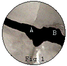
teeth, but things look quite different when seen at 50x to 100x
magnification, as (Fig.1) shows. You can see the hair-line
crack in the enamel of the tooth above the reference point (A)
and the chipped edge to the right - B. In fact, this image
was taken of my front teeth just after I had finished having a
complete overhaul of my teeth and gums.
Most of us can face an occasional bit of 'drilling' and 'filling' but there is a single phrase which is guaranteed to strike terror into the hearts of the bravest... a root filling.
The final solution
There comes a time when there is little left to fill. The advance
of decay attacks the living heart of the tooth and it is doomed.
Or is it? After all - it's only the nerve screaming at us that
the rot has gone too far. Why not just take it out? The nightmare
begins! We stare at the ceiling waiting... searching for that
invading drill to hit the soft spot and touch the nerve .
Fortunately, modern methods practised by a good dentist, should result in little discomfort and no real pain at all. I have often wondered what was actually involved in this 'seemingly' final solution. Last time I went down the dentist, I took the time to find out. I bet you would love to know too. No..? Too late. I am about to show you!
A Microscope in disguise
Perhaps it's cheating a bit to call the dentist's x-ray camera a
disguised microscope but through combining digital sampling
techniques with computers, it is possible to do much more with
the tiny x-ray pictures. The next time your dentist sticks a mini
x-ray plate in your mouth and asks you to hold still while he
puts on his lead-lined apron, you may well remember the following
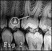
images.  To understand what a
root-filling is, we first need to study the nerve cavity (pulp)
and root of a comparatively healthy tooth. In (Fig.2) you
can see an x-ray image of several teeth. Most of them have been
capped with man-made material. Observe the pulp indicated by (A)
deep inside the healthy tooth. The dark patch along with the fine
dark lines running up into the root and jaw represents the
lifeline to the tooth. Here, the blood vessels and nerve endings
serve the whole tooth. It is when decay penetrates this region
beneath the outer enamel and dentine, that real pain begins. To
perform a root filling, the dentist must evacuate this area of
decayed tooth and the living nerves and vessels, then fill
the cavity with alloy, resin, or pure metal such as gold.
Getting to the point
 The dentist's drill should have
little difficulty removing the decay and entering the soft cavity
(pulp) but how are the fine canals containing the vessels and
nerves to be emptied of their contents without leaving debris
behind that could lead to problems? Easy - rake them out -
or said more accurately: put fine metal wire rasps down the nerve
canals and scrape them out. You can see in (Fig.3)
two such tiny tools inserted through the exposed pulp and up into
the two nerve canals. Although this image has been reduced in
size you can just see the zig-zagged edge of the tool indicated
by (A).
What's that you say? A bit too small to see properly. Okay,
here's another one then (Fig.4). The tooth here is a
different one. It has only  a single
root and nerve/blood vessel canal. The slender rasp, pushed deep
into the tooth, is indicated by (B). Above this you can
see another tooth which has been filled; evident by the bright
white chunk of alloy embedded in its crown (A). Below the
filling, a dark line runs centrally down to the root of the
tooth. This is a normal nerve/blood vessel canal still alive and
serving the tooth. As a final point to mention: once the pulp,
nerve (and the blood vessels) are destroyed or removed, the tooth
itself effectively dies but remains usable. In some cases it may
turn black; this discoloration is due to any remaining pulp
tissue (blood cells etc.) left in the tooth system. If this is
totally removed then discoloration should not ensue - (enamel
itself having no life of its own).
Note: my thanks to Jack Grennan B.D.Sc. for his kind help in preparing this article.
Comments to Maurice Smith.
Please report any Web problems
or offer general comments to the Micscape Editor,
via the contact on current Micscape Index.
Micscape is the on-line monthly
magazine of the Microscopy UK web
site at Microscopy-UK