Images of Pond-life from Video
by Mike Samworth
After reading about other people's exploits
with video and the microscope I couldn't resist having a play
myself. Though I don't have a video or camcorder of my own I am
fortunate in having access to a video camera without lens at
work. I have borrowed this on a few occasions and attached it to
my microscope. Each time I have learnt a little more about what
works best. The following still images are from those first
sessions.
Amoeba
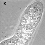
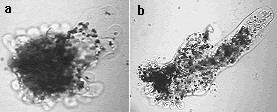 These two images are both of
good old Amoeba proteus. The first is from early in the
video sequence when the organism is still suffering the
consequences of being thrown onto a slide. the second shows the
creature putting out pseudopodia as it moves.
These two images are both of
good old Amoeba proteus. The first is from early in the
video sequence when the organism is still suffering the
consequences of being thrown onto a slide. the second shows the
creature putting out pseudopodia as it moves.
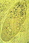
 Another shot of Amoeba, this time under higher
magnification and using phase contrast.The latter shows up the
highly granular nature of the cytoplasm. The video seems to cope
well with phase images. I have found however, that most times
using the video, all automatic circuitry should be turned off,
especially that for colour balance. Not that much colour balance
is needed for Amoeba! I have only had phase contrast
available on my microscope relatively recently. As in many things
in life, once you have something you wonder how you ever survived
without it. Phase contrast is no exception and is especially
useful for protozoa.
Another shot of Amoeba, this time under higher
magnification and using phase contrast.The latter shows up the
highly granular nature of the cytoplasm. The video seems to cope
well with phase images. I have found however, that most times
using the video, all automatic circuitry should be turned off,
especially that for colour balance. Not that much colour balance
is needed for Amoeba! I have only had phase contrast
available on my microscope relatively recently. As in many things
in life, once you have something you wonder how you ever survived
without it. Phase contrast is no exception and is especially
useful for protozoa.
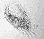
Paramecium
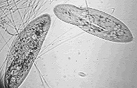 These two images are both of
the ciliate Paramecium. They both show the organism
rather flattened. This is due to the presence of silk fibres in
the water on the slide. I find that this slows down the movement
of ciliates quite well. Just tease out fibres from a piece of
silk with tweezers under a stereo mic' and put onto the slide.
The image on the right illustrates a common error with video,
that of having your lamp turned down. At first you do this
because of the high sensitivity of the camera, but of course it
adds a colour cast. It is better to turn the auto-circuitry off
and adjust manually. Turn your lamp up and use neutral density
filters if necessary.
These two images are both of
the ciliate Paramecium. They both show the organism
rather flattened. This is due to the presence of silk fibres in
the water on the slide. I find that this slows down the movement
of ciliates quite well. Just tease out fibres from a piece of
silk with tweezers under a stereo mic' and put onto the slide.
The image on the right illustrates a common error with video,
that of having your lamp turned down. At first you do this
because of the high sensitivity of the camera, but of course it
adds a colour cast. It is better to turn the auto-circuitry off
and adjust manually. Turn your lamp up and use neutral density
filters if necessary.
Euplotes
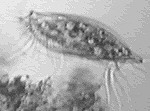
 These are two very different
views of a very different type of ciliate from the last one. They
are both views of Euplotes, a common and widespread
hypotrich ciliate. Hypotrichs have 'blocks' of cilia in groups or
rows called cirri. They often use these cirri to 'walk' over the
substrate. On left is a ventral view, the one normally seen when
the organism is free-swimming. On the left is the view from the
side of the ciliate.
These are two very different
views of a very different type of ciliate from the last one. They
are both views of Euplotes, a common and widespread
hypotrich ciliate. Hypotrichs have 'blocks' of cilia in groups or
rows called cirri. They often use these cirri to 'walk' over the
substrate. On left is a ventral view, the one normally seen when
the organism is free-swimming. On the left is the view from the
side of the ciliate.
It effectively illustrates how the ventral cirri that
characterise hypotrichs are used for movement over a substrate.
This is one of the advantages of video, you can take a sequence
and then select out the images that show particular behaviour.
Rotifers
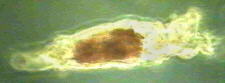 One problem with video is that
often the magnification is just too high. So for larger
multicellular organisms such as rotifers they more than fill the
screen.
One problem with video is that
often the magnification is just too high. So for larger
multicellular organisms such as rotifers they more than fill the
screen.
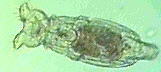 This can be avoided in two ways. Either you can use a
very low power objective, say 2.5x, or set up your camera at such
a position that you can dispense with the eyepiece altogether.
This is not optically a good idea but nevertheless has been
employed here for these two views of the rotifer Philodina.
Of course in professional video systems a supplementary lens
would be used instead of an eyepiece to compensate. The rotifer
on the left is shown under phase contrast.
This can be avoided in two ways. Either you can use a
very low power objective, say 2.5x, or set up your camera at such
a position that you can dispense with the eyepiece altogether.
This is not optically a good idea but nevertheless has been
employed here for these two views of the rotifer Philodina.
Of course in professional video systems a supplementary lens
would be used instead of an eyepiece to compensate. The rotifer
on the left is shown under phase contrast.
Please do not hesitate to contact
me if you have any comments or questions - Mike
Samworth.
© Microscopy UK or their
contributors.
Please report any Web problems
or offer general comments to the Micscape Editor,
via the contact on current Micscape Index.
Micscape is the on-line monthly
magazine of the Microscopy UK web
site at Microscopy-UK
WIDTH=1
© Onview.net Ltd, Microscopy-UK, and all contributors 1995 onwards. All rights
reserved. Main site is at www.microscopy-uk.org.uk with full mirror at www.microscopy-uk.net.