© R. Neumeyer 1996
This picture was taken with the Canon T90 and TL300 flash on Konica ASA 100 colour film. A Periplan 10x eyepiece combined with a Zeiss planapochromatic phase 40x (correction collar) and a Zeiss achromatic-aplanatic phase-contrast condenser supplied the optical magnification.
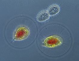
This genus of algae, Haematococcus, is often relatively abundant in ditches, bog-pools and rain-pools (the cell size ranges between 8- 30 microns). These particular specimens originated from snow melt in a bird bath. The cells frequently become brick-red in colour (as in the image) owing to the presence of haematochromin, a red pigment. The cell-body is connected to the cell wall by delicate strands of protoplasm, just visible in this picture (rather like being suspended in a ball of clear Jell-O!). The reddish chloroplast is bell-shaped. There are two flagella, fused together at the base, a portion of which is visible in the left specimen at about eight o'clock. The high objective magnification, and NA, resulted in a very shallow depth of field. Consequently the picture is really an "optical slice" through this ball, and hence the flagella are out-of-focus and cannot be seen.
I cannot be certain what genus the small ciliate in between the algae is, but it is a common member of spring pond life, possibly a Tetrahymena species. The thing to note is that the cell is in the final stages of asexual reproduction, referred to as "binary fission." Within 10 minutes of snapping this shot there were two daughter cells.
The Micscape and Microscopy UK Editors thank Ron Neumeyer for submitting this month's Image of the Month.
Comments to the author Ron Neumeyer welcomed.
Micscape is the on-line monthly magazine of the Microscopy
UK web
site at Microscopy-UK