Micscape Article: Stomata
Text and Images by Mike Morgan.
 Stomata are
microscopic pores found on the under-surface of leaves and on
stems. They occur in the epidermal tissue. Each stomatal pore is
bounded by two crescent shaped guard cells. The guard cell wall
closest to the pore is thicker than the remaining walls and
therefore less flexible.
Stomata are
microscopic pores found on the under-surface of leaves and on
stems. They occur in the epidermal tissue. Each stomatal pore is
bounded by two crescent shaped guard cells. The guard cell wall
closest to the pore is thicker than the remaining walls and
therefore less flexible.
Each guard cell contains chloroplasts, unlike the adjacent
epidermal cells. The glucose concentration of the the cells
changes with the photosynthetic activity and therefore it is the
guard cells that regulate the opening and closing of the stoma.
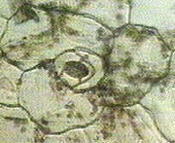
This is a low power view of a stoma.
 When there is an
increase in the osmotic potential, there is a decrease in the
water concentration and therefore water moves into the guard
cells by osmosis in response to the concentration gradient. As a
result the thin-cell wall bulges into the epidermal cell pulling
the thick wall with it and therefore opening the pore. The cells
are then turgid. Conversely when guard cells become less turgid
the pore closes.
When there is an
increase in the osmotic potential, there is a decrease in the
water concentration and therefore water moves into the guard
cells by osmosis in response to the concentration gradient. As a
result the thin-cell wall bulges into the epidermal cell pulling
the thick wall with it and therefore opening the pore. The cells
are then turgid. Conversely when guard cells become less turgid
the pore closes.
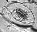
This is a high power view, clearly showing the chloroplasts in
the guard cells.
The alteration in the size of the stomata occur in response to
a variety of the external stimuli such as light, carbon dioxide
concentration and water.
The stomata are important for the exchange of gases by
diffusion between the outside air and intercellular spaces for
respiration and also for the evaporation of water by
transpiration.
Mike Morgan.
© Microscopy UK or their
contributors.
Please report any Web problems
or offer general comments to the Micscape Editor,
via the contact on current Micscape Index.
Micscape is the on-line monthly
magazine of the Microscopy UK web
site at Microscopy-UK
WIDTH=1
© Onview.net Ltd, Microscopy-UK, and all contributors 1995 onwards. All rights
reserved. Main site is at www.microscopy-uk.org.uk with full mirror at www.microscopy-uk.net.