| THE CHAETOGNATH, A STRANGE CREATURE |
by Jean-Marie Cavanihac, France |
 |
|
| THE CHAETOGNATH, A STRANGE CREATURE |
by Jean-Marie Cavanihac, France |
| |
|
| The first time I saw a chaetognath, it was in a sample I had taken from algae floating in the Mediterranean Sea and I was astonished by its formidable predatory behaviour. I didn't know its name then, and I had nicknamed it 'Voracious Fish'. It was caught earlier in Spring and at the time only a few specimens were present. Some months later, I have found many more of them but their sizes were the same; between two and four millimeters long. Since then, I have learned its common name: arrow worm, given because of the elongated shape. |
| For taking the pictures shown here I used a black and white CCD video camera with a resolution of 480 x 500 pixels (priced under £50), an image capture card (capture format - 384 x 288 pixels) and stored them on a PC. To give the images a more pleasant appearance, I colored them with Paint Shop Pro, to mimic their natural colors. (Note: for the same size of CCD: 1/4 ", B & W cameras have a better resolution than color cameras: video Web cameras are not so good ).The picture above is a montage made with four pictures, because the subject exceeds the objective field of view. |
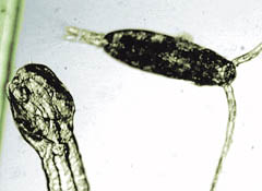
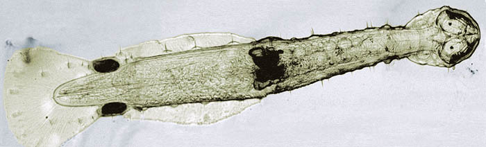
| Chaetognath bodies are very transparent like glass, and it's not easy to see them in a little jar. Brilliant lateral illumination can help. But another difficulty occurs: they are often glued to the walls of the container by little adhesive pads near the caudal fin (and perhaps at the middle of body). (See lefthand picture) |
|
|
|
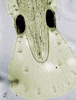
| The body is shown upright (like a snake on its tail) with the mouth facing up. It seems to be a hunting posture while waiting to grab prey. You will need to be careful if catching them with a pipette, because the chaetognath can glue itself to the inner tube especially if the pipette is made of glass: use plastic in preference. |
|
The adhesive pads are also
strange: during an observation on a slide, one of the pads and then the other became
empty, ejecting what appeared to be wriggling filaments. It's possible that chemical
residues on the glass may have triggered a defensive feedback. Note added March 2001: The author
thanks Peter Ahnelt who has kindly pointed out that 'the
"adhesive pads" you describe are actually sperm packages that are assembled to
be exchanged with partners of these hermaphroditic animals. So the "wriggling
filaments" are actually spermatozoa which may have been released due to capturing
stress. |
|
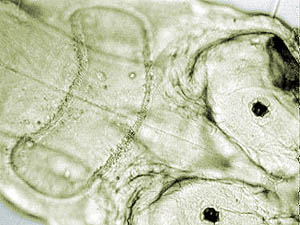
This picture shows a sort of ciliary crown behind the head. Indeed, some particles can be seen moving inside on a water flow. The crown may be a sense organ. |
|
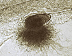
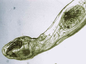
While I observed them in the jar
with a X8 hand-held magnifier, I saw a chaetognath quickly capture a copepod and I
immediately put the specimen on a slide. The picture shows the copepod bent into the
stomach of the chaetognath. This creature can quickly deploy what appear to be retractable
spines on the sides of its jaw which grasp prey and transfers them into the mouth. Click to see more detailed images. Unfortunately I couldn't take a picture of the copepod capture itself, but it's so fast that only a video sequence would be able to take it. |
The story continues on page 2 where you will find a not so happy ending for our arrow worm! |
Comments to the author Jean-Marie Cavanihac are welcomed.
All photographs © Jean-Marie Cavanihac 1999
Prepared for the web by Wim van Egmond
Published in the January 2000 edition of Micscape Magazine.
Please report any Web problems or offer
general comments to the Micscape
Editor,
via the contact on current Micscape Index.
Micscape is the on-line monthly magazine
of the Microscopy UK web
site at Microscopy-UK