What we can discover on a spider's
leg
by Wim van Egmond
the Netherlands
|
|
| During
the last summer I have been making many 3D images of
spiders and insects. Since I was curious how they would
look under the microscope (and I am rather clumsy with
chemicals as well as a bit lazy) I ordered a series of
prepared slides. Maurice Smith sent me some very
interesting slides he sells in his on-line shop. (For
more information, see the bottom of the page) One of the slides contained a couple
of legs of a garden spider (Areneus diadematus). I
knew that just one leg contains many interesting
features. In this article I like to show you some of the
things you can find on a single spider leg.
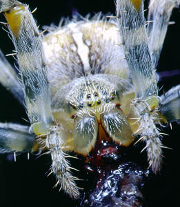
Garden spider
eating a fly
(Click
image to view large version)
|
| The garden
spider belongs to the orb-web spider family (Araneidae).
It catches its prey with a large web. It does not rely
that much on its eyesight (which is rather poor) but more
on organs that can sense the slightest trembling of the
web. It is not strange that the legs contain many sensors
that can detect such vibrations in the web. It is easy to
demonstrate this by holding a tuning fork against the
web. The spider will leave its retreat thinking its the
beating of the wings of an insect. |
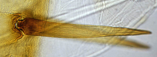
Tactile
hair, showing its texture The wide strand on the
background is a tendon (photographed with 25X obj.)
|
| But
what do these receptors look like? The spider's leg is
obviously very hairy. Almost all these hairs are sensory
hairs (in fact they are more like bristles), and act as
receptors that can detect touch and vibration. There are several types of sensory
hairs. You can identify them by the way they stand in
their socket. The large tactile hair above emerges
obliquely. There are also much smaller so called
trichobothria (they project vertically from their
socket). They are extremely sensitive to air currents or
low frequency air vibrations. Spiders can sense a flying
insect from several centimetres. Some spiders are able to
detect and catch a flying butterfly in mid-air.
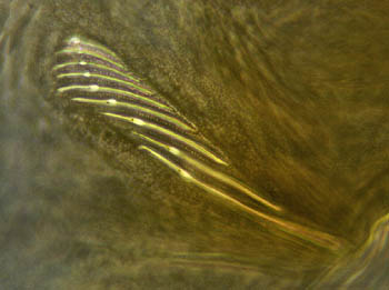
There are also
chemosensitive hairs, the tip of the leg contains a
series of these hairs the spider uses to taste.
(Image
taken with 40X phase-contrast obj.)
|
|
|
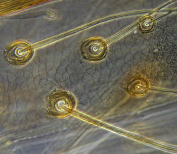
There is also a whole variety of
other receptors. So-called slit sensilla and lyriform
sensilla can be found all over the leg (most lyriform
organs are situated close to the leg joints.) With these
sensilla the spider senses it's own movements but they
also enable it to detect sound.
Each slit has
a thin membrane in which we can see a small dot. This is
a dendrite. This nerve cell detects deformation of the
slit.
(Image
taken with 40X phase contrast obj.)
|
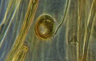
Rather difficult to find is the
tarsal organ. It is a small spherical pit situated near
the end of the leg (the last segment called the tarsus).
They are supposed to detect odor as well as humidity.
(Image
of tarsal organ taken with 40X obj.
Cropped to show more detail)
Within the legs I could
also see the long tendons for the movement of the limbs.
The legs are stretched because they are under pressure of
body fluids. The spider only has to pull the cord to bend
the leg.
|
|
| But I saved the most spectacular
part of the spiders leg for last. The foot of a spider is
a very interesting object to study under the microscope.
You can see how the garden-spider is able to cling to the
threads of its web. The foot possesses two claws, one
downward pointing hook and several serated hairs. The
hooked claw grabs the thread and the serated hairs hold
the thread in the hook. There is a difference between web
building spiders and spiders that stalk their prey like
wolf-spiders and jumping-spiders. These don't possess the hooked claw.
But they often have tufts of hairs on their foot that
enable firm adhesion to a surface by means of capillary
force.
|
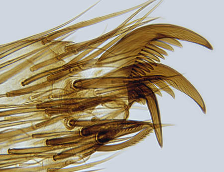
(Image
taken with 25X obj.)
|
| Detailed illustrations of how the
spider walks in it's web can be found in the Micscape
article Why a garden spider does not get
stuck in it's own web Since spiders are easy to find and
abundant it is not difficult to study these interesting
creatures. If you like to study a prepared slide like I
did, go to the OnView Shop to order spider legs or whole
mounts of spiders and insects.
|
Further reading:
Rainer F. Foelix, Biology
of Spiders (Harvard University Press)
Michael J. Roberts,
Spiders of Britain and Western Europe.
Spider websites:
Venomous
spiders
The slide was a Northern
Biological Supplies slide (N.B.S.) prepared by Eric
Marson
Comments to the
author Wim van Egmond are welcomed.
or visit Wim's HOME PAGE
Copyright
all material: Wim van Egmond 1999-2000
©
Microscopy UK or their contributors.
Published
in the January 2000 edition of Micscape Magazine.
Please
report any Web problems or offer general comments to the Micscape Editor,
via the contact on current Micscape Index.
Micscape is
the on-line monthly magazine of the Microscopy UK web
site at Microscopy-UK
WIDTH=1
© Onview.net Ltd, Microscopy-UK, and all contributors 1995 onwards. All rights
reserved. Main site is at www.microscopy-uk.org.uk with full mirror at www.microscopy-uk.net.