Camera 'auto portrait' (using a mirror!)
A CMOS video camera and white led lighting |
by Jean-Marie Cavanihac, France |
||||
| Microscopic
direct observation is always interesting, but we often
want to preserve images of our most beautiful specimens.
One solution is to make mounted slides, but this way is
not always satisfying because the object must be fixed
and this process makes colors difficult to maintain. This
method is particularly unusable when we observe living
and moving critters, because movement can add to our
knowledge of micro-organisms: For example protozoa
motion, bryozoans, even diatoms ... very quickly, the
need for a video camera appears. A black and white camera
is a good choice, but some subjects are more beautiful
with their own colors. This topic has been already been treated (see the Micscape Library) and many approaches has been made. Probably web cameras (using a USB port) are a good compromise between picture resolution and low cost. If the camera has a limited performance, the capture software sold with the device can often correct them or enhance details or interpolate lines. But not all microcomputers have this type of port. In my case, I have only a capture card (MIRO) used with external camera having analogue (TV - PAL compatible) video output. (A web camera has a digital output and is not TV compatible). Note that an analogue camera can be directly connected to your home TV or videotape recorder (using the composite video input socket) if you want to show your 'live' observations on a large screen for some people. (To interest children for example: If you have some live critters, no doubt you will have great success!) |
|
I have bought, from an electronic parts and kits supplier, a color video camera, built with CMOS technology (CAMCOL manufactured by VELLEMAN). Its first advantage; cost less than 80$, second; size and weight: 28 x 28 mm and only 30g ! Third, the device is normally powered by 12 volts DC, but in fact a 5 volts regulator is integrated on the printed board and a voltage around 7 volts is sufficient for a current of 20 mA: A single small 9 volt battery would be used! See picture at left with battery giving the scale. |
Camera 'auto portrait' (using a mirror!) |
| It is also possible to obtain a 'long focal
length' (12 mm!) video lens in place of a standard wide
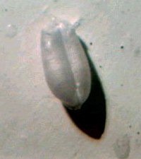
angle (3,6 mm) lens, which is very useful to take macro
views. The image right shows picture a X8 view of a wheat seed obtained with its 12 mm lens. This video camera uses cheap technology and the economies of scale used for making microprocessors. |
|
| In the
usual CCD devices, each image cell is used to store
electrical charge generated by photons and transfers it
to an adjacent cell. With CMOS, the internal circuitry is
more complex and each sensitive pixel has an associated
amplifier improving signal/noise ratio and reducing
distortion. An integrated co-processor chip performs gain
control, automatic exposure (electronic shutter or
electronic iris), and electronic built in white balance. On my model, the sensor has 682 x 582 (390 000) pixels, providing horizontal resolution of around 380 lines. Minimum light sensitivity is 4 lux. A capture format of 384 x 288 pixels is well matched to the line resolution of the camera. Larger formats (640 x 400) are not usable as lines appear on the image. (Web cameras can capture up to 640 x 400 pixel images, but probably, one line among two is interpolated by software). The automatic shutter is very convenient, as it adapts automatically to image brightness (reaction time is around 2 or 3 seconds), according to MEAN brightness of the subject. Constrast, brightness and color saturation can to be adjusted by capture software: a mid range setting works well. Yet at very low light levels, a little noise (snow) appears in images. |
|
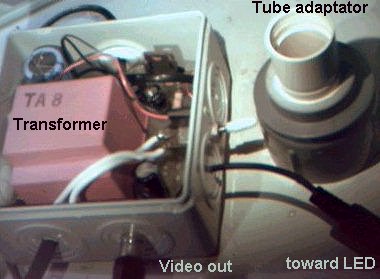
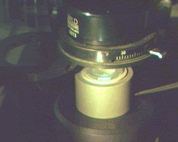
It's easy to fit such a small camera in place of an eyepiece; as shown on my Wild M20 using PVC tubes and splice assembly (in upper right corner of left picture). The camera is used without its lens. The normal microscope field of view is reduced, because the sensor size is only 1/4". On the left side of the adaptor you can see the home made 'camera control box' including 12 Volts DC power supply for the sensor and the illuminator, (but 8 Volts work as well) and video output socket. |
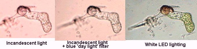
| But if
you use the camera as is, on your microscope, you will be
a bit disappointed because colors are not natural and we
have to consider an important feature: LIGHTING. The pictures below show a germinating fern spore: First image is taken with the incandescent bulb (6 volts - 15 watts) normally powered, included in the microscope illuminator; the 2nd same with blue filter in condenser and the 3rd with white led's as lighting (0,08 watts only!!). (Colors are not modified by software). Thus the sensor is very sensitive to infrared light of the tungsten light. |
 |
| If we
are using color cameras, it's to obtain true color
pictures: The light color temperature is thus very
important, because sensor sensitivity for wavelengths
present in light is NOT the same as the human eye's
sensitivity. In this case color rendering is good, only
when I am using a cold white light. (A fiber optic
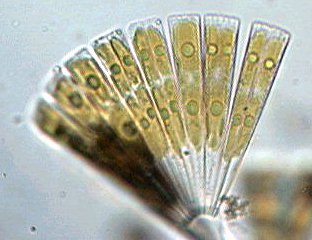
illuminator can also supply this type of cold lighting.) I have made an illuminator with a high efficiency white LED ** (see articles using the "LED" keyword in the Micscape Library Search) which works very well with the condenser. The LED current is only 20mA (according to manufacturer data sheets for THIS model) for a light flux reaching 2 Candela! Note that white LED's are made by combining blue, green and red LEDs, (picture below) into a diffusing medium, and LEDs primary colors can be sometimes be seen in pictures, giving a nice sort of RHEINBERG lighting. In the diatom (Licmophora) images below, chlorophyl appears with its true color. You can also obtain a slight pseudo 3D effect when the LED is off-center. White LED's don't produce light at IR wavelengths. |
 |
 |
| Inside the white LED in true colors. Note the two connexion wires . |
| Pictures below show the LED in a 32 mm diameter PVC tube, perfectly matched to the diameter of an M20 illuminator glass (right). |
 |
|
| White LED centered into 32 mm diameter PVC tube and assembly placed under M20 condenser. |
| **
LED is an acronym of Light Emiting Diode, an electronic
component used as indicator. LEDs exist which emit in red
(650 - 660 nm vawelength light), yellow (585 - 590 nm),
green (565 nm), and now in blue (430-470 nm) light. A
high efficiency green LED illuminator works as you would
use a green filter in the condenser filter holder, to
remove chromatic aberrations. A white LED is obtained by
combining in one chip of different colors LEDs, each
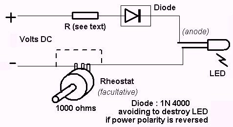
powerd by carefully computed current. For readers interested in experimenting with HIGH efficiency white LEDs, the minimum value resistor to place in serial with the LED is calculated below: V = voltage DC battery (DON'T use AC voltage), 0,02 = 20 mA = current into LED, R resistor value in ohms: R= (V -3,6)/0,02 . See schematic below. For example if V=9 volts, R = (9 - 3,6)/0,02 = 270 ohms. You can increase this resistor value (adding a 1000 ohms rheostat in SERIAL with the LED) to decrease light BUT color temperature will remain the same, which is very convenient. Don't forget that LEDs are a polarized component and the shortest wire is the cathode (must be connected to negative " -- " of the battery). The price of a white LED is around 5$' more than the usual red or green devices because it is a more sophisticated component. White LED illuminators can probably be used with 35 mm 'daylight' films to avoid an orange/yellow dominant color when taking pictures with incandescent bulb lighting.
|
Comments to the author Jean-Marie Cavanihac are welcomed. Microscopy UK Front Page All photographs © Jean-Marie Cavanihac 2001 Published in the January 2001 edition of Micscape Magazine. Please report any Web problems or offer general comments to the Micscape Editor, via the contact on current Micscape Index. Micscape is the
on-line monthly magazine of the Microscopy UK web |
|||