BIG DIATOMS

Photographing Coscinodiscus

by Wim van Egmond, The Netherlands


Coscinodiscus granii, one side of the cell is higher than the other. The top specimen has undergone cell division - 10X obj. (D.I.C.) 
The study of marine phytoplankton isn't bound to any season. Of course the bloom of diatoms during spring is really spectacular but marine diatoms can be found throughout the year. In this article I like to portray a genus of diatoms that can be found all year: Coscinodiscus.

This winter I collected plankton from the North Sea using a small plankton net. One jar of plankton was enough for days of observation through the microscope. 


The only disadvantage is that these marine water samples don't last that long. Only the most robust types last more than one or two days. And they will only last when the water is kept in a shallow dish stored at a cold temperature. This is relatively easy during the winter. During the plankton bloom you have to store your material in the fridge and work very fast because the diatoms are so plentiful; their concentration will ensure they deteriorate very fast. (Dilute the catch!)

When you observe plankton more often, you learn to know the fragile organisms and the robust ones. I always start working with the fragile ones and leave the tough ones for a couple of days later. Coscinodiscus always comes later in the week, just before the copepods, the 'die hards' of the plankton.

At the height of the phytoplankton bloom in April/May it is easy to see if the catch was successful since the sample will be yellow-brown because of the enormous quantities of diatoms. During winter it is much more difficult to see if it is a good catch.
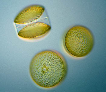
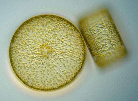
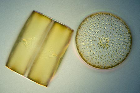
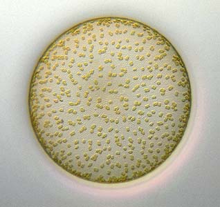
But there are some diatoms that are quite big. In this article I like to show one of those big diatoms, of the genus Coscinodiscus. Not all species are big but some can be more than half a millimeter in diameter. (Diatoms of the related genus Ethmodiscus can even be 2 millimeter!). So when you are shortsighted like I am, you can spot these diatoms with the naked eye when observing a plankton catch in a jar.

So it is quite easy to pick up a couple of these diatoms with a pipette and transfer them onto a slide for further examination under the microscope.


If you see a picture of the front side of a diatom you don't have a clue of the three-dimensional shape of the organism. Therefore it is nice to see the diatom from different views. 

TIP! For the images in this article I used a single hair from a big brush. (Just cut a couple of hairs from a normal house paint brush). With some (often a lot of) patience, the 5 centimeter long hairs can be worked under the coverslip. Under a dissecting (stereo) microscope you manipulate the organisms. In this case I tried to show a front view (valve side) as well as side view (girdle side) of the cell in one image. I toppled one of the cells. In this case the plane of focus shows the lines on the girdle.

 
Since my aim is to photograph organisms as lifelike as possible I look carefully to see if they are undamaged. When the coverslip is lowered on the glasslike shells of the diatoms they are easily destroyed. The aid of dots of Vaseline under the corners of the coverslip prevents this. Since you will get the best resolution in the area just under or against the coverslip you should try to lower the coverslip as much as possible, almost against the specimen. But you have to be very careful. The trouble with Coscinodisus is that it is big. The cell is often as deep as wide, especially when it is undergoing cell division.
- 16X obj.
In all the images in this article the granular chloroplasts are clearly visible. In this image you can see the nucleus and the cytoplasmic strands that hold the nucleus.


Click image if you like a bigger version in a new window!

If you'd like to have a closer look at the cells texture. Zoom in to see that big diatoms have amazingly fine details!

CLICK TO ZOOM-IN!

When the diatoms are under stress the chloroplasts will withdraw. This 'plasmolysis' is a form of protection. You often see pictures where this happens. Which is not a problem but I would prefer the natural state. But it is not easy to avoid this happening due to the stress of the coverslip and the strong light of the microscope lamp.

In this article I show some aspects of the diatoms. You can see on the zoom-in page that the diatoms are very fine structured. The way the cell is built can be seen in the two sided images, but there is much more to be discovered.

Just search for diatoms in Micscape's search engine and you will find several introductions to diatoms and many articles with images to show the wide variety of shapes.


All comments to the author Wim van Egmond are welcomed.

Visit Wims home page for links to his many web pages on microscopy

Microscopy UK Front Page
Micscape Magazine
Article Library


WIDTH=1

© Microscopy UK or their contributors.

Published in the January 2002 edition of Micscape Magazine.

Please report any Web problems or offer general comments to the Micscape Editor,
via the contact on current Micscape Index.

Micscape is the on-line monthly magazine of the Microscopy UK web
site at Microscopy-UK


© Onview.net Ltd, Microscopy-UK, and all contributors 1995 onwards. All rights reserved. Main site is at www.microscopy-uk.org.uk with full mirror at www.microscopy-uk.net.